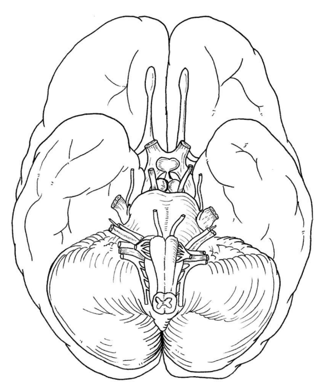
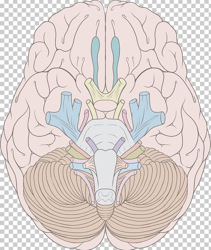
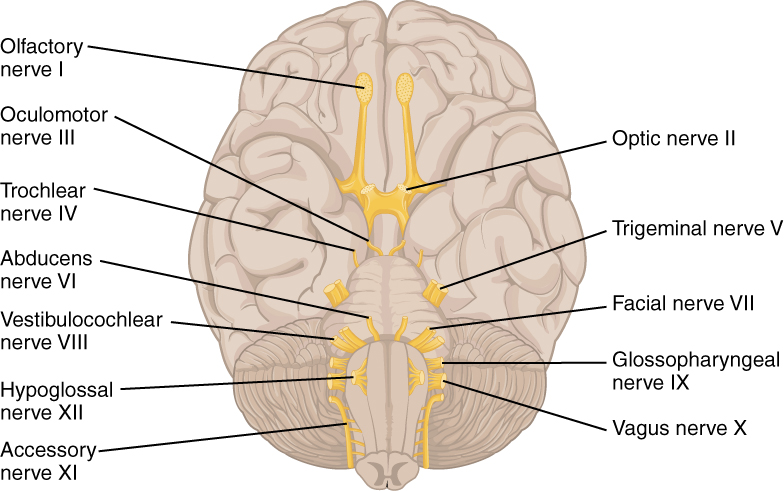
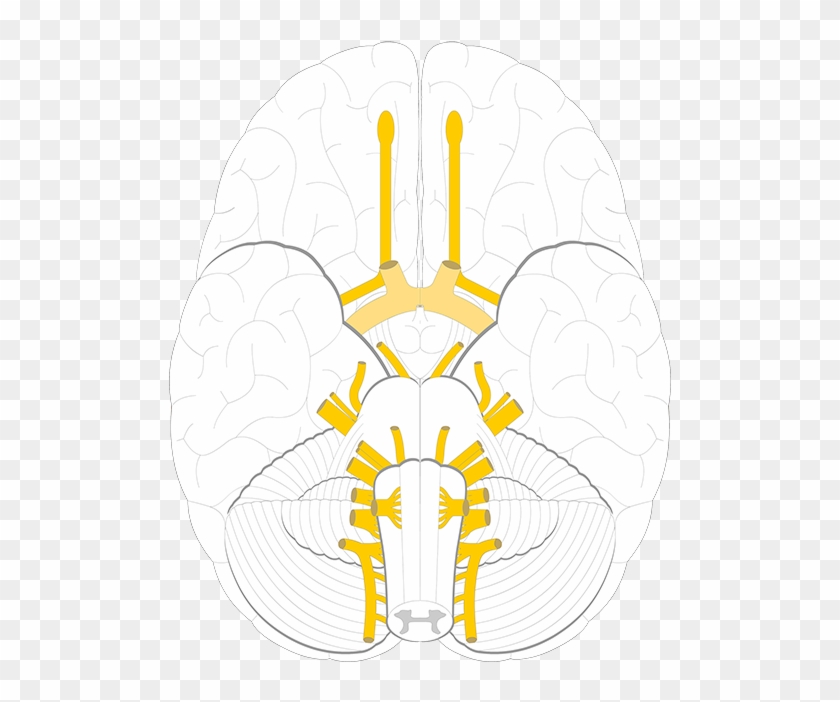
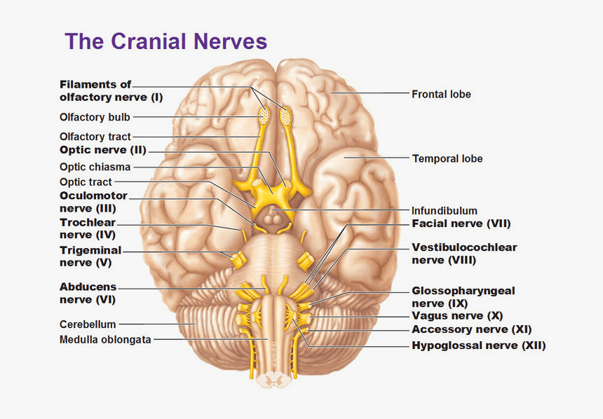
42 Cranial Nerves Diagram Unlabeled
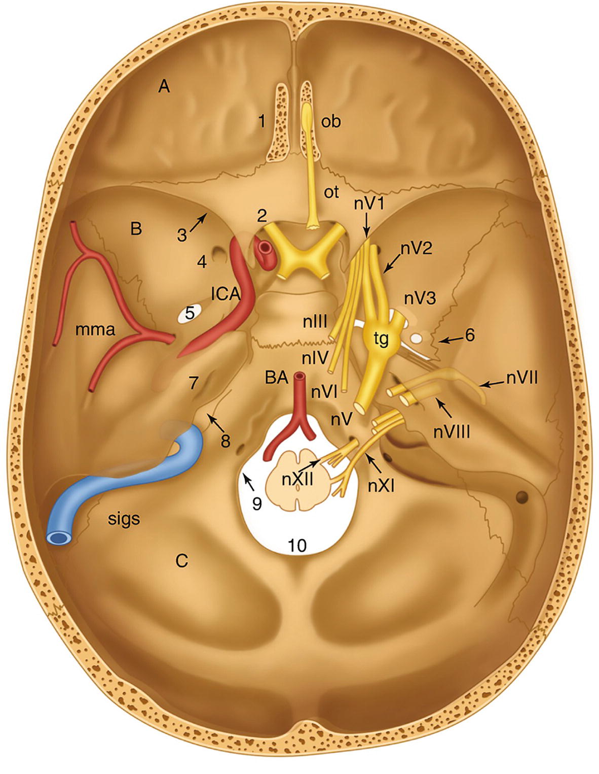
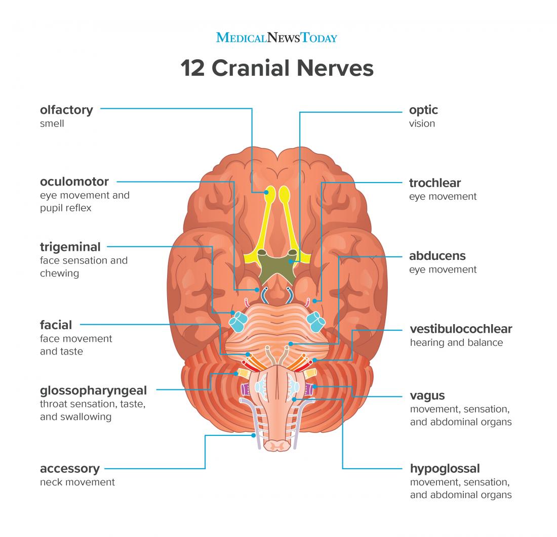
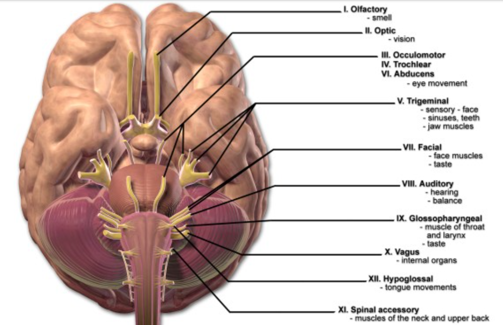
The 12 cranial nerves are pairs of nerves that start in different parts of your brain. Learn to explore each nerve in a 3-D diagram. This mnemonic helps to remember the cranial nerves in order of cranial nerve I to CN XII. Refer the following image for better understanding. It also contains the sensory, motor and mixed-function mnemonic for these nerves. Just remember both mnemonic and you are good to go! Source – Nursing Education Consultant, Inc 5.
With more related ideas as follows unlabeled pelvis bone anatomy vertebrae diagram unlabeled and cranial nerves labeling quiz. All cranial nerves originate from nuclei in the brain. Circle the correct underlined term. While some cranial nerves contain only sensory neurons most cranial nerves and all spinal nerves.
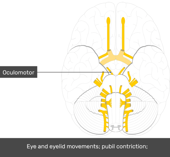
Cranial nerves diagram unlabeled
Cranial nerves III, IV, and VI a. carry sensory information from the retina to the occipital lobe. b. carry sensory information from the organ of Corti to the primary auditory cortex. c. innervate the extrinsic eye muscles. d. are the motor nerves involved in the corneal reflex. Brain And Cranial Nerves Labeled Diagrams 1/4 Read Online Brain And Cranial Nerves Labeled Diagrams Human Brain in 1969 Pieces-Wieslaw L. Nowinski 2013-12-20 The Human Brain in 1969 Pieces, version 2.0 is a highly sophisticated, visually stunning 3D neuroanatomy atlas. Professional academic writers. Our global writing staff includes experienced ENL & ESL academic writers in a variety of disciplines. This lets us find the most appropriate writer for any type of assignment.
Cranial nerves diagram unlabeled. Time 4 Learning Anatomy Of Brain The Brain Is Presented In Three Views Lateral Coronal And Midsaggital Brain Anatomy Brain Diagram Neuroscience Brain Human Normal Inferior View With Labels En 2 Cranial Nerves Wikipedia The Free Encyclopedia Craniale Zenuwen Het Menselijk Lichaam Menselijk Lichaam Brain Unlabeled Study Sheet Ear Anatomy Human Anatomy And Physiology Anatomy […] We provide solutions to students. Please Use Our Service If You’re: Wishing for a unique insight into a subject matter for your subsequent individual research; Oct 28, 2021 · Try to understand and memorize what you can from the labeled diagram, then, try to label the cranial nerves yourself with our cranial nerves labeling quiz exercise available to download below. This is a great way to start to get the cogs turning and warm up your memory before you take our other cranial nerve quizzes (but one thing at a time. oxygen to the spinal cord also use these spaces. You have 8 pairs of cervical nerves, 12 thoracic, 5 lumbar and 6 sacral. Near the waist, the nerves continue in a bundle called the cauda equina. This is commonly called the 'horses tail' as that's what it looks like. Spinal nerves transmit and receive messages to and from the brain.
Cranial Nerves and Brainstem Unlabeled. Nora Guerrero. 133 followers. Human Brain Anatomy. Gross Anatomy. Human Anatomy Drawing. Unlabeled Muscle Diagram Worksheets. Posterior Muscles Unlabeled. Diane. School. Human Skeleton Anatomy. Human Body Anatomy. Human Anatomy And Physiology. Anatomy Practice. Cranial Nerves Quiz for Anatomy & Physiology Class. This cranial nerves exam will test your knowledge on all the cranial nerves that you will have to know for an exam in Anatomy & Physiology. This cranial nerves quiz will ask you about the function and name of each nerve. 1. There are 14 pairs cranial nerves. *. True. False. Brain Anatomy Diagram Unlabeled angelo. November 4, 2021.. Free Human Body Worksheets For Class 3 2021 In 2021 Anatomy And Physiology Cranial Nerves Human Anatomy And Physiology. Occipital Lobe Damage Https Www Facebook Com Anoxicbraininjury Ref Hl Neuroscience Brain Occipital Lobe Somatosensory Cortex . Oct 07, 2020 · 48 Likes, 2 Comments - College of Medicine & Science (@mayocliniccollege) on Instagram: “🚨 Our Ph.D. Program within @mayoclinicgradschool is currently accepting applications! As a.
A foramen (pl. foramina) is an opening that allows the passage of structures from one region to another.. In the skull base, there are numerous foramina that transmit cranial nerves, blood vessels and other structures - these are collectively referred to as the cranial foramina. In this article, we shall look at some of the major cranial foramina, and the structures that pass through them. This atlas of human anatomy describes the spinal cord through 18 anatomical diagrams with 270 anatomical structures labeled. It was designed particularly for physiotherapists, osteopaths, rheumatologists, neurosurgeons, orthopedic surgeons and general practitioners, especially for the study and understanding of medullary diseases. Get your assignment help services from professionals. All our academic papers are written from scratch. All our clients are privileged to have all their academic papers written from scratch. Paul Rea, in Clinical Anatomy of the Cranial Nerves, 2014. The Anatomy—in More Detail. As a long slender structure, the trochlear nerve arises from the posterior (dorsal) aspect of the midbrain. It lies immediately caudal to the nucleus of the oculomotor nerve. The fibers of the trochlear nerve pass around the periaqueductal grey matter decussating at the level of the superior medullary velum.
Cranial skeleton (Neurocranium) Calarvia Frontal, Temporal, Parietal, Occipital Cranial base Facial skeleton (Viscerocranium). vestibulocochlear nerve (8 th) and facial nerve (7 th) Skull - 41 Facial canal: internal acoustic meatus; stylomastoid foramen. Skull - 42 Temporal bone External surface
In the mean time we talk related with Facial Bones Worksheet, we already collected particular related images to complete your references. unlabeled skeleton worksheet, cranial nerves labeling quiz and unlabeled vertebral column diagram are some main things we want to show you based on the gallery title.
This human anatomy module is about the cranial nerves. It consists of 15 vector anatomical drawings with 280 anatomical structures labeled. It is intended for the use of medical students working on human anatomy, student nurses, physiotherapists, electro-radiological technicians and residents – especially those working in neurology, neurosurgery, otolaryngology – and for any physician.
Cranial Nerves. Spinal Cord. Spinal Nerves. Textbook Reference: See Chapte. r 11 for histology of nerve tissue and spinal cord See Chapter 12 for brain and spinal cord anatomy See Chapter 13 for cranial nerves and spinal nerves What you need to be able to do on the exam after completing this lab exercise: Be a
Start studying Bio 121: Cranial Nerves Unlabeled. Learn vocabulary, terms, and more with flashcards, games, and other study tools.
Choose a cranial nerve to discuss in detail and describe its function including the origination in the brain, the path it follows through the skull, its innervation (what body part it serves), and whether its function is sensory, motor, or both. Cranial Nerves Presentation Assignment.
Anterior view of spinal cord showing meninges and spinal nerves. For this view, the dura and arachnoid membranes have been cut longitudinally and retracted (pulled aside); notice the blood vessels that run in the subarachnoid space, bound to the outer surface of the delicate pia mater. Ventral root of sixth cervical nerve Dorsal root of
Get 24⁄7 customer support help when you place a homework help service order with us. We will guide you on how to place your essay help, proofreading and editing your draft – fixing the grammar, spelling, or formatting of your paper easily and cheaply.
Diagram of human nervous system anatomy with brain, spinal cord , spinal nerves and plexuses, unlabeled. Pituitary gland, medical drawing. Illustration of the pituitary gland, anterior and posterior lobes, unlabeled version.
Professional academic writers. Our global writing staff includes experienced ENL & ESL academic writers in a variety of disciplines. This lets us find the most appropriate writer for any type of assignment.
Oct 28, 2021 · Take a look at the labeled diagram of the respiratory system above. As you can see, there are several structures to learn. Spend a few minutes reviewing the name and location of each one, then try testing your knowledge by filling in your own diagram of the respiratory system (unlabeled) using the PDF download below. Respiratory system unlabeled
Nervous System with Labels. Nervous System Unlabeled. Nervous System Diagram Blank
Cranial nerves III, IV, and VI a. carry sensory information from the retina to the occipital lobe. b. carry sensory information from the organ of Corti to the primary auditory cortex. c. innervate the extrinsic eye muscles. d. are the motor nerves involved in the corneal reflex.
Unlabeled diagram showing the muscles of the leg (download free PDF below!). look no further than our library of free quiz guides on tricky exam topics like the cranial nerves, bones of the skull and reproductive systems. Sources. Layout: Molly Smith.
The spinal cord is the central nervous system part that extends into the axial skeleton and provides the two-way traffic required to interact with our environment. During pregnancy, early development of the spinal cord is influenced by the maternal dietary requirement for folate for closure of the neural tube. Later development requires the contribution of neural crest associating with the.
Brain And Cranial Nerves Labeled Diagrams 1/4 Read Online Brain And Cranial Nerves Labeled Diagrams Human Brain in 1969 Pieces-Wieslaw L. Nowinski 2013-12-20 The Human Brain in 1969 Pieces, version 2.0 is a highly sophisticated, visually stunning 3D neuroanatomy atlas.
See 12 Best Images of Anatomy Practice Worksheets. Inspiring Anatomy Practice Worksheets worksheet images. Skull Bones Worksheet Anatomy Directional Terms Worksheet Skull Axial Skeleton Labeling Worksheet Unlabeled Vertebral Column Diagram Cranial Nerves Labeling Quiz
122 Exercise 9 9 T he axial skeleton (the green portion of Figure 8.1 on p. 108) can be divided into three parts: the skull, the ver-tebral column, and the thoracic cage. This division of the skeleton forms the longitudinal axis of the body and protects
The sensory cranial nerves are involved with the senses, search as sight, smell, hearing, and touch. Whereas the motor nerves are responsible for controlling the movements and functions of muscles and glands, cranial nerves supply sensory and motor information to areas of the head and neck. One nerve, the vagus nerve, extends beyond the neck to.
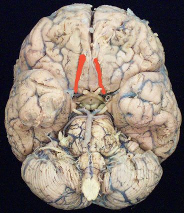
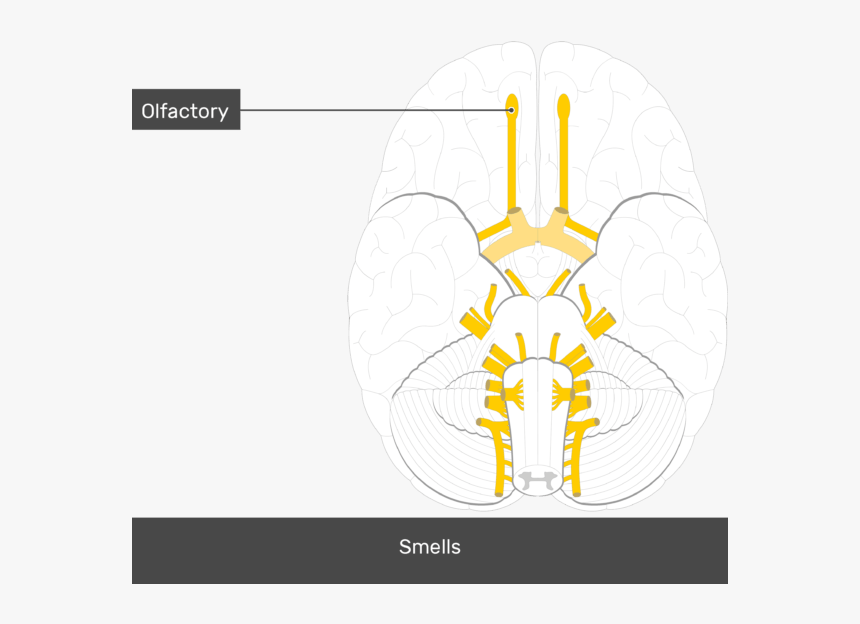
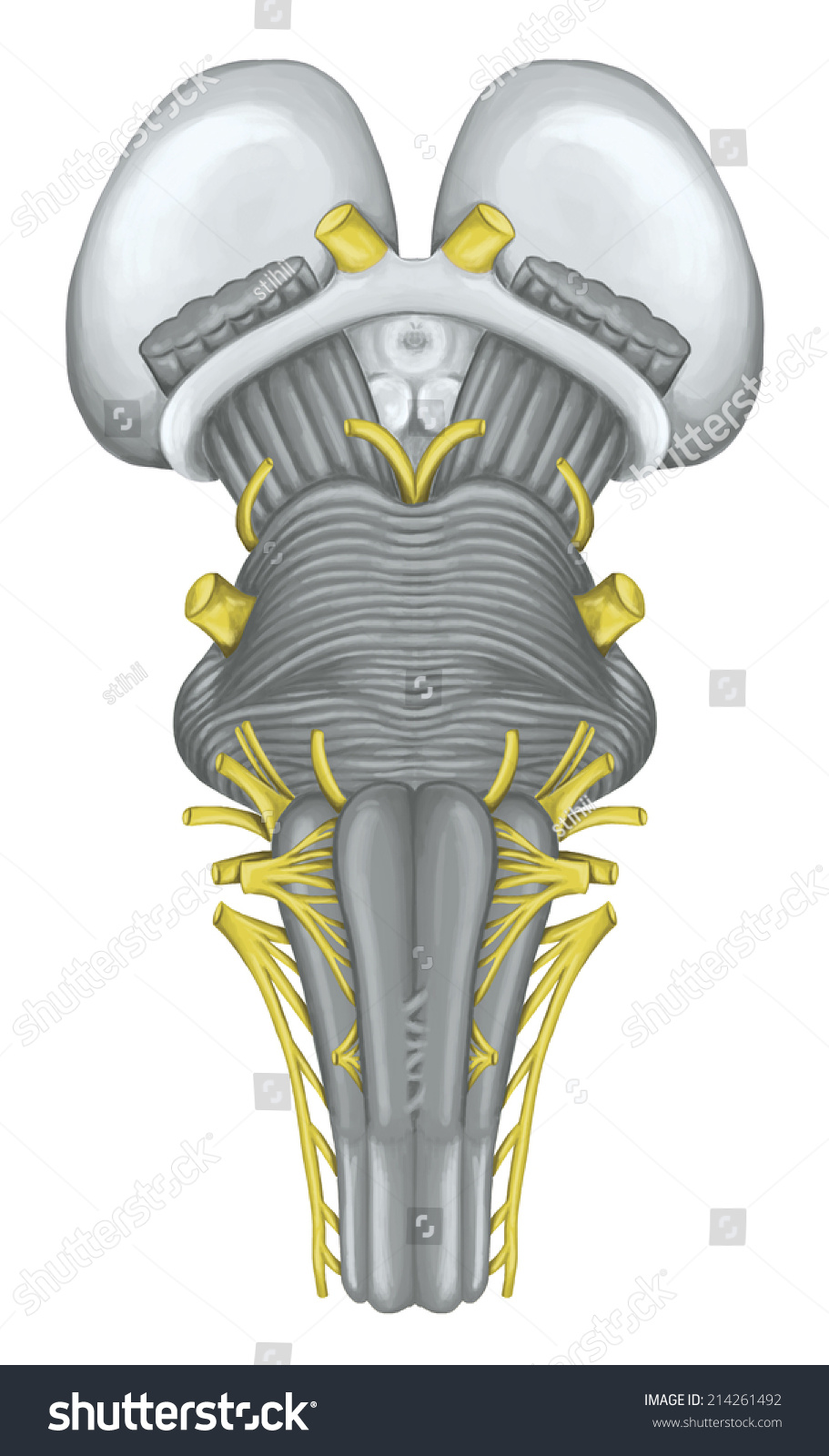
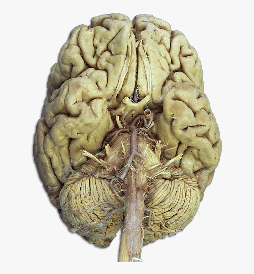
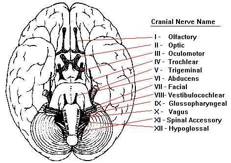
Cranial Nerves. Extending from the inferior side of the brain are 12 pairs of cranial nerves. Each cranial nerve pair is identified by a Roman numeral 1 to 12 based upon its location along the anterior-posterior axis of the brain. Each nerve also has a descriptive name (e.g. olfactory, optic, etc.) that identifies its function or location.
The Edinger–Westphal nucleus (accessory oculomotor nucleus) is the parasympathetic pre-ganglionic nucleus that innervates the iris sphincter muscle and the ciliary muscle.. Alternatively, the Edinger–Westphal nucleus is a term often used to refer to the adjacent population of non-preganglionic neurons that do not project to the ciliary ganglion, but rather project to the spinal cord.
21-0960C-3 Cranial Nerve Conditions 21-0960C-4 Diabetic Sensory-Motor Peripheral Neuropathy 21-0960C-5 Central Nervous System and Neuromuscular Diseases (except TBI, ALS, PD, MS, Headaches, TMJ, Epilepsy, Narcolepsy, Peripheral Nerves, Sleep Apnea, Cranial Nerves, Fibromyalgia, and CFS)
Olfactory nerve optic nerve oculomotor nerve trochlear nerve trigeminal nerve abducens nerve facial nerve vestibulocochlear nerve glossopharengeal nerve vagus nerve spinal accessory nerve and hypoglossal nerve. 12 cranial nerves of the brain worksheet unlabeled bones of the head and face and blank head and neck muscles diagram are three of main.
Learn the major cranial bone names and anatomy of the skull using this mnemonic and labeled diagram. Sutures connect cranial bones and facial bones of the skull. Develop a good way to remember the cranial bone markings, types, definition, and names including the frontal bone, occipital bone, parieta
2. The 2nd cranial nerve that carries nervous impulses from the retina of the eye to the brain.... 3. The largest nerve in the body serving the muscles of the leg...... 4. The 1st cranial nerve that carries impulses from the organ of smell in the nose to the brain. ..... 5. The 10th cranial nerve that supplies the pharynx, lungs, heart,
Get 24⁄7 customer support help when you place a homework help service order with us. We will guide you on how to place your essay help, proofreading and editing your draft – fixing the grammar, spelling, or formatting of your paper easily and cheaply.
Assessment of Cranial Nerves I-XII Below you will find descriptions of how to perform a neurological exam for cranial nerves. All tests are performed bilaterally: Cranial Nerve I (Olfactory Nerve): Sensory for Smell Always begin by asking patient if he/she has had any decrease in ability to smell.
its unlabeled, so that your practce better. carotid canal coronal suture ethmoid bone external occipital protuberance foramen lacerum foramen magnum foramen

:background_color(FFFFFF):format(jpeg)/images/library/11648/overview_cranial_nerves_diagram.jpg)




:max_bytes(150000):strip_icc()/trigeminal_nerve-cef33b116607464fbfe8213e4a4d2a4e.jpg)








0 Response to "42 Cranial Nerves Diagram Unlabeled"
Post a Comment