41 The Diagram Below Shows A Bacterial Replication Fork And Its Principal Proteins.
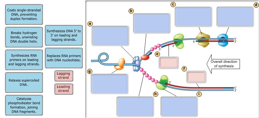
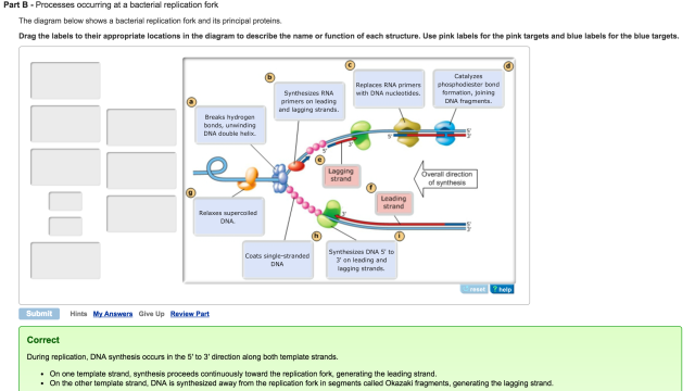
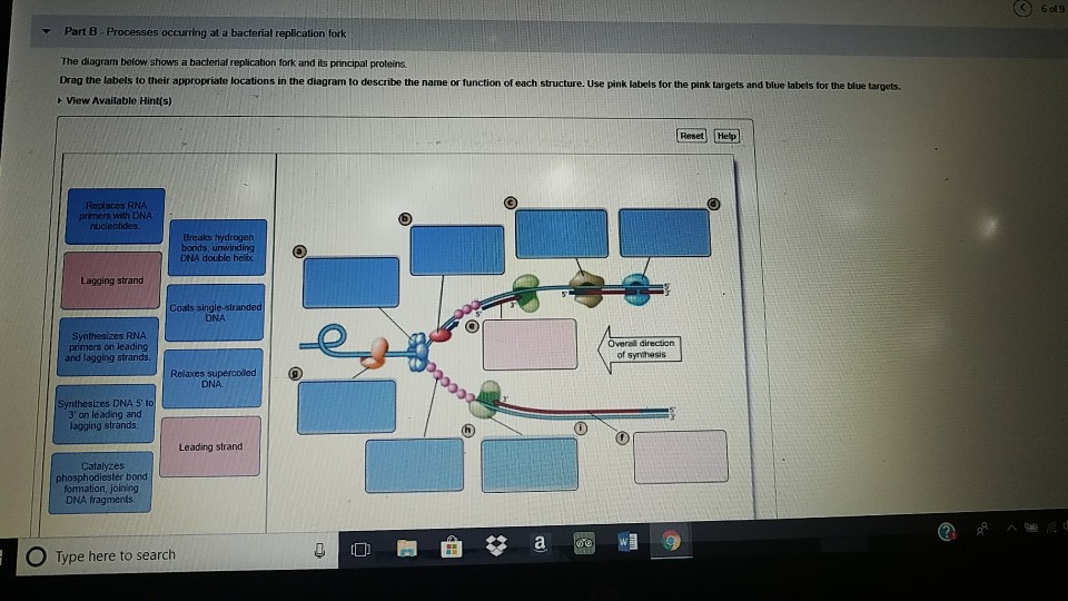
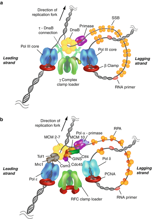
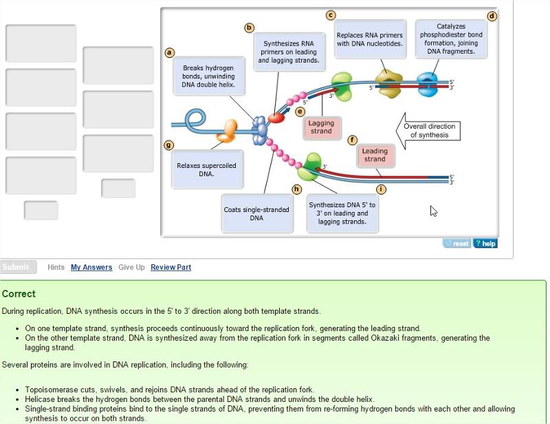
The diagram below shows a bacterial replication fork and its principal proteins. Drag the labels to their appropriate locations in the diagram to describe the name or function of each structure. Use pink labels for the pink targets and blue labels for the; Question: The diagram below shows a bacterial replication fork and its principal proteins. The diagram below shows a bacterial replication fork and its principal proteins. Drag the labels to their appropriate locations in the diagram to describe the name or function of each structure. Fragment a is the most recently synthesized okazaki fragment.
The diagram below shows a bacterial replication fork and its principal proteins. The diagram below shows a bacterial replication fork and its principal proteins. The newly synthesized dna strands are not shown but the polymerase on each parental strand is shown labeled 1 and 2. Drag the labels to their appropriate locations in the diagram to.
The diagram below shows a bacterial replication fork and its principal proteins.
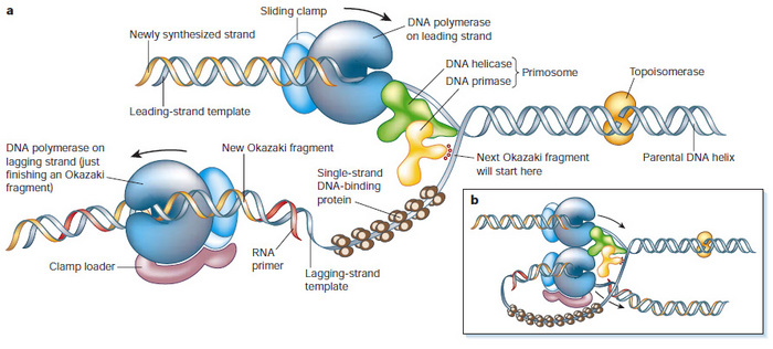
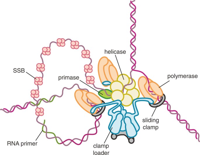
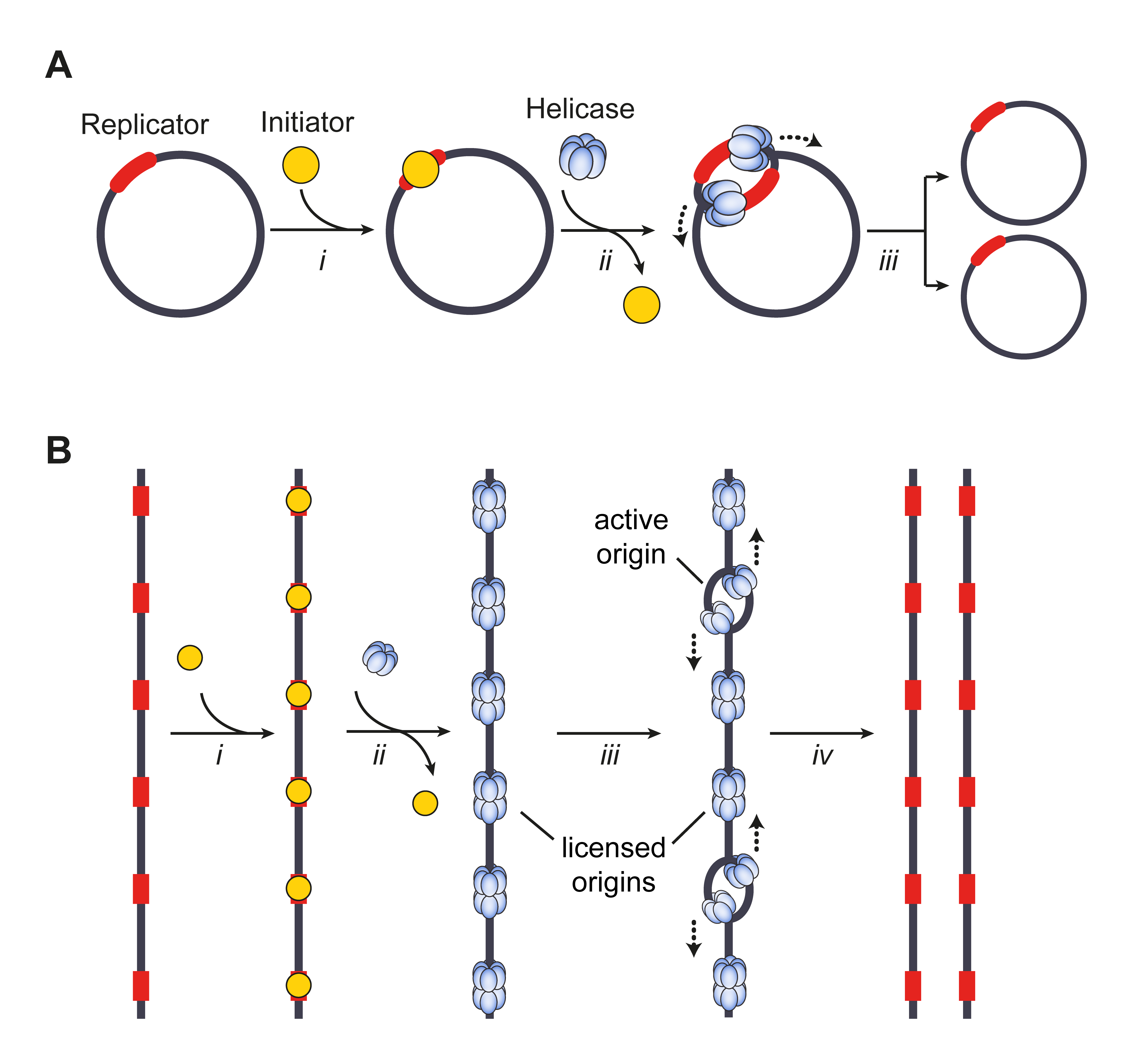
Gentex 341 With Homelink And Temperature Wiring Diagram; Fisher Plow Wiring Diagram Minute Mount 2; The Diagram Below Shows A Bacterial Replication Fork And Its Principal Proteins. We4m527 Wiring Diagram; 2jz Maf Sensor Wiring Diagram; Clipsal 2 Way Switch Wiring Diagram; Recent Comments. Peng X. on 175 watt metal halide ballast wiring diagram Kawasaki Bayou 220 Carb Diagram; Eager Beaver Trailer Wiring Diagram; Procraft 1780v Wiring Diagram; Wiring Diagram Of Transfer Case Range Position Sensor 06 For Fusion 2.3; The Diagram Below Shows A Bacterial Replication Fork And Its Principal Proteins. Eincar Wiring Diagram; 3415s Simplicity Wiring Diagram; Chevy 454 Firing Order Diagram Helicase can move along the double helix structure right at the front of the replication fork. There are specific regions for replication to get started, also known as replication origins. The replication process always happens in 5’ to 3’ direction, which means that a new nucleotide is added to the 3’-OH group of the raising DNA strand.
The diagram below shows a bacterial replication fork and its principal proteins.. Academia.edu is a platform for academics to share research papers. Helicase can move along the double helix structure right at the front of the replication fork. There are specific regions for replication to get started, also known as replication origins. The replication process always happens in 5’ to 3’ direction, which means that a new nucleotide is added to the 3’-OH group of the raising DNA strand. Transcribed text From Image: Part B Processes occurring at a bacterial replication fork The diagram below shows a bacterial replication fork and its principal proteins. Drag the labels to their appropriate locations in the diagram to describe the name or function of each structure. Use pink labels for the pink targets and blue labels for the blue targets. The diagram below shows a bacterial replication fork and its principal proteins. In arcmap you can use labeling or annota. Passive Margin Wikipedia For example the vajont dam was constructed at monte toc italy in the early 1960s. Label each target on the map below with the name of the appropriate geological feature.. Drag the labels to their.
Show transcribed image text the diagram below shows a bacterial replication fork and its principal proteins. The diagram below shows a double stranded dna molecule parental duplex. Nucleic acids are made up of chains of many repeating units called nucleotides see bottom left of figure 1 below. In the labels the original parental dna is blue and. The diagram below shows a bacterial replication fork and its principal proteins. Drag the labels to their appropriate locations in the diagram to describe the name or function of each structure. Use pink labels for the pink targets and blue labels for the blue targets. View Available Hint (s) Reset Help Synthesizes RNA primers on leading and. The diagram below shows a bacterial replication fork and its principal proteins. Drag the labels to their appropriate locations in the diagram to describe the name or function of each structure. (a) Gentex 341 With Homelink And Temperature Wiring Diagram; Fisher Plow Wiring Diagram Minute Mount 2; The Diagram Below Shows A Bacterial Replication Fork And Its Principal Proteins. We4m527 Wiring Diagram; 2jz Maf Sensor Wiring Diagram; Clipsal 2 Way Switch Wiring Diagram; Recent Comments. Peng X. on 175 watt metal halide ballast wiring diagram
The diagram below shows a bacterial replication fork and its principal proteins. The parental dna is shown in dark blue the newly synthesized dna is light blue and the rna primers associated with each strand are red. Use pink labels for the pink targets and blue labels for the blue targets. The diagram below shows a bacterial replication fork and its principal proteins. Ch 16 mastering biology. The Diagram Below Shows A Bacterial Replic Clutch Prep Drag the labels to their appropriate locations in the diagram to describe the name or function of each structure. The diagram below shows a double stranded dna molecule parental dna. The diagram below shows a replication bubble with synthesis of the leading and lagging strands on both sides of the bubble. Show transcribed image text the diagram below shows a bacterial replication fork and its principal proteins. In the labels the original parental dna is blue and the dna synthesized during replication is red. The diagram below shows a bacterial replication fork and its principal proteins. The diagram below shows a double stranded dna molecule parental dna. Please help dna replication is the mechanism by which dna is copied. Helicase is the enzyme which cause the dna molecule to unzip.
Drag the correct labels to the appropriate locations in the diagram to show the composition of the daughter duplexes after one and two cycles of dna replication. Mar 19 the diagram below shows a bacterial replication fork and its principal proteins. 16 which nitrogenous bases tend to pair with each other in a double stranded molecule of dna.
Kawasaki Bayou 220 Carb Diagram; Eager Beaver Trailer Wiring Diagram; Procraft 1780v Wiring Diagram; Wiring Diagram Of Transfer Case Range Position Sensor 06 For Fusion 2.3; The Diagram Below Shows A Bacterial Replication Fork And Its Principal Proteins. Eincar Wiring Diagram; 3415s Simplicity Wiring Diagram; Chevy 454 Firing Order Diagram
The diagram below shows a bacterial replication fork and its principal proteins. Drag the labels to their appropriate locations in the diagram to describe the name or function of each structure. Use pink labels for the pink targets and blue labels for the blue targets
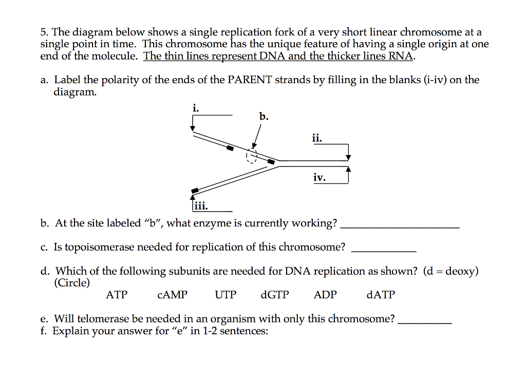
The diagram below shows a replication fork with the two parental DNA strands labeled at their 3' and 5 4/4 (5). The diagram below shows a bacterial replication fork and its principal proteins. Drag the labels to their appropriate locations in the diagram to describe the name or function of each structure. Use pink labels for the pink targets.
Transcribed image text: Part B Processes occurring at a bacterial replication fork The diagram below shows a bacterial replication fork and its principal proteins. Drag the labels to their appropriate locations in the diagram to describe the name or function of each structure. Use pink labels for the pink targets and blue labels for the blue targets.
Feb 17, 2017 · The diagram below shows a bacterial replication fork and its principal proteins. Drag the labels to their appropriate locations in the diagram to describe the name or function of each structure. Use pink labels for the pink targets and blue labels for the blue targets.
When two tuning forks A and B are sounded together, X beats/sec are heard. Frequency of A is n. Now when one prong of fork B is loaded with a little wax, the no. of beat/sec decreases. The frequency of fork B is (a) n + X (b )n-X (c) n - X2 (d) n - 2X 23.
Problem: The diagram below shows a bacterial replication fork and its principal proteins. Drag the labels to their appropriate locations in the diagram to describe the name or function of each structure. Use pink labels for the pink targets and blue labels for the blue targets. FREE Expert Solution. play-rounded-fill.
7-184949-22 Wiring Diagram. Welcome to A.O. Smith's line of Century®. Motors. This pocket manual is designed for one purpose — to make it simple for you to install, maintain and. This manual is designed for one purpose - to make it easier for you to install, maintain, troubleshoot and service Century pool, spa and jet pump motors. All you.
The diagram below shows a bacterial replication fork and its principal proteins. Drag the labels to their appropriate locations in the diagram to describe the name or function of each structure. Use pink labels for the pink targets and blue labels for the blue targets.
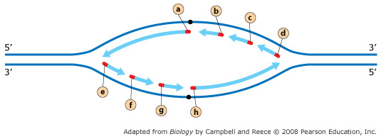
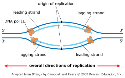
The diagram below shows a replication bubble with synthesis of the leading and lagging strands on both sides of the bubble. This imposes several restrictions on dna replication. Dna separates into two strands then two new complementary strands are generated following the rules of base pairing. The diagram shows the process of dna replication.
The origin of replication is indicated by the black dots on the parental strands. Show transcribed image text the diagram below shows a bacterial replication fork and its principal proteins. Drag the correct labels to the appropriate locations in the diagram to show the composition of the daughter duplexes after one and two cycles of dna.
The origin of replication is indicated by the black dots on the parental strands. In the labels the original parental dna is blue and the dna synthesized during replication is red. Show transcribed image text the diagram below shows a bacterial replication fork and its principal proteins. The structure of dna.
Academia.edu is a platform for academics to share research papers.
The diagram below shows a bacterial replication fork and its principal proteins. Drag the labels to their appropriate locations in the diagram to describe the name or function of each structure. Use pink labels for the pink targets and blue labels for the blue targets
Leads to unstable rna dna duplex. The diagram below shows a bacterial replication fork and its principal proteins. Genes Function Makeup Human Genome Project And Research Same sequence as rna transcript except for having u instead of t. The diagram below shows a length of dna containing a bacterial gene. Only a small percentage of plasmids.















0 Response to "41 The Diagram Below Shows A Bacterial Replication Fork And Its Principal Proteins."
Post a Comment