40 Dogs Ear Canal Diagram
Dr Chris Gleeson from Bayside Mobile Vet explain some basic anatomy of the dogs ear and how to clean the ear. Oscar allows us to demonstrate this process.Dr. Ear Infection in Bulldog and Otitis In French Bulldogs is common and is mostly due to the breed inherently abnormal narrow ear canal due to selective breeding. Your and other dog breeds ear canal cell migration and wax movement is a normal upward motion, in contrast, bulldogs and French bulldogs lack migration or even suffer from an abnormal.
The outer ear includes the pinna (the part you see that is made of cartilage and covered by skin, fur, or hair) and the ear canal. The pinna is shaped to capture sound waves and funnel them through the ear canal to the eardrum. In dogs, the pinnae are mobile and can move independently of each other. The size and shape of the pinnae vary by breed.
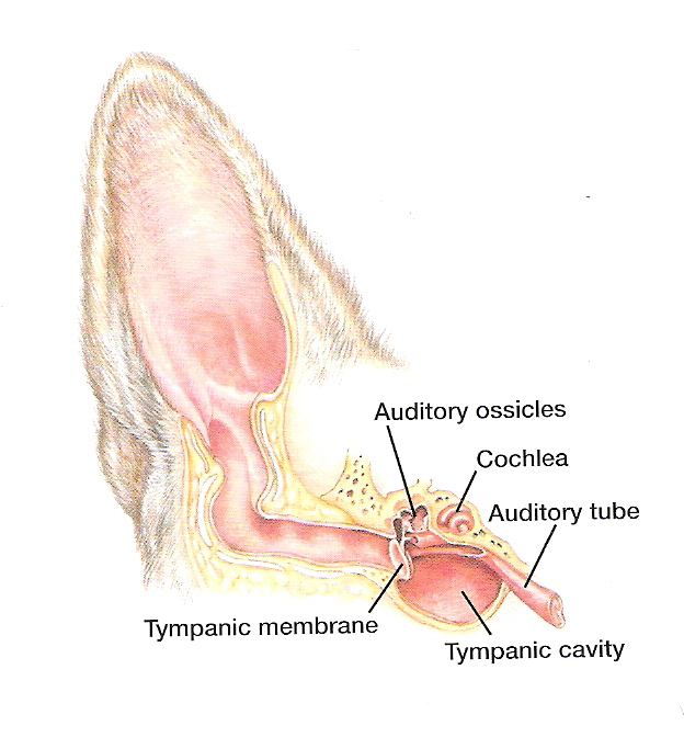
Dogs ear canal diagram
The external ear consists of the prominent earflap or pinna (also called the auricle) and the external ear canal (also called the auditory canal or meatus). The pinna is a funnel-shaped structure that collects sound and directs it into the external ear canal. The pinna is covered by skin, and the outer or posterior aspect is covered by fur. Infection of the external ear canal (outer ear infection) is called otitis externa and is one of the most common types of infections seen in dogs. Some breeds, particularly those with large, floppy or hairy ears like Cocker Spaniels, Miniature Poodles, or Old English Sheepdogs, appear to be more prone to ear infections, but ear infections may occur in any breed. Ear infections are painful. Sometimes a dog's ear can be filled with debris or some foreign object can get lodged in the ear canal causing a variety of ear issues. Ticks can also be the cause of dog ear issues.
Dogs ear canal diagram. We've gathered our favorite ideas for Dog Ear Anatomy Diagram, Explore our list of popular images of Dog Ear Anatomy Diagram and Download Photos Collection with high resolution While holding your dog's ear flap, gently but firmly with one hand, hold the ear cleaning solution in your other hand. Squeeze some ear cleaning solution into your dog's ear. Use enough cleaner to completely fill the ear canal. It is fine if some of the cleaner spills out of the ear canal. DO NOT put the tip of the bottle into the ear. Once the ear canal is exposed to foreign allergens, microbes, stress or if it is not lubricated with an adequate amount of ear wax, different dog ear problems may develop. Similarly, if the immune system of the dog is suppressed or if the dog is experiencing any generalized illness, there is a chance that this will lead to increased microbial. Next, reach for the ear, lift the flap, and briefly manipulate the ear as you would to gain better access to the ear canal. Mark and reward. Repeat three to five times. 2. Add the solution bottle. If all is going well, repeat the previous steps, this time while holding the closed bottle in your hand.
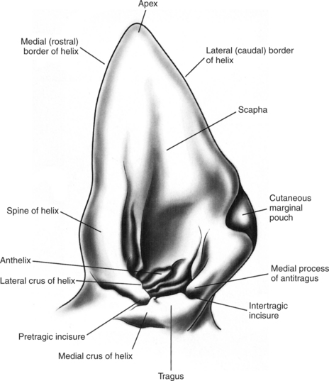
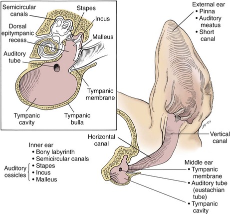
The external ear consists of the prominent earflap or pinna (also called the auricle) and the external ear canal (also called the auditory canal or meatus). The pinna is a funnel-shaped structure that collects sound and directs it into the external ear canal. The pinna is covered by skin, and the outer or posterior aspect is covered by fur. Diagram showing the external, middle and inner ears of the dog. The external ear canal and tympanic cavity are normally air-filled, while the cavities of the inner ear (in black) are normally. never use a cotton swab. When cleaning the ear canal of a australian labradoodle you need to be confident that you will not hurt your dog's ear. A dog's ear canal is shaped like an l the sensitive no touch parts are at the far end of the l, this means that you cannot hurt your dog's hearing or ear by cleaning the part you can see. start by dog ear canal. March 12, 2017 March 12, 2017 Cathy. a diagram of the dogs ear canal. Share on Facebook. Tweet. Follow us. Share. Share. Share. Share. Share. permalink. About Cathy Animals are my passion in life. I am here to help you take care of your pet/pets. Educating yourself in pet care will lead to a happier healthier pet and that is.
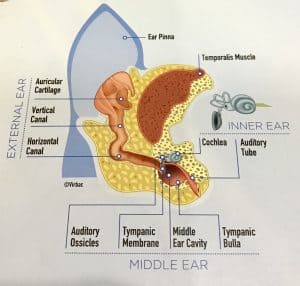
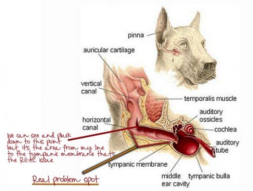
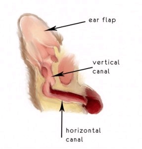
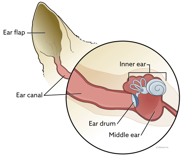
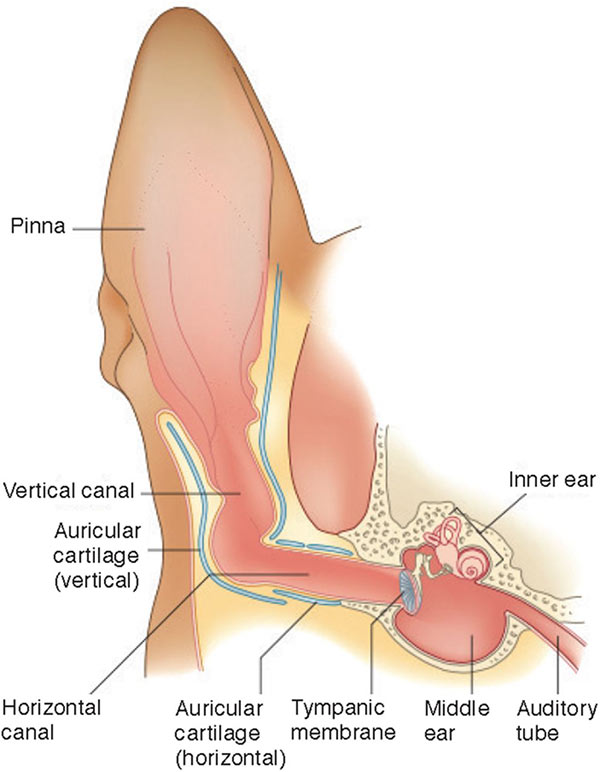
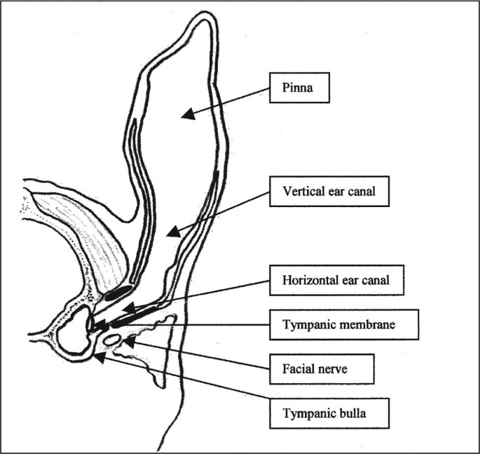
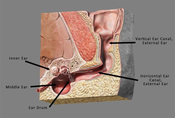
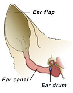
A Visual Guide to Understanding Dog Anatomy With Labeled Diagrams.. They can hear sounds that are undetectable to the human ear. As compared to the 2 to 3 million scent glands that humans possess, dogs have between 200 to 300 million. The tail set is from where the tail begins. Some dogs have high-set tails, while some have low-set tails. The canine ear consists of the pinna, external ear canal, middle ear and inner ear. The external ear is composed of auricular and annular cartilage. The auricular cartilage of the pinna becomes funnel shaped at the opening of the external ear canal. The vertical ear canal runs for about 1 inch, then. The ear has 3 major parts: outer ear; middle ear; inner ear; The outer ear consists of the ear flap (also called the pinna) which is usually upright in cats with the exception of specific breeds such as the Scottish fold cat whose ears are folded over. The ear flap funnels sound into the ear canal. Unlike humans that have a very short ear canal, dogs and cats have a long narrow ear canal that. Diagram of Dogs Ear. In this sketch of a dogs' ear canal, please notice that dogs have a short vertical canal and a longer horizontal canal. The eardrum separates the external ear from the middle ear. Unlike people, a dog ear infection starts in the external ear, not the middle ear or inner ear.
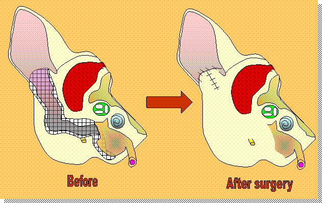
Total ear canal ablation in dogs is a procedure veterinarians use to treat ruptured ear drums due to chronic ear infections, cancer, and congenital imperforate ear canals. This form of surgery is a delicate procedure as the ears have a number of close facial nerves, which is why an experienced and licensed veterinary surgeon is required to.
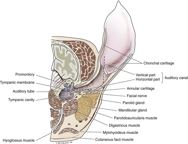
The external ear is composed of three cartilages: annular, auricular, and scutiform. The ear canal is formed proximally (near the skull) by the annular cartilage and distally (away from the skull) by the auricular cartilage, which fans out to form the pinna ( Fig. 1.3 ). Figure 1.3 Auricular and annular cartilage of the right ear of a dog.
Dog ear anatomy diagram. Again, I will show the summary of the dog ear anatomy with the labeled diagram. In this diagram, I tried to show you the most important structure from a dog ear's outer, middle, and inner parts. If you need a more labeled diagram on the dog ear, you may get it on social media of the anatomy learner.
The outer ear consists of the ear flap (also called the pinna) which can be upright (a prick ear) or floppy. The ear flap funnels sound into the ear canal. Unlike humans that have a very short ear canal, dogs have a long narrow ear canal that makes almost a 90 degree bend as it travels to the deeper parts of the ear.
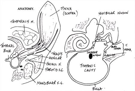
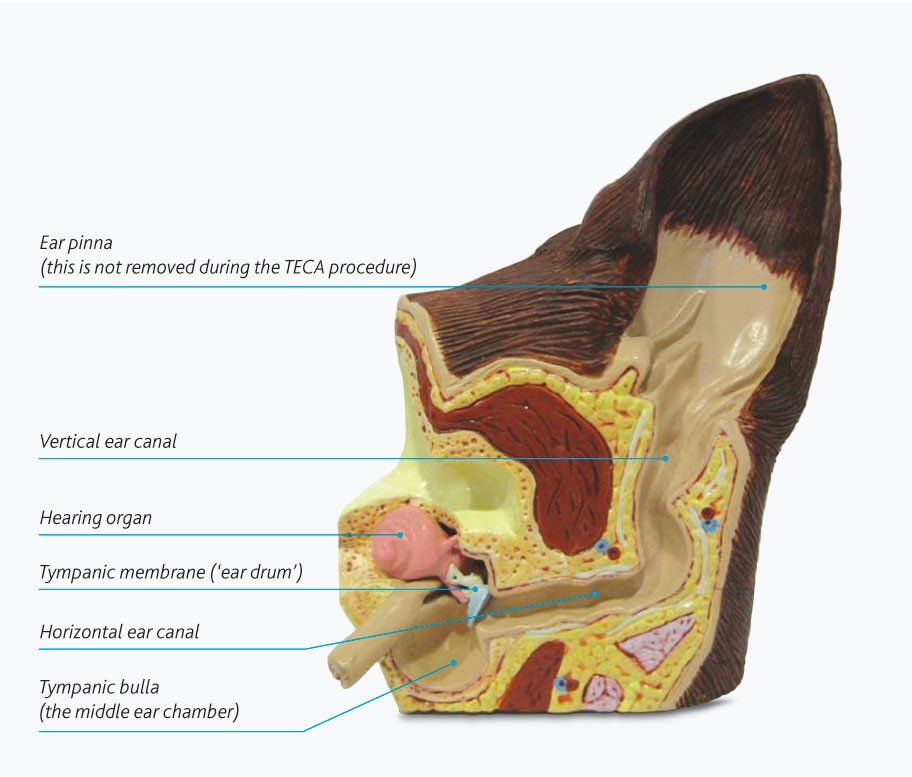
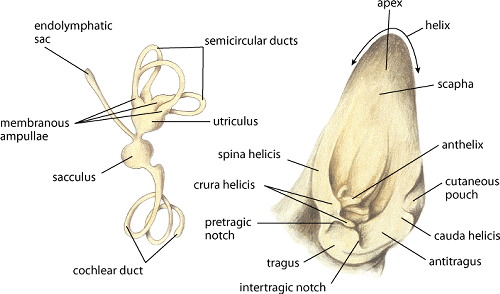
Dog ear anatomy. Dog ear anatomy. In this image, you will find dog ear, dog ear anatomy, pinna, auricular cartilage, vertical canal, horizontal canal, temporalis muscle, auditory ossicles, cochlea, auditory tube in it. You may also find tympanic membrane, middle ear cavity, tympanic bulla, dog ear structure, dog internal ear anatomy, dog middle.
Infection of the external ear canal (outer ear infection) is called otitis externa and is one of the most common types of infections seen in dogs. Some breeds, particularly those with large, floppy or hairy ears like Cocker Spaniels, Miniature Poodles, or Old English Sheepdogs, appear to be more prone to ear infections, but ear infections may occur in any breed. Ear infections are painful.
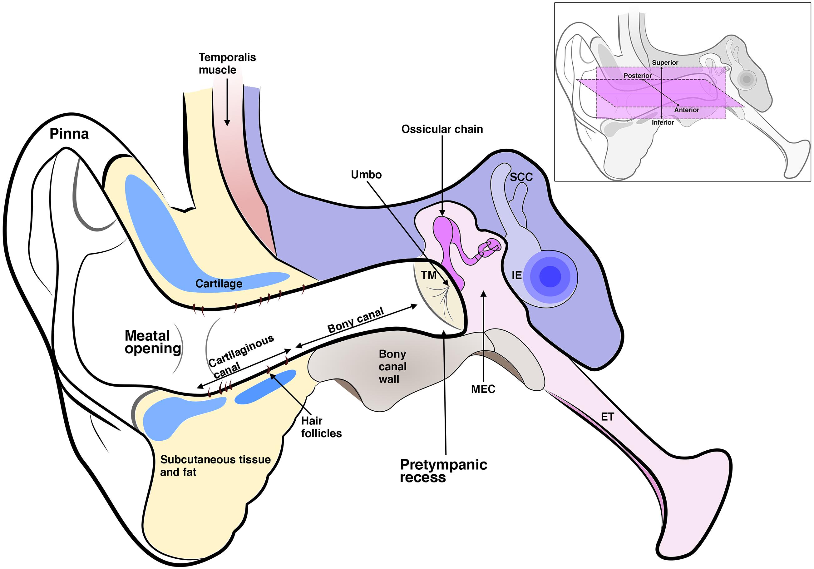
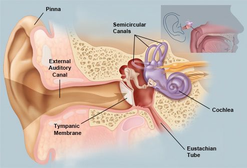
Anatomically, the ear can be looked at in three parts: 1. Outer ear - pinna and auditory canal down to the level of the tympanic membrane. 2. Middle ear - contains the malleus, incus and stapes bones - known as the ossicles. 3. Inner ear - contains the membranous and bony labyrinths, and the cochlea.
The external ear canal in the dog is 5 to 10 cm long and 4 to 5 mm wide (see Figure 1-5). The ear canal consists of an initial vertical part, which may extend an inch. The vertical canal runs ventrally and slightly rostrally before bending to a shorter horizontal canal that runs medially and forms the horizontal part of external ear canal.
Dog Ear Canal Diagram Ear Canal Diagram, Dog Anatomy, Anatomy And Physiology, Pet. yahoolifestyle. Yahoo Life. 332k followers. Few dog s tolerate anything being poked into the external ear canal, and dog s with painful ear s (from infections or foreign bodies) almost never allow adequate examination without anesthesia.
Dog birth canal diagram. 01.11.2020 · He inched his way into the Birth Canal head first, wiggling forward using his hips, stomachs and fingers. But within minutes he realised he was stuck, with no room to turn around or even go backwards. His only option was to keep moving forward, and exhaled the air from his chest so he could fit through the.
A dog ear has three main parts: Outer ear: The outer ear of a dog consists of two different components. The ear flap or outer covering is called the pinna and works to protect the ear. The inner surface of the pinna at the base contains cartilage which not only concentrates different sounds, but also acts as a shock absorber.
Ear diagram dog. The external ear canal in the dog is 5 to 10 cm long and 4 to 5 mm wide see figure 1 5the ear canal consists of an initial vertical part which may extend an inch. Unlike our ear canal the dogs external ear canal is l shaped. The horizontal canal lies deeper in the canal and terminates at the eardrum.
Sometimes a dog's ear can be filled with debris or some foreign object can get lodged in the ear canal causing a variety of ear issues. Ticks can also be the cause of dog ear issues.
Few dogs tolerate anything being poked into the external ear canal, and dogs with painful ears (from infections or foreign bodies) almost never allow adequate examination without anesthesia. The eardrum separates the external ear from the middle ear, and it is the area where vibrations sent from the external ear are focused and amplified.


















0 Response to "40 Dogs Ear Canal Diagram"
Post a Comment