38 What Part Of The Brain Is Highlighted In The Diagram Below?
WebMD's Brain Anatomy Page provides a detailed diagram and definition of the brain including its function, parts, and conditions that affect it. What part of the brain is highlighted in the diagram below?. What are the responsibilities of the region of the brain highlighted below? Coordinating movement and balance by using information from sensory nerves, including hand-eye coordination... What part of the brain is highlighted in the diagram below?
Brain diagram highlighting various parts of the human brain.. The hypothalamus is a small and essential part of the brain, located precisely below the thalamus. It is considered the primary region of the brain, as it is involved in the following functions: Receives impulses;
What part of the brain is highlighted in the diagram below?
Coronal sections of the brain Author: Lorenzo Crumbie MBBS, BSc • Reviewer: Walter Muruet Last reviewed: October 25, 2021 Reading time: 13 minutes In clinical practice, the nervous system is usually visualised in sections that cut through one of the three main orthogonal planes: sagittal, coronal or horizontal.Each of these planes provides the clinician with information that allows the. The falx separates the right and left half of the brain and the tentorium separates the upper and lower parts of the brain. Arachnoid: The second layer of the meninges is the arachnoid. This membrane is thin and delicate and covers the entire brain. There is a space between the dura and the arachnoid membranes that is called the subdural space. SAY: The brain is a dense organ with various functional units. Understanding the anatomy of the brain can be aided by looking at it from different organizational layers. (Purves 2012/p717/para1) In this lesson, we'll explore these organizational layers by discussing the principle brain regions, layers of the brain, and lobes of the brain.
What part of the brain is highlighted in the diagram below?. Brain structures are highlighted on the G2C Brain as you study each structure, and parts are labeled when "View Labels" is clicked.. On the brain diagram below, label the parts that are visible. (If you did not do Part 1, write the functions of the parts too.) Student The cranium (skull) is the skeletal structure of the head that supports the face and protects the brain.It is subdivided into the facial bones and the brain case, or cranial vault (Figure 1).The facial bones underlie the facial structures, form the nasal cavity, enclose the eyeballs, and support the teeth of the upper and lower jaws. Back to The Brain and Learning. Support for LabX programming is generously provided by the Marian E. Koshland Endowment Fund PLAY. What are the responsibilities of the region of the brain highlighted below? Coordinating movement and balance by using information from sensory nerves, including hand-eye coordination. Which of the following structures or systems is correctly paired with its function? Nice work!
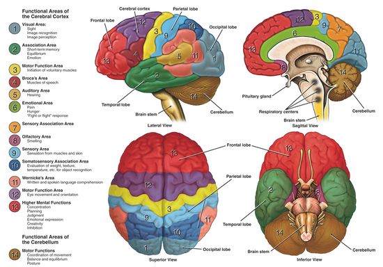
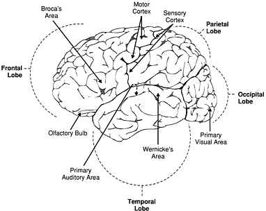
The cerebrum, the foremost part of the brain, is the largest part of the brain in humans comprising about 83% of total brain mass. Identify the parts of the spinal cord shown in the diagram below: 1 = Anterior horn, 5= Lateral column, 9 = Posterior median sulcus, 13= DRG.... For example, what nerve is highlighted in blue, below: Phrenic nerve. The brain is a 3-pound organ that contains more than 100 billion neurons and many specialized areas. There are 3 main parts of the brain include the cerebrum, cerebellum, and brain stem.The Cerebrum can also be divided into 4 lobes: frontal lobes, parietal lobes, temporal lobes, and occipital lobes.The brain stem consists of three major parts: Midbrain, Pons, and Medulla oblongata. Lobes of the Brain. The four lobes of the brain are the frontal, parietal, temporal, and occipital lobes (Figure 4). The frontal lobe is located in the forward part of the brain, extending back to a fissure known as the central sulcus. The frontal lobe is involved in reasoning, motor control, emotion, and language. The cerebellum ("little brain") is a fist-sized portion of the brain located at the back of the head, below the temporal and occipital lobes and above the brainstem. Like the cerebral cortex, it has two hemispheres. The outer portion contains neurons, and the inner area communicates with the cerebral cortex.
SAY: The brain is a dense organ with various functional units. Understanding the anatomy of the brain can be aided by looking at it from different organizational layers. (Purves 2012/p717/para1) In this lesson, we'll explore these organizational layers by discussing the principle brain regions, layers of the brain, and lobes of the brain. Brain Injury: How the brain functions November 13, 2019 June 19, 2010 by Dr. Matthew E. Bain To help you better understand your loved one's injury, print this out this page and ask members of the health care team to mark this picture to show you the location of your loved one's injury or the areas of the brain that are affected. Biology questions and answers. VueSUI CUTII Jlatus. QUESTION 33 The diagram below shows DNA of a hypothetical fruit fly gene ken (called ken and barbie, for my favorite gene). Several elements are highlighted by numbers 1-4. In wildtype fly larvae, the gene product (protein) is expressed in cells that will eventually become excitatory neurons. Want to learn more about the parts of the brain? Try our free brain diagrams and quizzes! Cerebrum. The cerebral cortex, which is the area of the cerebrum seen at a lateral view of the brain, is about 2-5 mm thick and accounts for about 80% of the brain's totalling mass. Its total area has been estimated to be about 2000 cm².
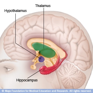
Slide Show How Your Brain Works Mayo Clinic
Click here 👆 to get an answer to your question ️ WILL MARK BRAINIEST What part of the brain is highlighted in the diagram below? Occipital lobe Temporal lob… Brainly User Brainly User 05/19/2020 Biology High School answered WILL MARK BRAINIEST What part of the brain is highlighted in the diagram below? Occipital lobe Temporal lobe

The Development Of Brain Functional Connectome During Text
Lobes of the Brain. The four lobes of the brain are the frontal, parietal, temporal, and occipital lobes (Figure 2). The frontal lobe is located in the forward part of the brain, extending back to a fissure known as the central sulcus. The frontal lobe is involved in reasoning, motor control, emotion, and language.

The Four Csf Filled Ventricles In The Adult Mouse Brain
In vertebrate anatomy, the brainstem is the posterior part of the brain adjoining, and structurally continuous with, the spinal cord. Though small, the brainstem is an extremely important part of the brain, as the nerve connections from the motor and sensory systems of the cortex pass through it to communicate with the peripheral nervous system.
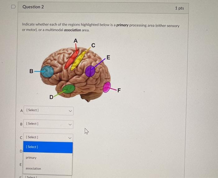
Solved Question 2 1 Pts Indicate Whether Each Of The Regions
What part of the brain is highlighted in the diagram below? (4 points) Frontal lobe Occipital lobe Temporal… Get the answers you need, now! kyliebruner kyliebruner 04/27/2021 Biology High School answered 3. What part of the brain is highlighted in the diagram below? (4 points) Frontal lobe Occipital lobe Temporal lobe Parietal lobe 1 See.
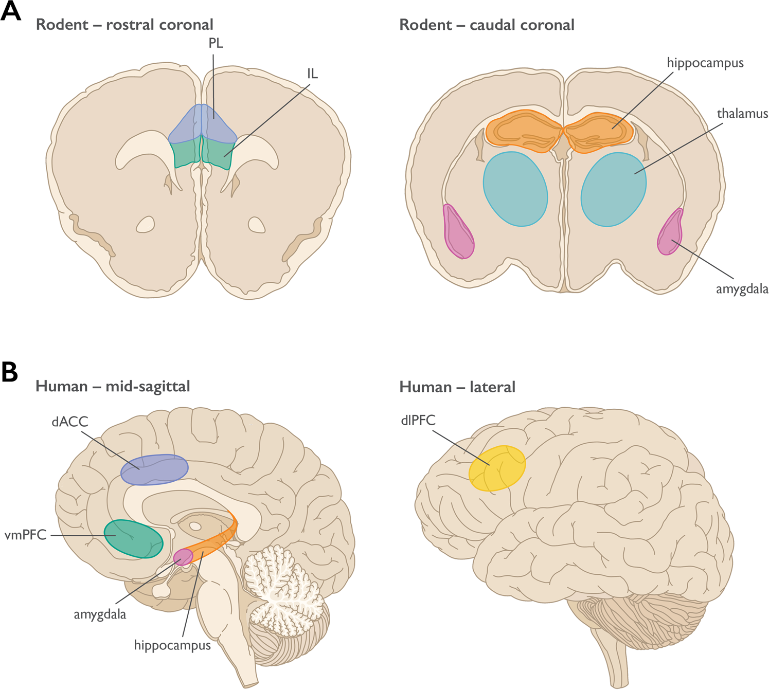
Prefrontal Cortex Amygdala And Threat Processing
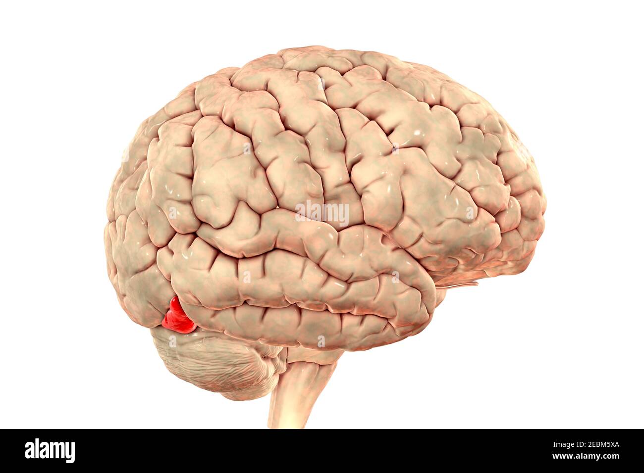
The word 'cerebellum' literally means little brain. It is the second largest part of the brain, and is located at the back, below the occipital lobe, beneath the cerebrum and behind the brain stem. It contains an outer gray cortex and an inner white medulla, and has horizontal furrows, which makes it look different from the rest of the brain.

Basal Ganglia And Cerebellum Contributions To Vocal Emotion
Identify the highlighted structure. (three words). What part of the brain houses autonomic centers involved in the control of heart rate, respiratory rhythm, and blood pressure as well as involuntary centers involved in vomiting, swallowing and so on?... This extension of the dura mater separates the cerebrum from the cerebellum below ...

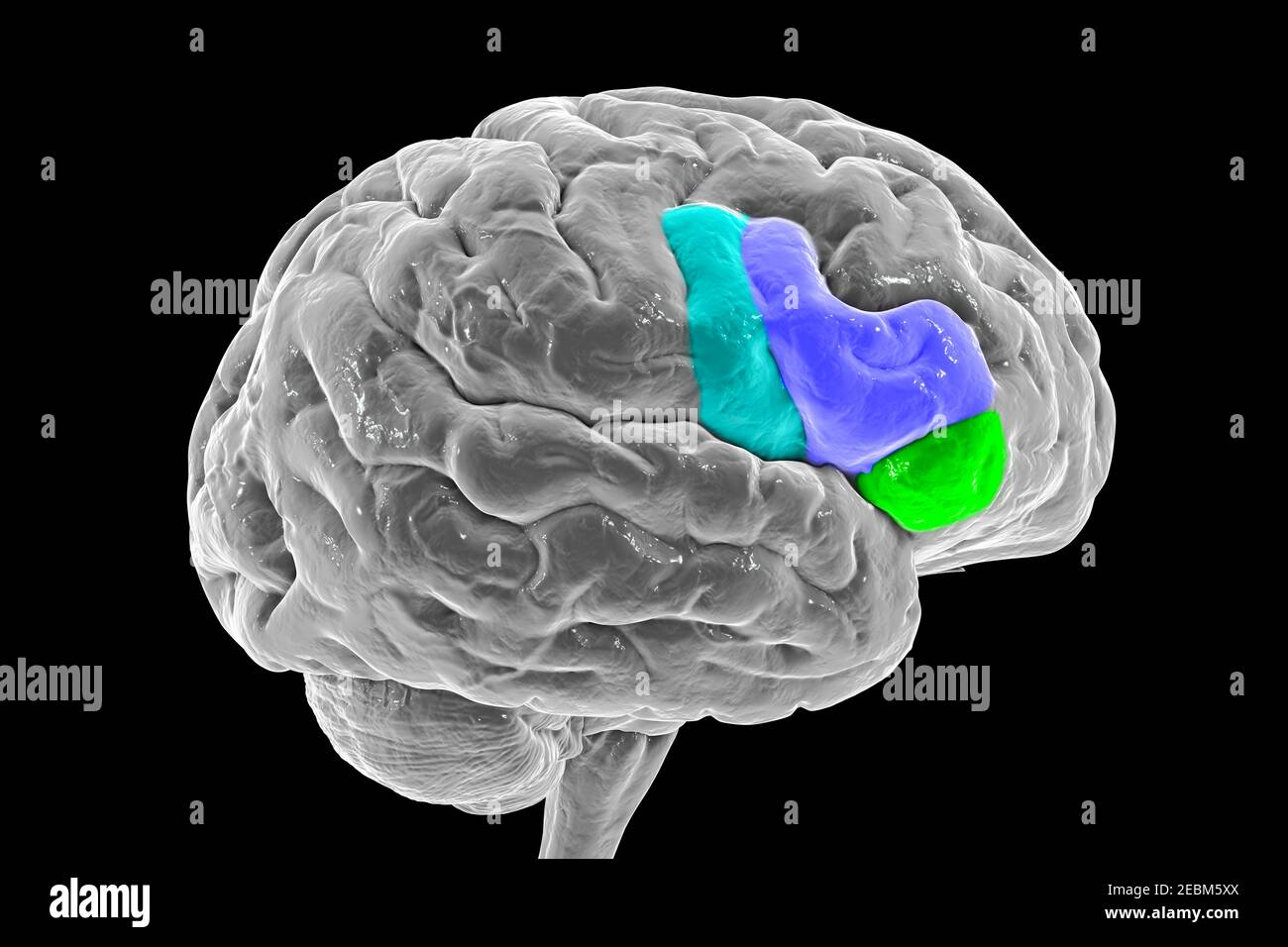
Broca S Area Stock Illustrations 13 Broca S Area Stock
What part of the brain is highlighted in the diagram below? Highlighter portion is at the bottom of the brain, forming a connection between the center of the brain and the spinal cord. Brain stem Pituitary gland Cerebrum Cerebellum

What Part Of The Brain Is Highlighted In The Diagram Below A
The highlighted part you are talking about is certainly the cerebellum. it is located in the posterior cranial fossa, below and at the back of the two hemispheres, it looks like a little brain. Its most important roles consist in the handling of the equilibrium of the body and learning ability.

Brain And Testis More Alike Than Previously Thought Open
The falx separates the right and left half of the brain and the tentorium separates the upper and lower parts of the brain. Arachnoid: The second layer of the meninges is the arachnoid. This membrane is thin and delicate and covers the entire brain. There is a space between the dura and the arachnoid membranes that is called the subdural space.

Brain And Cognitive Development Stiles Major Reference
Coronal sections of the brain Author: Lorenzo Crumbie MBBS, BSc • Reviewer: Walter Muruet Last reviewed: October 25, 2021 Reading time: 13 minutes In clinical practice, the nervous system is usually visualised in sections that cut through one of the three main orthogonal planes: sagittal, coronal or horizontal.Each of these planes provides the clinician with information that allows the.
Frontiers Targeting Mycn In Molecularly Defined Malignant
embryologically, the brain arises from the rostral end of a tubelike structure that quickly becomes divided into three major regions. group of structures that develop from the embryonic brain are listed below. designate the embryonic origin: the diencephalon, including the thalamus, optic chiasma, and hypothalamus

Illustration Of Neuroanatomy Of Study The Highlighted Region
What part of the brain is highlighted in the diagram below? The highlighted portion is at the rear base of the brain, behind the brain stem. Pituitary gland
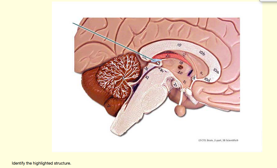
Solved Yo 06 T C15 Brain 2 Part 3 Sclentifice Identify The
Brain. The brain is one of the most complex and magnificent organs in the human body. Our brain gives us awareness of ourselves and of our environment, processing a constant stream of sensory data. It controls our muscle movements, the secretions of our glands, and even our breathing and internal temperature.
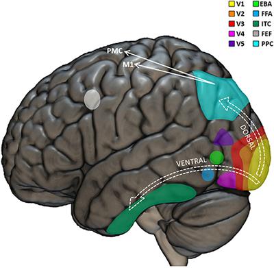
Frontiers Visual Neuropsychology In Development Anatomo
Parts of the brain. 22 terms. prism36. OTHER SETS BY THIS CREATOR. Skeletal System 1. 31 terms. daniculver TEACHER. Skeletal System. 42 terms.
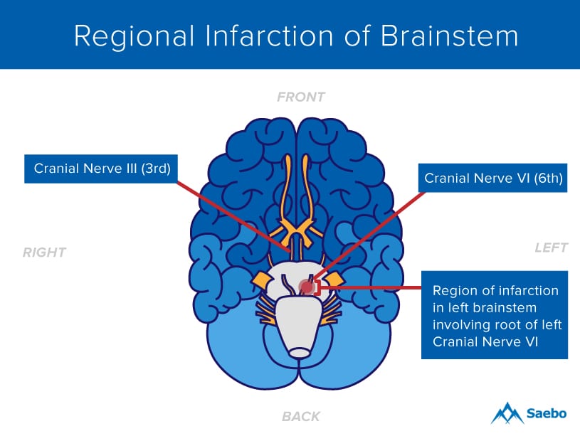
What Is A Brainstem Stroke Saebo
What part of the brain is highlighted in the diagram below? is it brain stem? brain stem pituitary gland cerebrum cerebellum - 10471101 sskibi5950 sskibi5950 06/14/2018
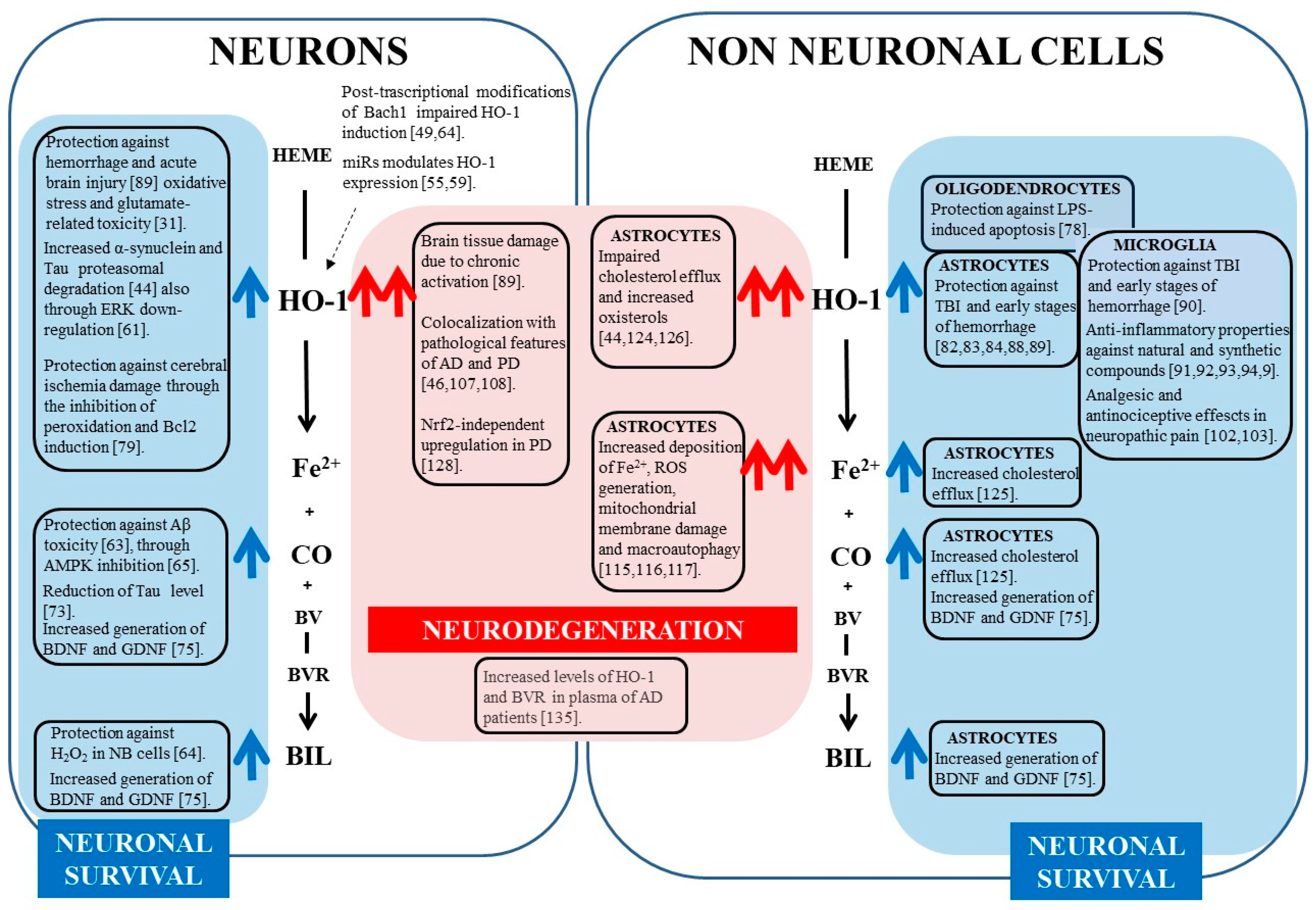
Ijms Free Full Text Heme Oxygenase 1 In The Nervous
Cerebral Cortex Physiopedia

Brain Sciences Free Full Text Dynamical Role Of Pivotal

What Part Of The Brain Is Highlighted In The Diagram Below

Interoceptive Accuracy And Its Impact On Neuronal Responses

A Amp P 1 Lab Unit 15 Pal Cns Flashcards Quizlet

A Amp P 1 Lab Unit 15 Pal Cns Flashcards Quizlet
Inferior View Brain High Resolution Stock Photography And
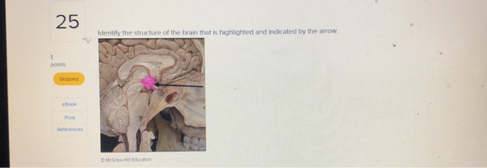
Solved Identify The Structure Of The Brain That Is Chegg Com

Wearable Scanners Will Be Able To Read Our Minds Financial
2 Major Structures And Functions Of The Brain Discovering

3 What Part Of The Brain Is Highlighted In The Diagram Below
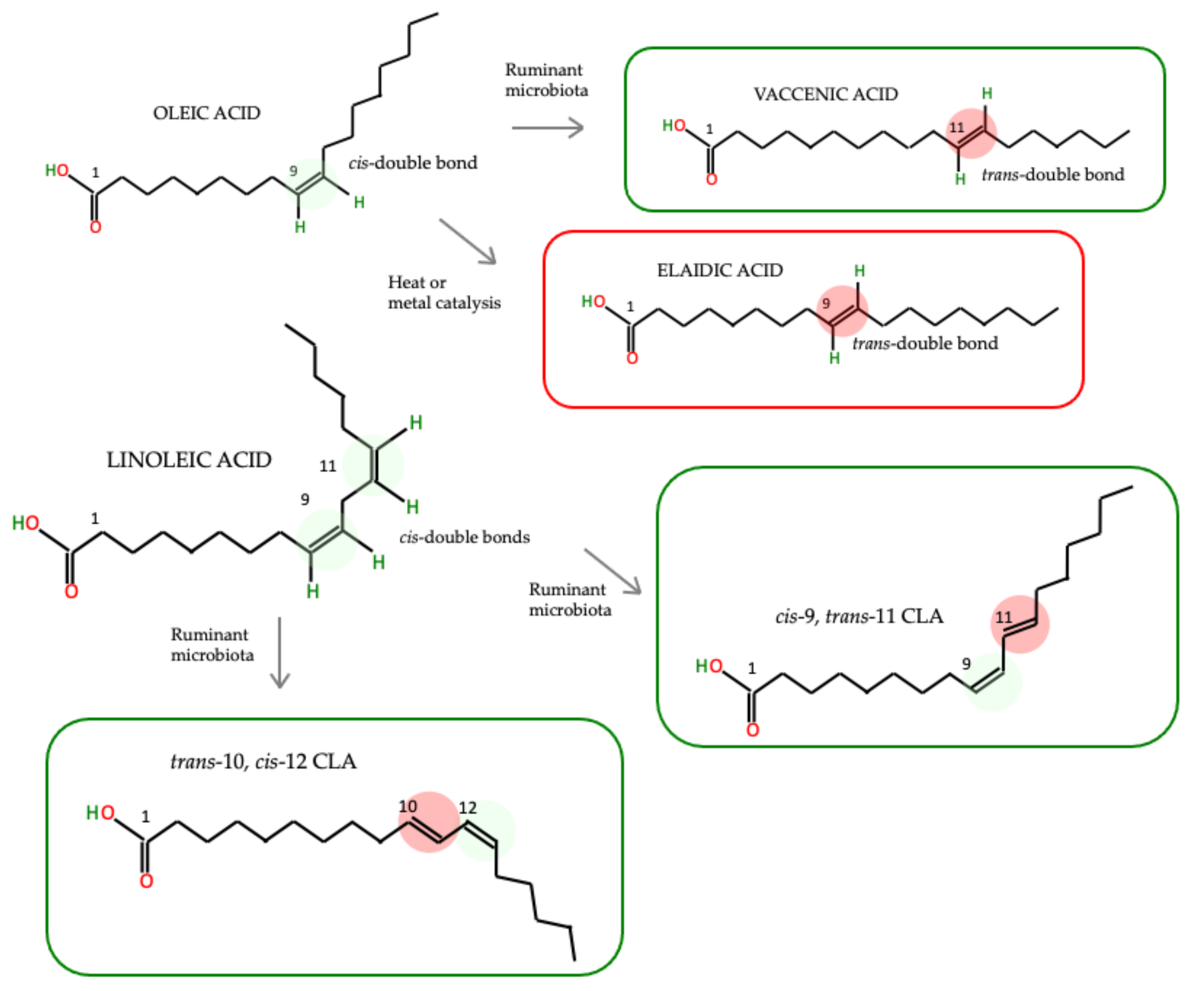
Foods Free Full Text The Effect Of Trans Fatty Acids On
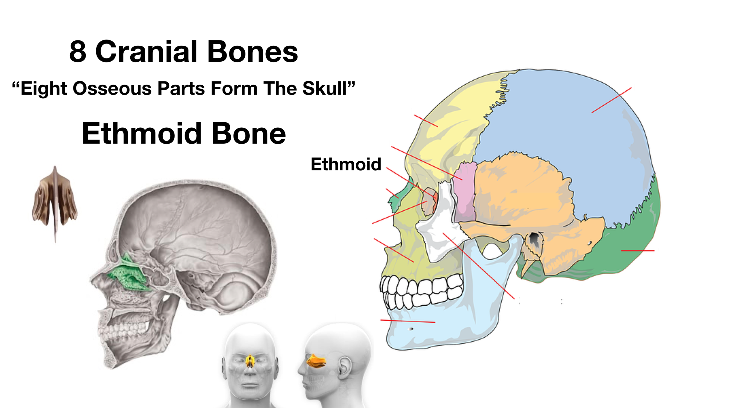
Skull Anatomy Cranial Bone And Suture Labeled Diagram

Occipital High Resolution Stock Photography And Images Alamy
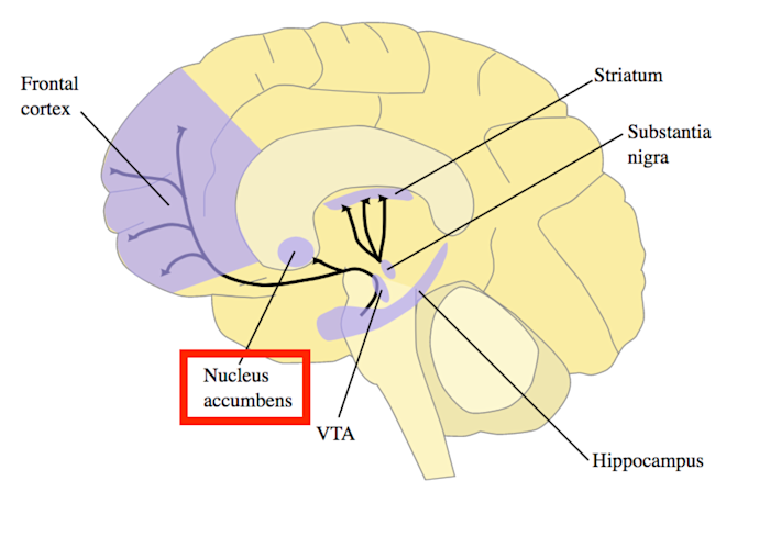
The Science Of Motivation
Brain Areas That Correspond With Regions Highlighted In The
Frontal Gyri High Resolution Stock Photography And Images Alamy

The Four Csf Filled Ventricles In The Adult Mouse Brain
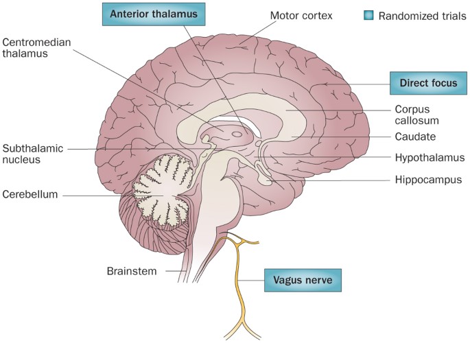
Electrical Brain Stimulation For Epilepsy Nature Reviews

What Part Of The Brain Is Highlighted In The Diagram Below

Blood Brain Barrier Crossing Renin Angiotensin Drugs And

Mind Matters Drugs And The Brain Nida For Teens
0 Response to "38 What Part Of The Brain Is Highlighted In The Diagram Below?"
Post a Comment