37 Diagram Of Sheep Brain
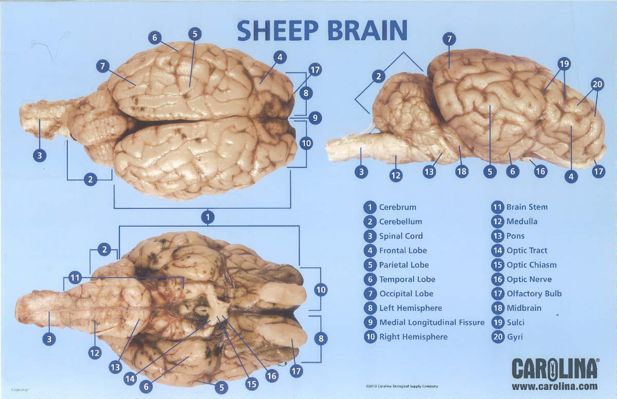
Diagram of Sheep Brain - Inferior view Sheep Brain Dissection Key. Image of the brain showing its major features for students to practice labeling. Answers are included. Pretty good picture of the sheep brain labeled. Shows pictures of a sheep and a human brain. Each of the 12 cranial nerves is represented, students color and number each nerve in both brains.
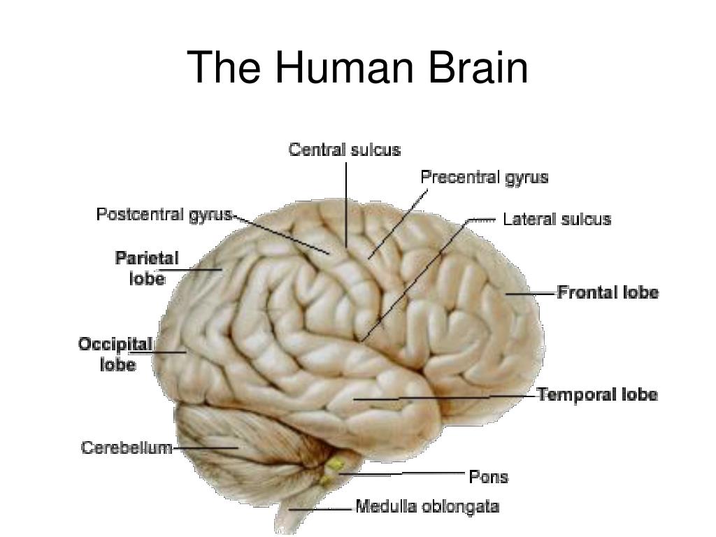
The brain is divided into three layers that are interconnected. Let us have a look at these three layers and learn about the human brain diagram and functions. Human Brain: Diagram and Functions. The human brain is an astonishing organ that takes care of each function and action of the body.

Diagram of sheep brain
Sheep Brain Anatomy Lab Manual. Based on original material by R. N. Leaton, Dartmouth College. Contributors to this version: Al Sorenson, Lisa Raskin, Sarah Turgeon, Steve George, and JP Baird. I. Introduction. The brain of the sheep is useful for study because its anatomy is similar to human brain anatomy. Although exact proportions (and names. Sheep Brain Dissection The purpose of this exercise is to introduce you to the mammalian brain; you will use a sheep's brain. While the sheep brain differs from the human brain in many details, they both have the same basic anatomy, and, it is larger than the rat brain. Work in teams of four students. Brain. The brain is one of the most complex and magnificent organs in the human body. Our brain gives us awareness of ourselves and of our environment, processing a constant stream of sensory data. It controls our muscle movements, the secretions of our glands, and even our breathing and internal temperature.
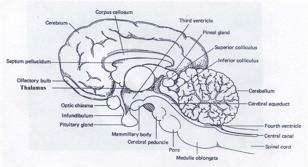
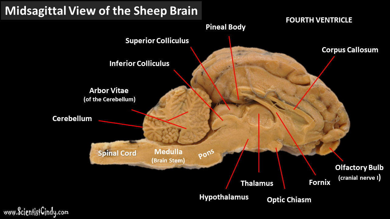
Diagram of sheep brain. BI 335 - Advanced Human Anatomy and Physiology Western Oregon University Figure 4: Mid-sagittal section of brain showing diencephalon (includes corpus callosum, fornix, and anterior commissure) Marieb & Hoehn (Human Anatomy and Physiology, 9th ed.) - Figure 12.10 Exercise 2: Utilize the model of the human brain to locate the following structures / landmarks for the Jun 5, 2014 - Learn the external and internal anatomy of sheep brains with HST's Learning Center science lesson and guide! Diagram worksheets also included. View now! Sheep Brain Dissection The purpose of this exercise is to introduce you to the mammalian brain; you will use a sheep's brain. While the sheep brain differs from the human brain in many details, they both have the same basic anatomy, and, it is larger than the rat brain. Work in teams of four students. Sheep Brain Dissection Guide Planes of Orientation In addition to the direction, the brain as a three dimensional object can be divided into three planes. There is the frontal or coronal planes which divides front from back. It can divide the brain and any location as long as it divides the brain from front to back.
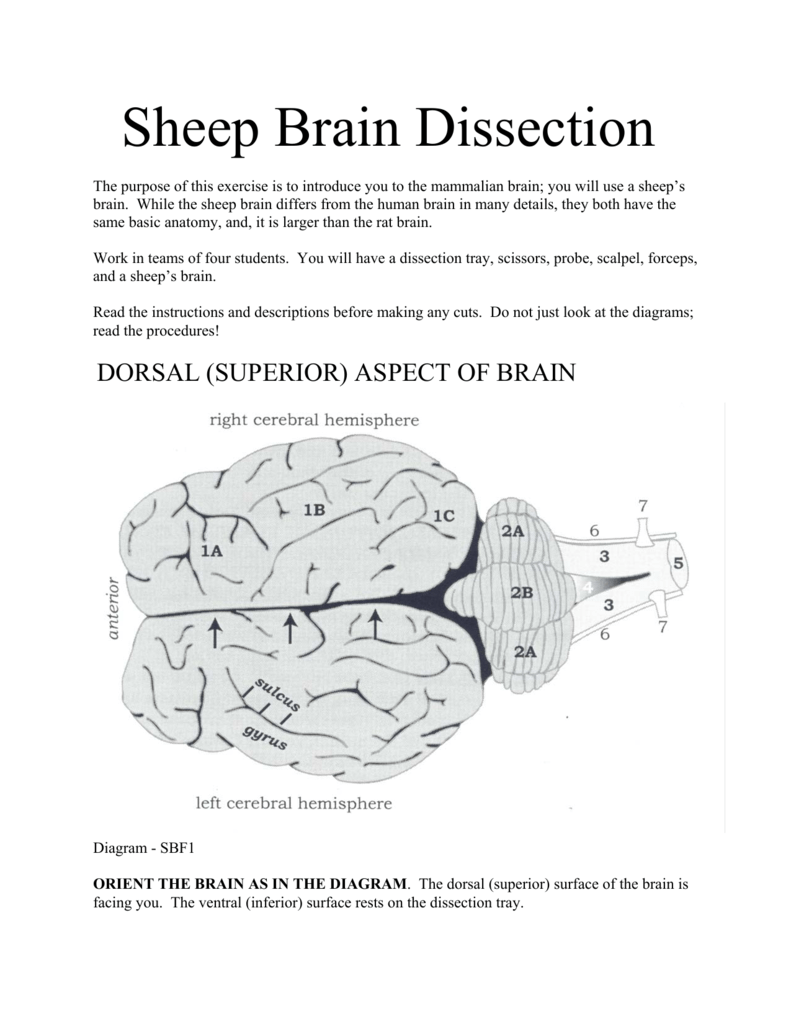
5 3 11 6 22 16 18 1. Gray Matter 2. White Matter 3. Corpus Callosum 4. Lateral Ventricle 5. Caudate Nucleus 6. Septum Pellucidum 7. Fornix 8. The sheep brain is quite similar to the human brain except for proportion. The sheep has a smaller cerebrum. Also the sheep brain is oriented anterior to posterior whereas the human brain is superior to inferior. 1. The tough outer covering of the sheep brain is the dura mater, one of three meninges (membranes) that cover the brain. You will. The lobes of the brain are visible, as well as the transverse fissure, which separates the cerebrum from the cerebellum. The convolutions of the brain are also visible as bumps (gyri) and grooves (sulci). Use the diagram below to help you locate these items. Dorsal View of the Sheep Brain. 8. Sheep Brain. Images taken from the dissection of the sheep's brain, some structures have been labeled. Anatomy Corner resource site for teachers and students of Anatomy and Physiology. Find quizzes, diagrams, and slide presentations on structures, functions, and systems.
Sheep Brain Neuroanatomy Online Self-Test. Use each diagram as a reference, and selected the correct answer for each lettered structure. You may find it useful to open the diagrams in a separate window to review while answering each question. Dorsal Surface. Sheep Brain Anatomy Lab Manual. Based on original material by R. N. Leaton, Dartmouth College. Contributors to this version: Al Sorenson, Lisa Raskin, Sarah Turgeon, Steve George, and JP Baird. I. Introduction. The brain of the sheep is useful for study because its anatomy is similar to human brain anatomy. Although exact proportions (and names. Use the online sheep brain guide to help you find structures. There is a link to this site on the syllabus and list of class related sites. It should be up in the lab when you come in. For the most part, except for 1, the numbers refer to the images on the online sheep brain guide. Play this game to review Human Anatomy. Name this part of the brain. Preview this quiz on Quizizz. Name this part of the brain. Sheep Brain Dissection DRAFT. 6th - 12th grade. 193 times. Biology, Other Sciences. 77% average accuracy. a year ago. mrsturmscience. 0. Save. Edit. Edit. Sheep Brain Dissection DRAFT. a year ago. by mrsturmscience.
Sheep Brain Diagram Blank angelo. October 2, 2021. Pin By Amy Pena On A P Brain Anatomy Anatomy Brain Images. Pretty Good Picture Of The Sheep Brain Labeled Basic Anatomy And Physiology Human Anatomy And Physiology Anatomy And Physiology .
Diagram Worksheets. Label the Parts of a Sheep Brain. Print out these diagrams and fill in the labels to test your knowledge of sheep brain anatomy. Internal anatomy: label the right side (.pdf) External anatomy: label the top view (.pdf) External anatomy: label the bottom view (.pdf) What other users say: Fun and Educational.
The sheep brain is exposed and each of the structures are labeled and described in a sequential manner, in the same way that a real dissection would occur. Sheep Brain Dissection. 1. The sheep brain is enclosed in a tough outer covering called the dura mater. You can still see some structures on the brain before you remove the dura mater.
Sheep Brain Labeled Diagram. angelo. August 10, 2021. Labeled Brain Brain Anatomy Anatomy And Physiology Anatomy. Image Result For Sheep Brain Labeled Nursing Study Guide Anatomy And Physiology Nursing Study In 2021 Nursing Study Guide Nursing Study Human Brain Anatomy.
function, and pathology. Those students participating in Sheep Brain Dissections will have the opportunity to dissect and compare anatomical structures. At the end of this document, you will find anatomical diagrams, vocabulary review, and pre/post tests for your students. The following topics will be covered: 1.
Lab: Sheep Brain Dissection 1 **Before starting this lab, open the "Brain Parts and Functions" document. Refer to images, descriptions, and functions of parts of the brain as you proceed through this lab. Sheep brains, although much smaller than human brains, have similar features and can be a valuable addition to anatomy studies.
Drag the labels onto the diagram to identify the parts of the dissected sheep brain, median section (part 1 of 2). Drag the labels onto the diagram to identify the structures. Drag the labels onto the diagram to identify the structures. Drag the labels onto the diagram to identify the cranial nerves.
Examining the external sheep brain. The tough outer covering of the sheep brain is the dura mater, the outermost meninges membrane covering the brain.Remove the dura mater to see most of the structures of the brain, but remove it carefully, so as to leave all the other structures beneath it intact. Removing the dura mater from the cerebellum at the back of the brain can be tricky.
Sagittal Sheep Brain Part 2. 7 terms. ambrose426. Ventral View of Sheep Brain With Dura. 8 terms. ambrose426. Upgrade to remove ads. Only $2.99/month.
The human brain is 15 centimetres long and a sheep's brain is only about a third of that length. Both have three divisions namely, cerebrum, cerebellum and the brainstem. The cerebellum and brain stem are located behind the cerebrum in sheep as they have a horizontal spine. While they are located below the cerebrum in humans as they have vertical spines and are upright.
Brain. The brain is one of the most complex and magnificent organs in the human body. Our brain gives us awareness of ourselves and of our environment, processing a constant stream of sensory data. It controls our muscle movements, the secretions of our glands, and even our breathing and internal temperature.
Diagram of Sheep Brain - Lateral view
The primary function of the meninges and of the cerebrospinal fluid is to protect the central nervous system. Dura Mater-encases the prain and is the first layer of the brain. Gyrus - a ridge or fold between two clefts on the cerebral surface in the brain. Sulcus - a groove or furrow, especially one on the surface of the brain.
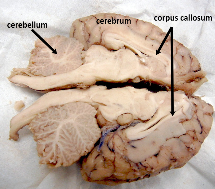
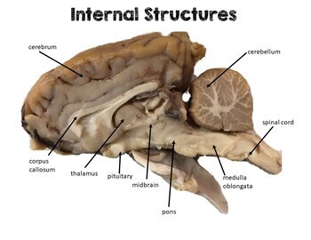
BRAIN ANATOMY CH 11 NAME: Physiology Use this website to label the internal view of the sheep brain. Use all of the terms from given at this site to label the brain. Label both the sketch and the actual image. Two - parts can be divided between the 2 diagrams..












0 Response to "37 Diagram Of Sheep Brain"
Post a Comment