39 sheep brain diagram labeled
11.7: Sheep Brain Dissection - Biology LibreTexts Jul 8, 2021 — Background Information: The sheep brain is remarkably similar to the human brain. One major difference, however, is in proportion. sheep brain labeling Quiz - PurposeGames.com This is an online quiz called sheep brain labeling. There is a printable worksheet available for download here so you can take the quiz with pen and paper. Your Skills & Rank. Total Points. 0. ... Label Parts of the Fingertip 14p Image Quiz. side of the skull markings and bones 35p Image Quiz. Superficial Muscles Anterior View 39p Image Quiz.
Sheep Brain Dissection labeled Diagram | Quizlet Only $2.99/month Sheep Brain Dissection labeled STUDY Learn Write Test PLAY Match Created by AllieKlinger Terms in this set (8) Corpus Collosum Lateral Ventricle Fornix Hypothalamus Cerebral Aqueduct Central Canal Inferior Collicuious Transverse Fissure THIS SET IS OFTEN IN FOLDERS WITH... Sheep Brain Dissection labeled 2 8 terms AllieKlinger

Sheep brain diagram labeled
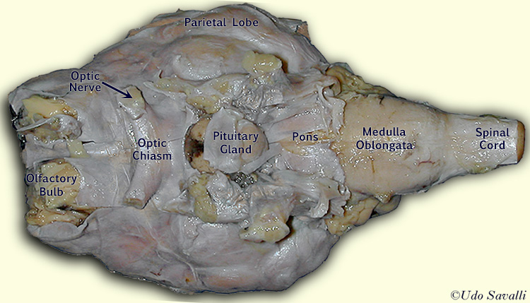
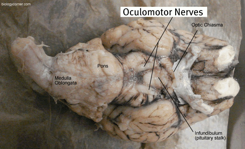
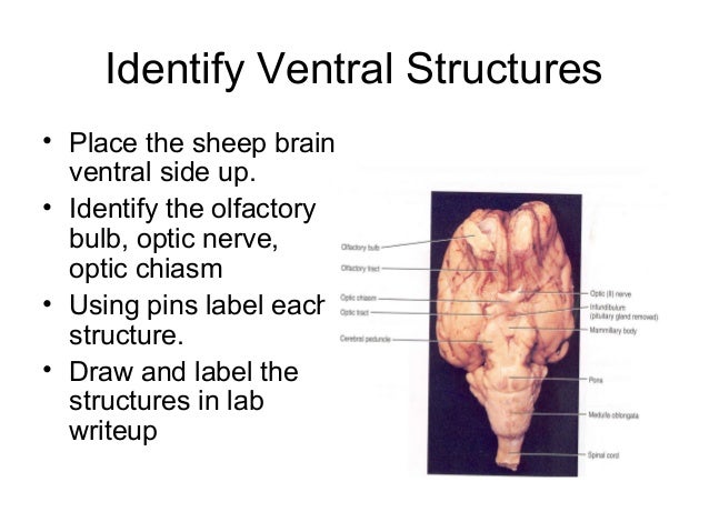
DOC Sheep Brain Anatomy Lab Manual - amherst.edu The cruciate fissure (labeled ansate sulcus in your photo atlas) is known in the human brain as the fissure of Rolando or central sulcus, and intersects the medial longitudinal fissure to mark off the anterior third of the cortex. Anatomy Sheep Brain Diagram | Quizlet Anatomy Sheep Brain STUDY Learn Flashcards Write Spell Test PLAY Match Gravity + − Created by quizlette331614 Terms in this set (19) Frontal 1 Parietal 2 Occipital 3 Cerebral Hemisphere 4 Spinal Cord 5 Medulla Oblongata 6 Nerve 7 Pons 8 Mid Brain 9 Inferior Collicus 10 Pituitary Gland 11 Optic Chiasma 12 Hypothalamus 13 Thalmus 14 Pineal Body 15 Sheep brain dissection | Human Anatomy and Physiology Lab ... The most prominent structure visible on the ventral side of the sheep brain is half of the optic chiasma, which is where the two optic nerves cross over each other and form an "X" shape. You will only see half the structure. Find the optic chiasma half on your brain. You may have removed the optic removed the chiasma with the dura mater.
Sheep brain diagram labeled. PDF Neuroanatomy: Dissection of the Sheep Brain Examine the sheep brain with the membranes intact. You should be able to identify and use the following directional terms: Anterior / Posteriorfront / back Rostral / Caudal towards the beak / towards the tail Medial / Lateral towards the middle / towards the side Dorsal / Ventral top / bottom (on the CNS of a quadruped) PDF DISSECTION OF THE SHEEP'S BRAIN - Hanover College DISSECTION OF THE SHEEP'S BRAIN Introduction The purpose of the sheep brain dissection is to familiarize you with the three-dimensional structure of the brain and teach you one of the great methods of studying the brain: looking at its structure. One of the great truths of studying biology is the saying that "anatomy precedes physiology". Sheep Brain Label - The Biology Corner Sheep Brain Label. Publisher: Biologycorner.com; follow on Google+. This work is licensed under a Creative Commons Attribution-NonCommercial 3.0 Unported License . Brain Label Answer Key. Image adapted from a photograph of the sheep brain. Sheep Brain Anatomy Quiz - ProProfs Quiz 1. What is the outer covering of the Sheep brain? A. Arachnoid Mater B. Pia Mater C. Medulla Oblongata D. Dura Mater 2. Which amongst the following is the largest structure in dorsal view? A. Pons B. Medulla Oblongata C. Cerebral cortex D. Hippocampus 3. What are the large folds that surround the cerebrum? A. Gyri B. Sulci C. Fissures D. Colliculus
Sheep Brain Dissection Project Guide | HST Learning Center Use the labeled picture to identify the corpus callosum, medulla, pons, midbrain, and the place where the pituitary gland attaches to the brain. (In many preserved specimens the pituitary gland is no longer present. It is not pictured.) Use your fingers or a teasing needle to gently probe the parts and see how they are connected to each other. Solved Art-labeling Activity: Midsagittal Section of the ... Anatomy and Physiology. Anatomy and Physiology questions and answers. Art-labeling Activity: Midsagittal Section of the Sheep Brain (Diagram, 2 of 2) Reset Help Fomix Infundibulum Olfactory bulb Optic chiasm Mosencephalon Pituitary gland Marillary body Medulla oblongata Pons Spinal cord Corpus callosum Art-labeling Activity: Midsagittal Section ... Sheep Brain Dissection Guide with Pictures | Nervous ... Pretty good picture of the sheep brain labeled. Dissection guide with instructions for dissecting a sheep brain. Checkboxes are used to keep track of progress and each structure that can be found is described with its location in relation to other structures. An image of the brain is included to help students find the structures. Sheep Brain Dissection Lab Use the picture below as a way to see how your sheep brain should look after you cut it in half. 10. In the image below, a probe indicates the location of the lateral ventricle. 11. Once the brain is cut this way, the colliculi can also be seen from the inside and the pineal gland is revealed only if you made a very careful incision.
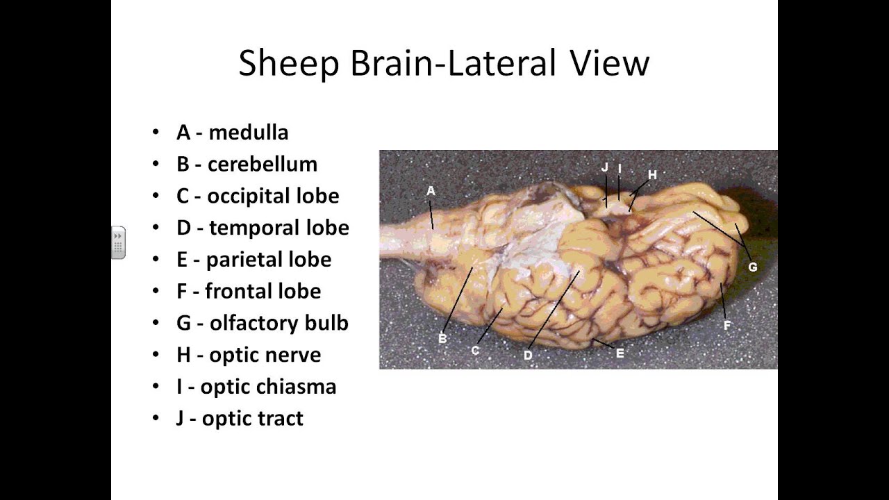
PDF Sheep Brain Midsagittal Section - Dr. Scott Croes' Website Sheep Brain -Parasagittal Section 1. Gray Matter 2. White Matter 3. Corpus Callosum 4. Lateral Ventricle 5. Caudate Nucleus 6. Septum Pellucidum 7. Fornix 8. Optic Chiasma 9. Third Ventricle 10. Thalamus (Ovid Nuclear Mass of Thalamus) 11. Corona Radiata 12. Hippocampus 13. Cerebral Aqueduct (of Sylvius) 14. Pituitary Gland (hypophysis) 15. Sheep Brain Dissection labeled 2 Diagram - Quizlet Sheep Brain Dissection labeled 2 STUDY Flashcards Write Test PLAY Match Created by AllieKlinger Terms in this set (8) Superior Colliculus Pineal Gland Cerebrum Thalamus Hypothalamus Cerebral Peduncle Pons Medulla Oblongata OTHER SETS BY THIS CREATOR Test Results4 Terms AllieKlinger Lecture 8 Part 225 Terms AllieKlinger Lecture 8 Part 114 Terms Sheep Brain Dissection APBio.pdf - AP Biology Sheep Brain ... AP Biology Sheep Brain Dissection Lab (50 points) Directions: You will be making three (3) detailed drawings of the Sheep Brain in your lab notebook. List the complete scientific classification (domain species) of a sheep. (You will have to look this up on the internet.) DIAGRAM 1: DORSAL VIEW 1. Make a detailed drawing of the dorsal (superior) view of the sheep brain. PDF Sheep Neuroanatomy Lab- Labeling Worksheet Psychology 2315 ... Sheep Neuroanatomy Lab- Labeling Worksheet Psychology 2315- Brain and Behaviour Kwantlen Polytechnic University Figure 1: Dorsal view Cerebellum, Frontal lobe, Occipital lobe, Parietal lobe, and Temporal lobe. Temporal Parietal Lobe Frontal Lobe Cerebellum Occipital Lobe

Internal Parts of the brain - Deja Vu
PDF Sheep Brain Dissection Lab - Home Science Tools Diagram Worksheets Print out the diagrams on the following pages and fill in the labels to test your knowledge of sheep brain anatomy. • External anatomy: label the top view (.pdf) • External anatomy: label the bottom view (.pdf) • Internal anatomy: label the right side (.pdf) See our other free dissection guides with photos and printable ...
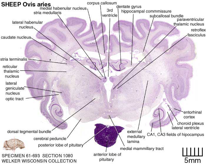
Atlas of the Sheep Brain > Section Image
PDF Lab: Sheep Brain Dissection - Mrs. Moretz's Science Site to anatomy studies. See for yourself what the . cerebrum, cerebellum, spinal cord, gray matter, white matter, and other parts of the brain look like! Observation: External Anatomy . 1. You'll need a . preserved sheep brain. for the dissection. Set the brain down so the flatter side, with the white . spinal cord. at one end, rests on the ...

Atlas of the Sheep Brain > Section Image
Week 7: Sheep Brains with Labeled Cuts | Northfield ... Week 7: Sheep Brains with Labeled Cuts. Sheep brains used in science classrooms such as ours are salvaged after sheep are slaughtered for their meat. Sheep brains are a good resource for teaching neuroscience because they have many similarities to human brains both in anatomy and function, and are more abundant. Side view.
Sheep Brain Dissection with Labeled Images
labeled brain - Pinterest labeled brain Brain Anatomy, Human Anatomy And Physiology, Medical Anatomy, ... Images taken from the dissection of the sheep's brain: cerebrum, cerebellum, ...
BIO201-Sheep Brain
Sheep Brain Label | Dissection, Human brain diagram, Brain ... Sheep Brain Label A drawing of the brain with the parts unlabeled. Students can practice naming the parts of the brain, then check their answers with the provided key. Biologycorner 17k followers More information unlabeled brain Find this Pin and more on A&P by Dijana Kovacevic. Brain Gym For Kids Human Brain Diagram Brain Anatomy And Function

On the Cutting Edge: Exploring Sheep Organs | Carolina.com
Sheep Brain Neuroanatomy Online Self-Test | KPU.ca ... Sheep Brain Neuroanatomy Online Self-Test. Use each diagram as a reference, and selected the correct answer for each lettered structure. You may find it useful to open the diagrams in a separate window to review while answering each question.
(294).jpg)
Sheep Brain Anatomy Quiz - ProProfs Quiz
Sheep brain labeling Quiz - PurposeGames.com The world - Fifteen islands 15p Image Quiz. Continue to count 15p Multiple-Choice. Midwest States and Capitals 12p Matching Game. Geometric Shapes 14p Image Quiz. Objects in ABBA songs - easy 2 15p Image Quiz. PG Lightning Game: Speed counting 20p Shape Quiz. States Without the Letter 'A' 14p Type-the-Answer. The Western States 11p Image Quiz.

sheep brain labeled 9880143 orig - Made By Creative Label
Labeled Diagrams of the Human Brain You'll Want to Copy ... Labeled Diagrams of the Human Brain. Central Core. The central core consists of the thalamus, pons, cerebellum, reticular formation and medulla. These five regions are the central areas that regulate breathing, pulse, arousal, balance, sleep and early stages of processing sensory information. The thalamus interprets the sensory information and ...
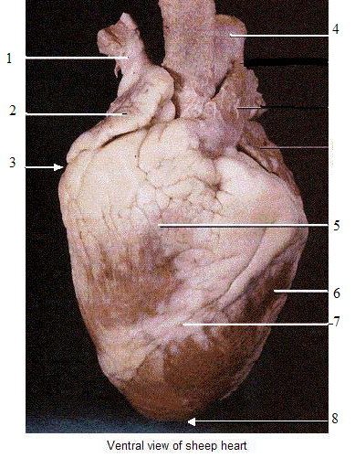
Heart Model Contiunued - ProProfs Quiz
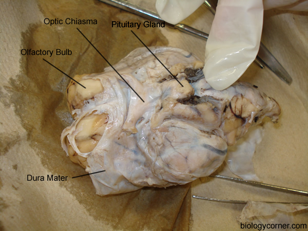
Sheep Brain Dissection with Labeled Images 1. The sheep brain is enclosed in a tough outer covering called the dura mater. You can still see some structures on the brain before you remove the dura mater. Take special note of the pituitary gland and the optic chiasma. These two structures will likely be pulled off when you remove the dura mater. Brain with Dura Mater Intact
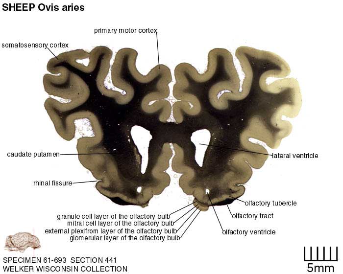
Atlas of the Sheep Brain > Section Image
Sheep Brain Quiz - PurposeGames.com About this Quiz. This is an online quiz called Sheep Brain. This quiz has tags. Click on the tags below to find other quizzes on the same subject. Anatomy. sheep brain.
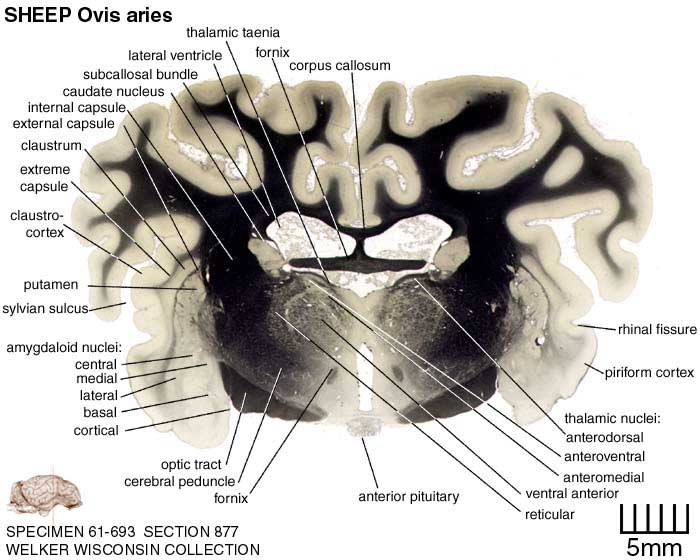
Atlas of the Sheep Brain > Section Image
sheep brain label Diagram | Quizlet sheep brain label Diagram | Quizlet sheep brain label STUDY Learn Write Test PLAY Match + − Created by Isabelhubb PLUS Terms in this set (6) left cerebral hemisphere ... right cerebral hemisphere ... superior colliculi of corpora quadrimina ... inferior colliculi of corpora quadiremina ... cerebellum ... spinal cord ...

diagram of the cow eye images how to guide and refrence | Cow eyes, Eye anatomy, Eyeball anatomy
Sheep Brain Images - San Diego Mesa College NERVOUS SYSTEM - SHEEP BRAIN IMAGES. Sheep Brain Unlabeled. Sheep Brain in Dura Mater · Dorsal Sheep Brain · Dorsal-Colliculi ... Sheep Brain Labeled.
Dr. Parker's A&P I Sheep Brain Dissection - YouTube
Sheep brain dissection | Human Anatomy and Physiology Lab ... The most prominent structure visible on the ventral side of the sheep brain is half of the optic chiasma, which is where the two optic nerves cross over each other and form an "X" shape. You will only see half the structure. Find the optic chiasma half on your brain. You may have removed the optic removed the chiasma with the dura mater.
Sheep Brain Dissection with Labeled Images
Anatomy Sheep Brain Diagram | Quizlet Anatomy Sheep Brain STUDY Learn Flashcards Write Spell Test PLAY Match Gravity + − Created by quizlette331614 Terms in this set (19) Frontal 1 Parietal 2 Occipital 3 Cerebral Hemisphere 4 Spinal Cord 5 Medulla Oblongata 6 Nerve 7 Pons 8 Mid Brain 9 Inferior Collicus 10 Pituitary Gland 11 Optic Chiasma 12 Hypothalamus 13 Thalmus 14 Pineal Body 15

Dorsal View of Sheep Brain (Figure 10.10) Quiz - By kschlittler
DOC Sheep Brain Anatomy Lab Manual - amherst.edu The cruciate fissure (labeled ansate sulcus in your photo atlas) is known in the human brain as the fissure of Rolando or central sulcus, and intersects the medial longitudinal fissure to mark off the anterior third of the cortex.
Sheep+brain+dissection
0 Response to "39 sheep brain diagram labeled"
Post a Comment