35 meiotic division beads diagram
Meiosis Questions and Answers. Get help with your Meiosis homework. Access the answers to hundreds of Meiosis questions that are explained in a way that's easy for you to understand. Diagram the corresponding images for each stage in the section titled "Trial 2 - Meiotic Division Beads Diagram". Be sure to indicate the number of chromosomes present in each cell for each phase. Also, indicate how the crossing over affected the genetic content in the gametes from Part1 versus Part 2. 95 Meiosis
Meiotic Division of Cell (With Diagram) In this article we will discuss about the meiotic division of a cell. The meiotic division includes two complete divisions of a diploid cell resulting into four haploid nuclei. The first meiotic division includes a long prophase in which the homologous chromosomes become closely associated to each other ...
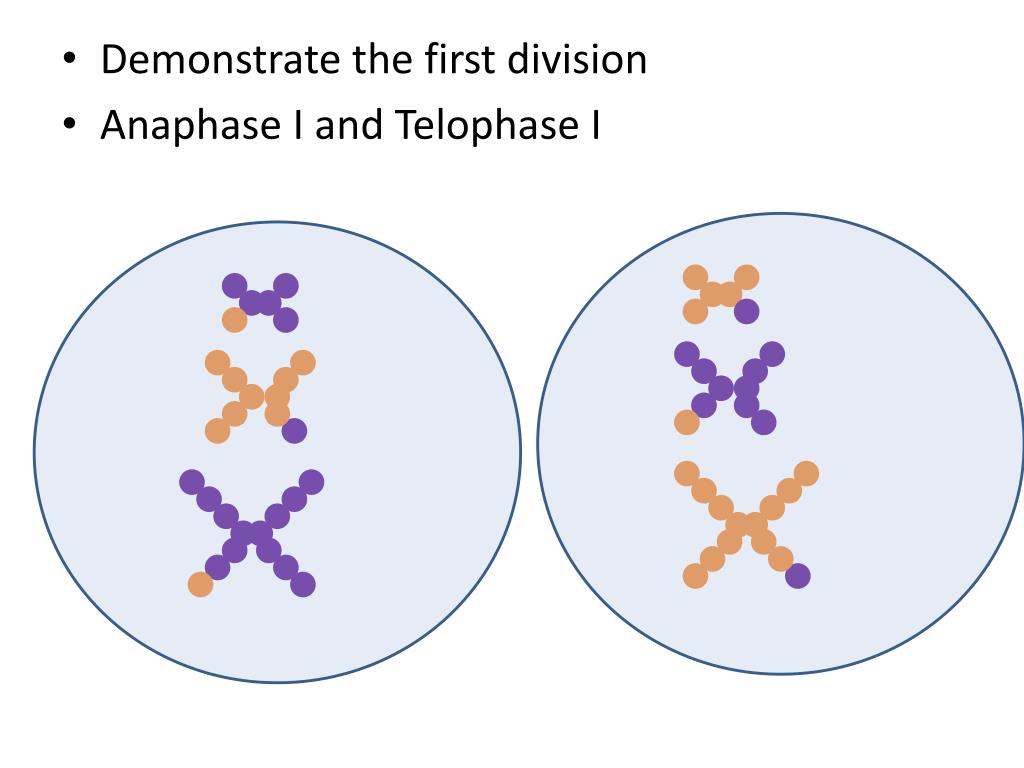
Meiotic division beads diagram
24.12.2014 · We examined meiotic division by ... segregation of heterozygous cen2–GFP during meiosis I. e, Schematic diagram of ... (∼ 8 × 10 7 cells) were … 5. Diagram the corresponding images for each stage in the sections titled "Trial 1 Meiotic Division Beads Diagram". Be sure to indicate the number of chromosomes present in each phase. 6. Disassemble the beads used in Part 1. You will need to recycle these beads for a second meiosis trial in Steps 8 - 13. Trial 1 - Meiotic Division Without Crossing Over Pipe Cleaners Diagram: This lab explores the processes of mitosis and meiosis through both physical and mathematical modeling. frankandmaven. Win challenges by examining genes on their chromosomes, recombining alleles and selecting the right gametes.
Meiotic division beads diagram. Diagram the corresponding images for each stage in the section titled "Trial 2 - Meiotic Division Beads Diagram". Be sure to indicate the number of chromosomes present in each cell for each phase. Take a picture of your results. Include a note with your name and date on an index card in the picture. Insert picture here. The male gonad is the testis (pl, testes).. The initial difference in male and female gonad development are dependent on testis-determining factor (TDF) the protein product of the Y chromosome SRY gene. Recent studies have indicated that additional factors may also be required for full differentiation. Diagram the corresponding images for each stage in the section titled "Trial 2 - Meiotic Division Beads Diagram". Be sure to indicate the number of chromosomes present in each cell for each phase. Also, indicate how the crossing over affected the genetic content in the gametes from Part1 versus Part 2. Meiosis is also a type of cell division – a reduction division of the nucleus of a germ cell because it reduces the amount of DNA in a cell.3 pages
Start studying BIO 351 FINAL EXAM !. Learn vocabulary, terms, and more with flashcards, games, and other study tools. stages of meiotic division (prophase I and II, metaphase I and II, anaphase I and II, telophase I and II, and cytokinesis). 12. Diagram the corresponding images for each stage in the section titled "Trial 2 Meiotic Division Beads Diagram". Be sure to indicate the number of chromosomes present in each cell for each phase. Also, indicate how ... Explore this photo album by Darrietta Lee on Flickr! 5. Diagram the corresponding images for each stage in the sections titled "Trial 1 - Meiotic Division Beads Diagram". Be sure to indicate the number of chromosomes present in each cell for each phase. 6. Disassemble the beads used in Part 1. You will need to recycle these beads for a second meiosis trial in Steps 7 - 12.
In experiment of Following chromosomal DNA movement through meiosis, what is the trial 1 and trial 2 meiotic division beads diagram for prophase l, metaphase l, anapahse l, telophase l, prophase ll, metaphase ll, anaphase ll, telophase ll, and cytokinesis? Start your trial now! First week only $4.99! arrow_forward. Diagram the corresponding images for each stage in the section titled "Trial 2 - Meiotic Division Bead Diagrams." Be sure to indicate the number of chromosomes present in each cell for each phase. Also, indicate how the crossing over affected the genetic content in the gametes from Part 1 versus Part 2. It also connects back to our work on the differences between cell division in cancer cells vs. normal cells. I typically keep the beads out in the classroom so that students can use them as we talk throughout the unit. I use these beads from the AP Biology class lab kit from Wards, but you can use any repeating unit bead/toy that you have on hand. Diagram the corresponding images for each stage in the section titled "Trial 2 - Meiotic Division Beads Diagram". Be sure to indicate the number of chromosomes present in each cell for each phase. Also, indicate how the crossing over affected the genetic content in the gametes from Trial 1 versus Trial 2. Trial 2 - Meiotic Division Beads ...
second meiotic division (meiosis II) that they finally are separated and distributed into separate ... In this diagram the chromosomes are shown as if they were visible, ... which clearly defined beads of local coiling (chromomeres) can be seen.
In cell biology, the spindle apparatus (or mitotic spindle) refers to the cytoskeletal structure of eukaryotic cells that forms during cell division to separate sister chromatids between daughter cells.It is referred to as the mitotic spindle during mitosis, a process that produces genetically identical daughter cells, or the meiotic spindle during meiosis, a process that produces …
Diagram the corresponding images for each stage in the sections titled "Trial 1 - Meiotic Division Beads Diagram". Be sure to indicate the number of chromosomes present in each cell for each phase. 6. Take a picture of your results. Include a note with your name and date on an index card in the picture. Insert picture here.
Diagram the corresponding images for each stage in the section titled "Trial 2 - Meiotic Division Beads Diagram". Be sure to indicate the number of chromosomes present in each cell for each phase. Also, indicate how the crossing over affected the genetic content in the gametes from Part1 versus Part 2. Part 2 - Meiotic Division Beads ...
Trial 1 - Meiotic Division Without Crossing Over Beads Diagram: Take pictures of your beads for each phase of meiosis I and II without crossing over. Include notes with your name, date and meiotic stage on index cards in the pictures. Please use the lowest resolution possible so that your file does not become too large to submit.
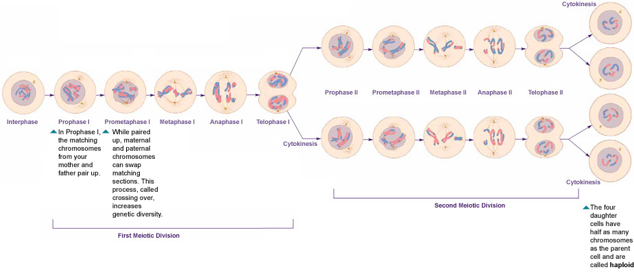
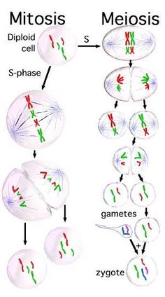
The second meiotic division is where sister (duplicated) chromatids separate. It resembles mitosis of a haploid cell. At the start of the second division, each cell contains 1N chromosomes, each consisting of a pair of sister chromatids joined at the centromere. Here is a simplified diagram illustrating the overall process and products of meiosis:
6. Diagram the corresponding images for each stage in the sections titled â Trial 1 - Meiotic Division Beads 186 Meiosis Diagramâ . Be sure to indicate the number of chromosomes present in each cell for each phase. 7. Disassemble the beads used in Part 1. You will need to recycle these beads for a second meiosis trial in Steps 8
Diagram the corresponding images for each stage in the section titled "Trial 2 - Meiotic Division Beads Diagram". Be sure to indicate the number of chromosomes present in each cell for each phase. Also, indicate how the crossing over affected the genetic content in the gametes from Part1 versus Part 2. Part 2 - Meiotic Division Beads Diagram:
Diagram the corresponding images for each stage in the sections titled "Trial 1 - Meiotic Division Beads Diagram". Be sure to indicate the number of chromosomes present in each cell for each phase. Disassemble the beads used in Trial 1. You will need to recycle these beads for a second meiosis trial in Steps 7 - 11.
Trial 2 meiotic division beads diagram prophase i 4. School Indiana University, Purdue University Indianapolis; Course Title BIOL MISC; Uploaded By ready2finish2022. Pages 14 Ratings 94% (18) 17 out of 18 people found this document helpful; This preview shows page 9 - 14 out of 14 pages. Students who viewed this also studied. Broward College • BSC1005L 1005. L12_Meiosis.docx. Mitosis ...
Meiotic Division Beads Diagram. For more information: 7activestudio [email protected] 7activemedical [email protected] sciencetuts 7activ. Cell division usually occurs as part of a larger cell cycle. all cells reproduce by splitting into two, where each parental cell gives rise to two daughter cells. these newly formed daughter cells could themselves divide and grow, giving rise to a new cell ...
stages of meiotic division (prophase I and II, metaphase I and II, anaphase I and II, telophase I and II, and cytokinesis). 12. Diagram the corresponding images for each stage in the section titled "Trial 2 Meiotic Division Beads Diagram". Be sure to indicate the number of chromosomes present in each cell for each phase. Also, indicate how ...
The meiotic board used includes six circles: one on top; two in the middle and four at the bottom. These circles represent the chromosomal make up of the seven nuclei involved in one meiotic event. The top circle represents the karyotype of mother nucleus, the center two of the two daughter nuclei, and the bottom four of the granddaughter nuclei.
Jun 02, 2019 · Biology I Lab Activity – Simulating Mitosis with “Pop Beads” Introduction: Mitosis is the process of one cell dividing to produce two new (daughter) cells (take a look at the diagram . Diagram the corresponding images for each stage in the section titled “Trial 2 - Meiotic Division Beads Diagram”.
Diagram the corresponding images for each stage in the sections titled “Trial 1 - Meiotic Division Beads Diagram”. Be sure to indicate the number of chromosomes present in each cell for each phase. Disassemble the beads used in Part 1. You will need to recycle these beads for a second meiosis trial in Steps 7 - 12. Trial 1 - Meiotic Beads Diagram:
Discussion on Meiotic Division Beads Diagram · Start with 20 beads of the same color to create your first sister chromatid pair. · Assemble a second pair of ...
A chromosome is a long DNA molecule with part or all of the genetic material of an organism. Most eukaryotic chromosomes include packaging proteins called histones which, aided by chaperone proteins, bind to and condense the DNA molecule to maintain its integrity. These chromosomes display a complex three-dimensional structure, which plays a significant role in …
Cell Cycle Division: Part 2 - Meiotic Bead Diagrams (With Crossing Over) Prophase I: One chromosome from mother one from father come. together and wrap around each other so closely that portions of one switch. with portions of the other. They are also lined up along the middle. Anaphase II: The chromosomes are split in half and move to the. side
Transcribed image text: Data She experi Lab 12 > Experiment 1 Data Sheet dels TRIAL 2 - MEIOTIC DIVISION BEAD DIAGRAMS: Prophase : LE Metaphase 1: Anaphase 1: Telophase 1: Prophase II: Metaphase II: Anaphase II: Telophase II: Cytokinesis: Data SheeExperia Lab 12 Experiment 1 Data Sheet TRIAL 2 - MEIOTIC DIVISION BEAD DIAGRAMS: Prophase 1 ...
View L12_Meiosis.docx from BSC1005L 1005 at Broward College. Meiosis EXPERIMENT 1: FOLLOWING CHROMOSOMAL DNA MOVEMENT THROUGH MEIOSIS Trial 1 - Meiotic Division Beads Diagram Prophase I Metaphase
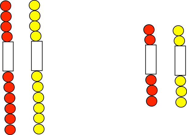
Part 1 – Meiotic Division Beads Diagram Prophase I Metaphase I Anaphase I Telophase I Prophase II Metaphase II Anaphase II Telophase II Cytokinesis Part 2: Modeling Meiosis with Crossing Over Build a pair of replicated, homologous chromosomes. 10 beads should be used to create each individual sister chromatid (20 beads per chromosome pair).

When we get to the river my heat sinks. It is wider than I've ever seen, flowing swift and strong. The water is a turbid brown from eroding the banks it usually passes by so softly. Branches have been blow in by the storm. The water eddies around them, but not that relaxed way water usually does, but harshly, more like mini vortexes. In the rain that still falls the surface is pitted so thickly that the radiating ripples cancel one another out. Adding to the torrent coming from the stone-grey sky is the April melt from the mountains to the East. This river is not what we prepared ourselves for
9.6.2021 · The beads were washed three times with wash buffer, once with high-salt wash buffer (5× PBS, 0.1% SDS, 0.5% NP-40 and 0.5% sodium deoxycholate) and once with PNK buffer (50 mM Tris-HCl, pH 7.4 ...
The meiotic board used includes six circles: one on top; two in the middle and four at the bottom. These circles represent the chromosomal make up of the seven nuclei involved in one meiotic event.Ninth grade Lesson Using a Simulation Activity to Explore and Compare Meiosis and MitosisMeiotic Division of Cell (With Diagram)
Trial 2 - Meiotic Division Beads Diagram: Prophase I. Metaphase I. Anaphase I. Telophase I. Prophase II. Metaphase II. Anaphase II. Telophase I. Cytokinesis. Post-Lab Questions. 1. Poloidy of the DNA at What is the the end of meiosis I? What about at the end of meiosis II?
Diagram the corresponding images for each stage in the section titled "Trial 2 - Meiotic Division Beads Diagram". Be sure to indicate the number of chromosomes present in each cell for each phase. Also, indicate how the crossing over affected the genetic content in the gametes from Part1 versus Part 2.
Trial 1 - Meiotic Division Without Crossing Over Pipe Cleaners Diagram: This lab explores the processes of mitosis and meiosis through both physical and mathematical modeling. frankandmaven. Win challenges by examining genes on their chromosomes, recombining alleles and selecting the right gametes.
5. Diagram the corresponding images for each stage in the sections titled "Trial 1 Meiotic Division Beads Diagram". Be sure to indicate the number of chromosomes present in each phase. 6. Disassemble the beads used in Part 1. You will need to recycle these beads for a second meiosis trial in Steps 8 - 13.
24.12.2014 · We examined meiotic division by ... segregation of heterozygous cen2–GFP during meiosis I. e, Schematic diagram of ... (∼ 8 × 10 7 cells) were …











0 Response to "35 meiotic division beads diagram"
Post a Comment