40 blood flow to the brain diagram
The fetal blood flow pathway occurs via a combination of various blood vessels and bypass shunts. Since the fetal lungs do not provide oxygen, one the main goals of fetal circulation is to be ... Oct 28, 2021 · The derivatives of the internal carotid arteries form the anterior blood supply (anterior circulation) of the brain, which includes the anterior and middle cerebral arteries. The subclavian artery is divided into three parts based on anatomical landmarks. The first part extends from its origin to the medial border of the scalenus anterior muscle.
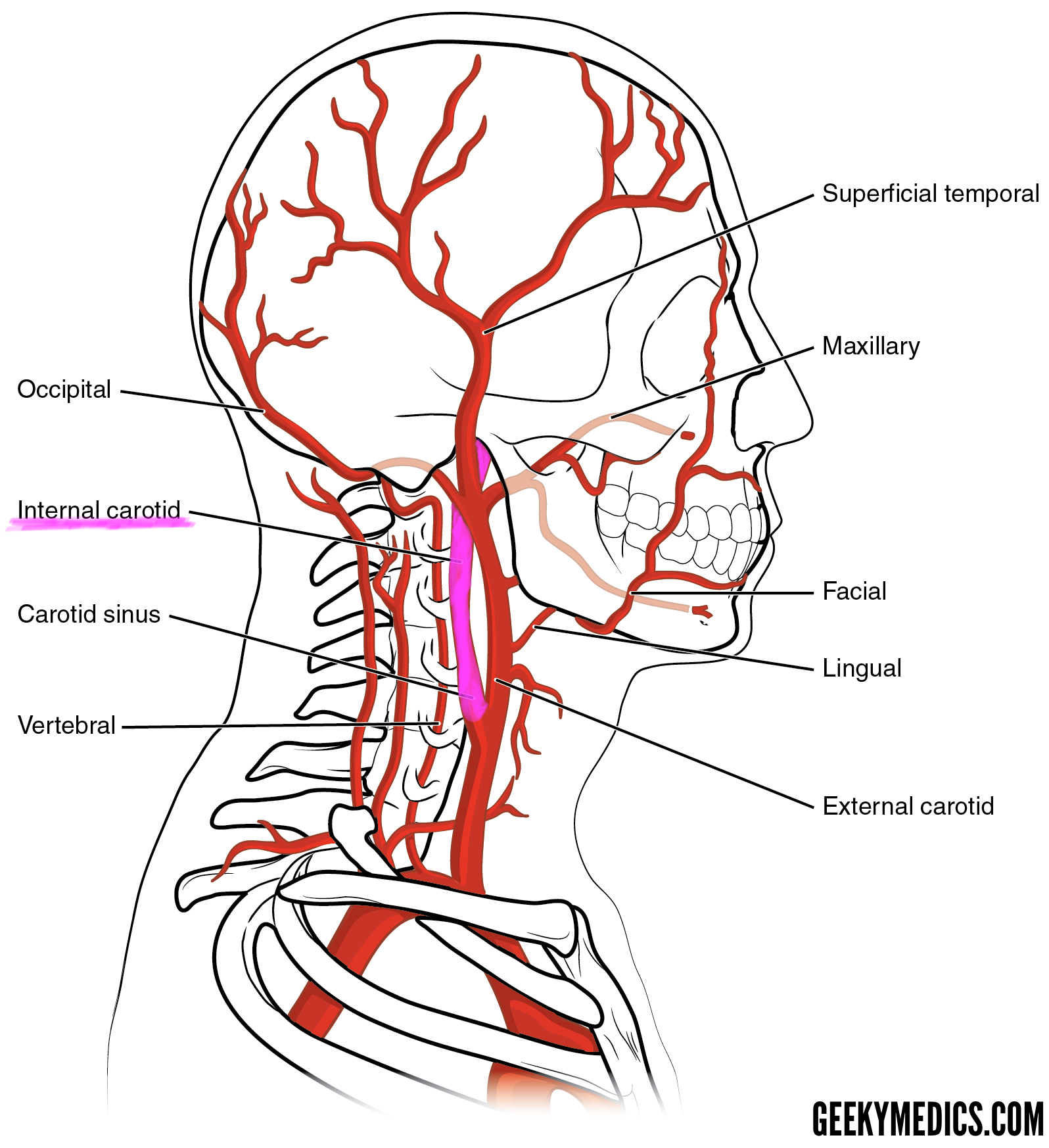
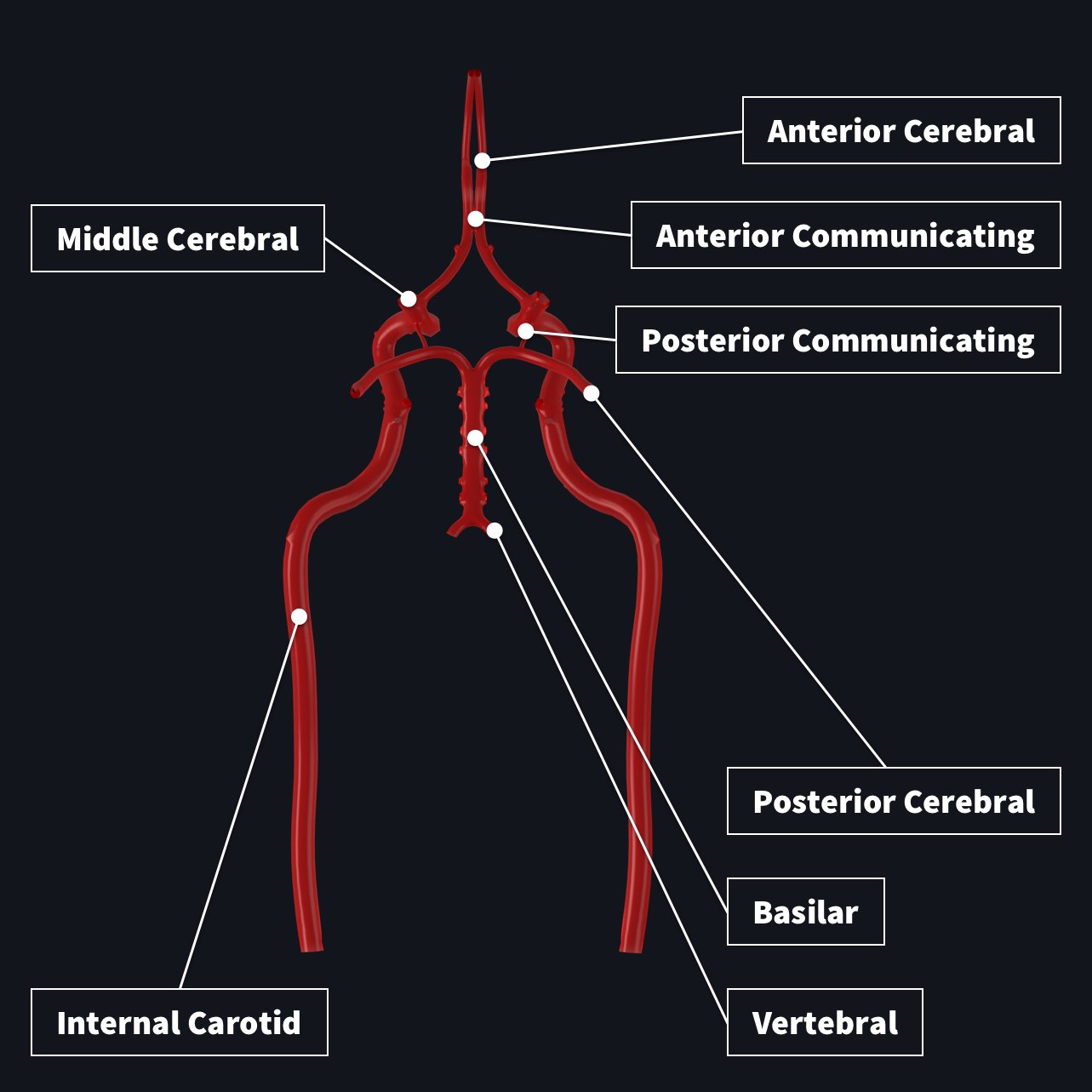
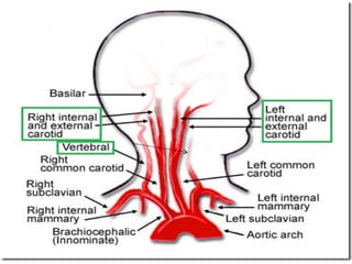
The entire blood supply of the brain and spinal cord depends on two sets of branches from the dorsal aorta. The vertebral arteries arise from the subclavian arteries, and the internal carotid arteries are branches of the common carotid arteries.
Blood flow to the brain diagram
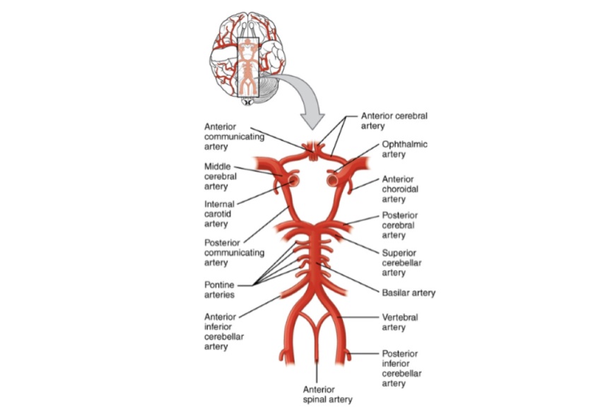
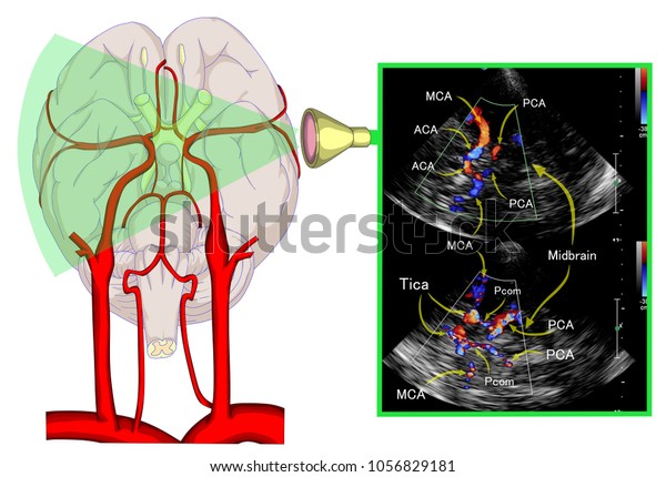
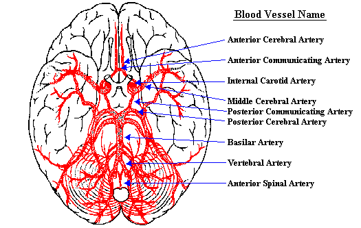
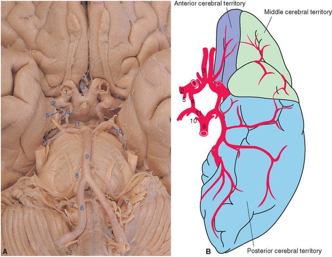
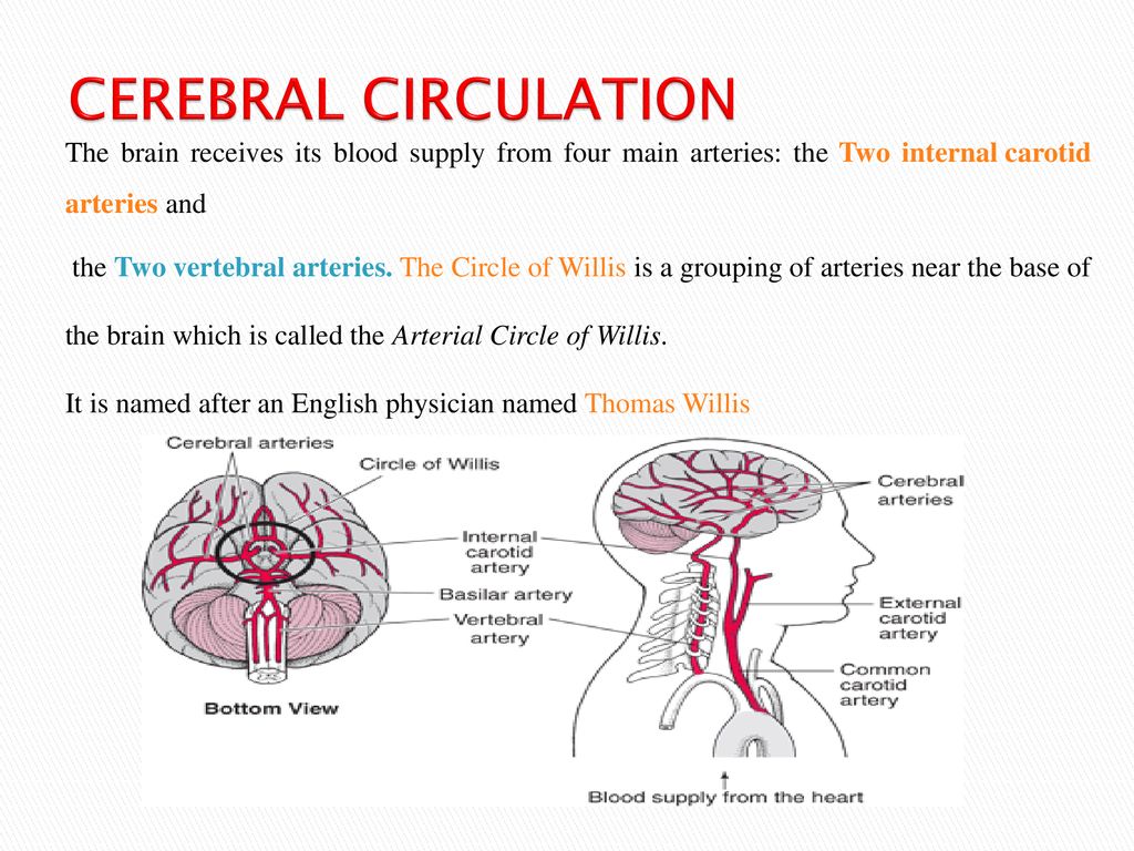
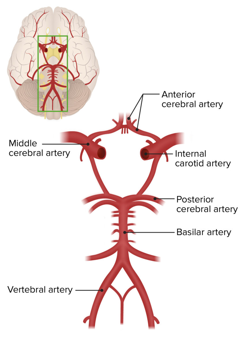
The main arteries that supply the brain with blood are the paired vertebral and internal carotid arteries. They begin in the neck and travel up to the cranium. Once in the cranial vault, the terminal branches form an anastomotic circle, commonly known as the Circle of Willis . Branches arise from the circle to supply most of the cerebrum. Blood Flow. All blood enters the right side of the heart through two veins: The superior vena cava (SVC) and the inferior vena cava (IVC) (see figure 3). The SVC collects blood from the upper half of the body. The IVC collects blood from the lower half of the body. Blood leaves the SVC and the IVC and enters the right atrium (RA) (3). When the ... This video is part of a playlist on blood vessel anatomy & physiology at my youtube channel drjahn41. Feel free to review the other videos in the playlist at...
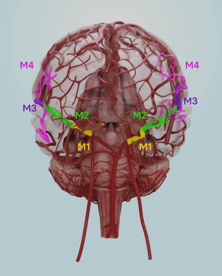
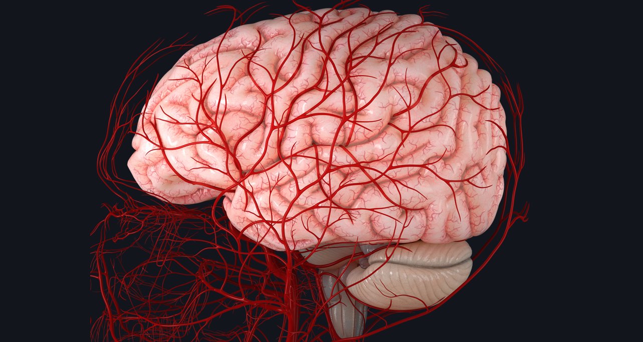
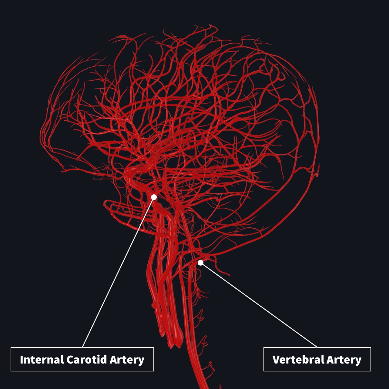
Blood flow to the brain diagram. Arterial blood supplies the brain with the necessary blood flow via four vessels. Near the pituitary gland, these four vessels converge to create one unified vessel. This can be located in the inferior surface of the brain. These four vessels are each one set of paired arteries, a set of internal carotid arteries and a pair of vertebral arteries. Reduced blood flow decreases the brain's supply of oxygen. Symptoms signaling a headache, such as distorted vision or speech, may then result, similar to symptoms of stroke. As these arteries constrict, the flow of blood to the brain is reduced. At the same time, blood-clotting particles called platelets clump together-a process which is ... The brain is one of the most highly perfused organs in the body. It is therefore not surprising that the arterial blood supply to the human brain consists of two pairs of large arteries, the right and left internal carotid and the right and left vertebral arteries (Figure 1). The internal carotid arteries principally supply the cerebrum, whereas the two vertebral arteries join distally to form ... Blood flow is a cycle that involves your lungs, heart chambers, valves, and blood vessels. Electrical pulses make your heart muscles squeeze and release. That action pushes blood through the two chambers on the right side of your heart and out to the lungs where it gathers oxygen.
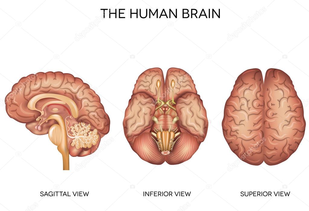
1. The Anatomy of Human Brain The human brain regulates nearly every part of the human body, from bodily processes to cognitive ability. This article will explore the anatomy of the human brain and visually depict it with a brain diagram.. The human brain contains functions of receiving and transmitting signals to various parts of the body through neurons. Blood is a body fluid in humans and other animals that delivers necessary substances such as nutrients and oxygen to the cells and transports metabolic waste products away from those same cells.. In vertebrates, it is composed of blood cells suspended in blood plasma.Plasma, which constitutes 55% of blood fluid, is mostly water (92% by volume), and contains proteins, glucose, mineral ions ... The vertebral arteries travel through the transverse foramina of the cervical vertebrae before entering the skull at the foramen magnum and joining at the base of the brain to form the basilar artery. From there the basilar artery provides blood to the posterior structures of the brain, including the brain stem, cerebellum, and cerebrum. The brain is particularly sensitive to oxygen starvation. A stroke is an acute development of a neurological deficit, due to a disturbance in the blood supply of the brain. There are four main causes of a cerebrovascular accident: Thrombosis - obstruction of a blood vessel by a locally forming clot.
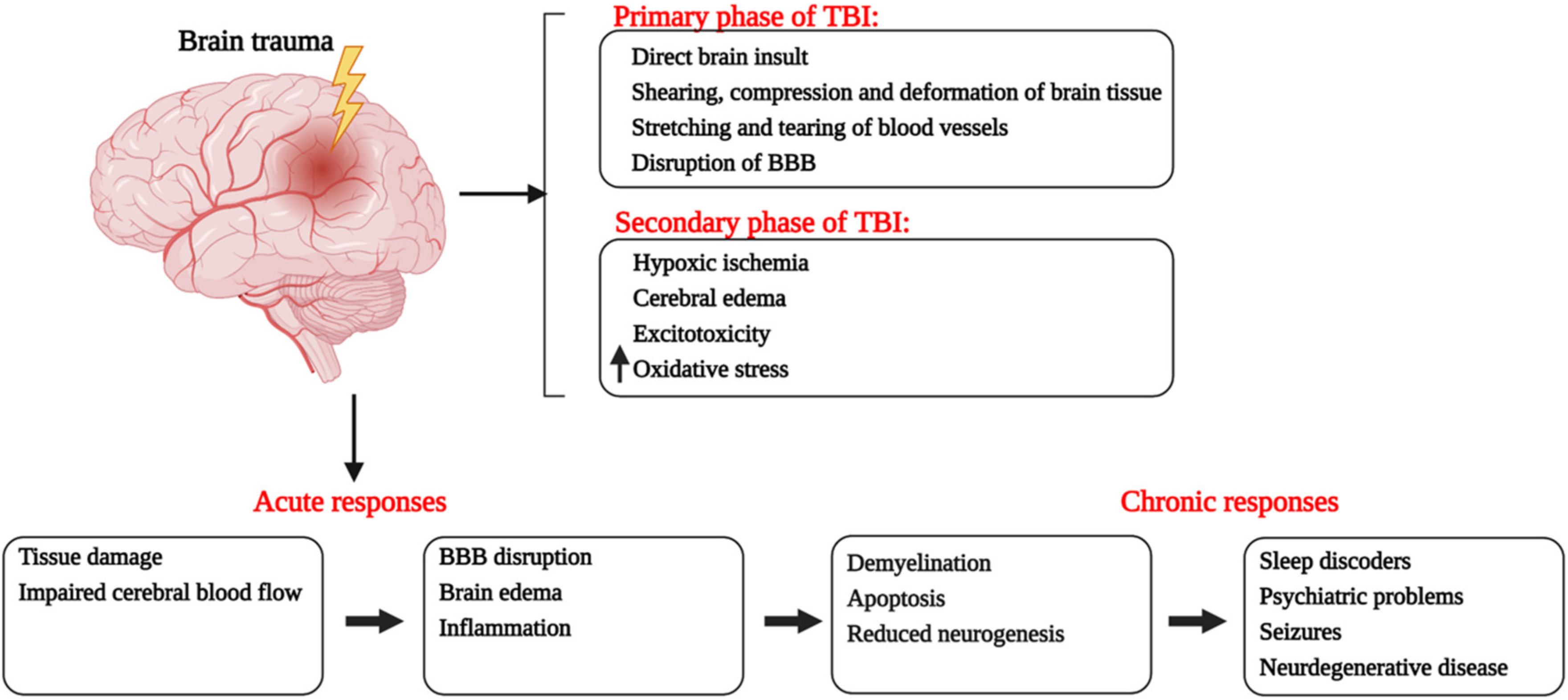
brain. Other medications increase blood flow into the brain which can help diminish the spiral effect caused by brain swelling. In some cases, removal of small amounts of fluids or from the brain or surgery may be beneficial. And in some cases, removal of damaged tissue may prevent further damage. Nov 04, 2021 · The rate of cerebral blood flow in an adult human is typically 750 milliliters per minute or about 15 of cardiac output. Blood flow through the brain diagram. This page has Four Sections. The left and right vertebral arteries and the left and right common carotid arteries. The brain sends a signal down a particular nerve fiber. This nerve fiber ends in a nerve cell in an artery, somewhere near the point where blood flow needs to change. These nerve cells -- called nonadrenergic-noncholinergic, or NANC -- produce nitric oxide and inject it into the blood and surrounding cells. Blood Flow Through the Heart. Beginning with the superior and inferior vena cavae and the coronary sinus, the flowchart below summarizes the flow of blood through the heart, including all arteries, veins, and valves that are passed along the way. 1. Superior and inferior vena cavae and the coronary sinus 2. Rt. atrium 3.
Arteries Arteries is a collection of sciencepics blood flow diagram stock pictures, royalty-free photos & images. circulation A simplified diagram showing blood circulation in the human body. blood flow diagram stock illustrations. 3d rendering of the human heart anatomy 3d rendering of the human heart anatomy blood flow diagram stock pictures ...
Arteries of the head, neck and brain 6p Image Quiz. Frontal heart section 24p Image Quiz. Arteries of the abdomen view 3 5p Image Quiz. Anatomy of the oral cavity 11p Image Quiz. Arteries of the body 3p Image Quiz. PurposeGames Create. Play. ... This online quiz is called The blood flow through the heart.
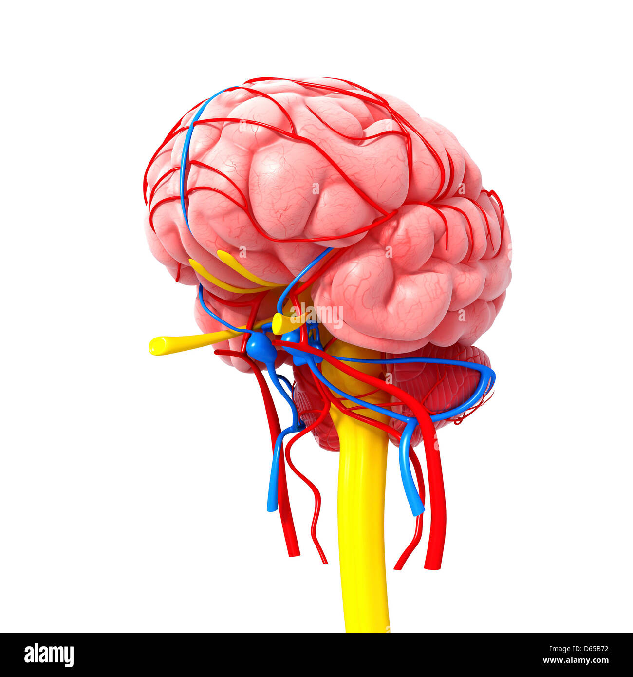
The right and left vertebral arteries come together at the base of your brain to form what is called the basilar artery in the vasculature of your head. As shown in the diagrams of the vasculature of the head, the vessels that carry oxygen-rich blood are colored red, and the vessels that carry oxygen-poor blood are colored blue. Share on.
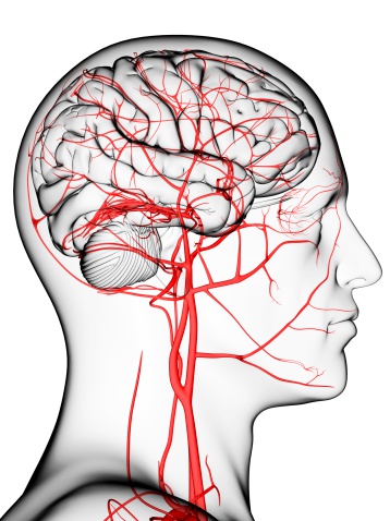
Every minute, about 600-700 ml of blood flow through the carotid arteries and their branches while about 100-200 ml flow through the vertebral-basilar system. The carotid and vertebral arteries begin extracranially, and course through the neck and base of the skull to reach the cranial cavity.
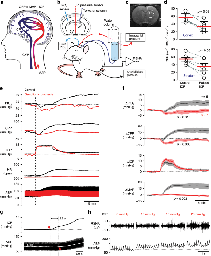
Hypoxic hypoxia (inspiratory hypoxia) stimulates an increase in cerebral blood flow (CBF) maintaining oxygen delivery to the brain. However, this response, particularly at the tissue level, is not well characterised. This study quantifies the CBF response to acute hypoxic hypoxia in healthy subjects …
Regulation of Blood Flow: 1. Increased carbon dioxide tension (increased pCO 2) is the most important factor. CO 2 is a power full vasodilator of the cerebral blood vessels. Increasing the CO 2 content of the inspired air (3-5%) almost doubles the blood flow to the brain. Voluntary hyperventilation decreases the pCO 2, and brings about ...
In general, if the heart stops beating, in about 4-6 minutes of no blood flow, brain cells begin to die and after 10 minutes of no blood flow, the brain cells will cease to function and effectively be dead. There are few exceptions to the above.
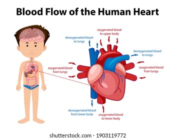
Diagram: Blue arrows demonstrate flow of deoxygenated blood through the right side of the heart. Red arrows demonstrate flow of oxygenated blood through the left side of the heart.
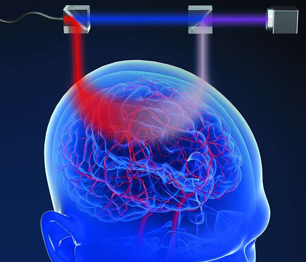
Cerebral circulation is the movement of blood through a network of cerebral arteries and veins supplying the brain.The rate of cerebral blood flow in an adult human is typically 750 milliliters per minute, or about 15% of cardiac output. Arteries deliver oxygenated blood, glucose and other nutrients to the brain. Veins carry "used or spent" blood back to the heart, to remove carbon dioxide ...
28.05.2021 · Brain size doesn't indicate a person's intelligence. ... the researchers injected pig brains with chemicals that mimicked blood flow and …
The brain comprises only 2% of total body weight, yet receives 15 to 20% of its total blood supply. There's a lot going on up there, so the brain requires a disproportionate amount of energy and nutrients.. Why Sufficient Blood Flow to the Brain Is Critical
Blood Flow • Blood flow is the volume of blood flowing through a vessel, an organ, or the entire circulation in a given period (ml/min). • If we consider the entire vascular system, blood flow is equivalent to cardiac output (CO), Dr. Naim Kittana, PhD 33
Aug 23, 2021 · Oxygenated blood supply is necessary for the brain to perform normal functions. Learn about the three arteries (internal carotid artery, anterior cerebral artery, and posterior cerebral artery ...
Oct 03, 2021 · The rate of cerebral blood flow in an adult human is typically 750 milliliters per minute or about 15 of cardiac output. The blood vessels and nerves enter the brain through holes in the skull called foramina. Draw the diagram of heart showing blood circulation.
23.01.2018 · Although the signal for erection originates in the brain, the dorsal nerve sends and receives signals to increase blood flow. Additionally, …
This video is part of a playlist on blood vessel anatomy & physiology at my youtube channel drjahn41. Feel free to review the other videos in the playlist at...
Blood Flow. All blood enters the right side of the heart through two veins: The superior vena cava (SVC) and the inferior vena cava (IVC) (see figure 3). The SVC collects blood from the upper half of the body. The IVC collects blood from the lower half of the body. Blood leaves the SVC and the IVC and enters the right atrium (RA) (3). When the ...
The main arteries that supply the brain with blood are the paired vertebral and internal carotid arteries. They begin in the neck and travel up to the cranium. Once in the cranial vault, the terminal branches form an anastomotic circle, commonly known as the Circle of Willis . Branches arise from the circle to supply most of the cerebrum.
:background_color(FFFFFF):format(jpeg)/images/library/14122/normal.png)











:watermark(/images/watermark_only.png,0,0,0):watermark(/images/logo_url.png,-10,-10,0):format(jpeg)/images/anatomy_term/circulus-arteriosus-cerebri/qNRQ5TN7dGARxv8Qr5ysNg_Circulus_arteriosus_cerebri_01.png)


0 Response to "40 blood flow to the brain diagram"
Post a Comment