38 uv vis block diagram
Another word for atomic spectroscopy is elemental analysis. 3 basic types Atomic spectroscopy, X-Ray Fluorescence, and ICP-MS. Look at these 4 spectra of chromium cations with 2 different ligands (NO 3 and Cl) in 2 different solvents. Figure 1: UV/Vis spectrum of Chromium cations with 2 different ligands (NO 3 and Cl) in water and ethanol. UV/UV-VIS detectors are most frequently used to measure components showing an absorption spectrum in the ultraviolet or visible region. A UV detector employs a deuterium discharge lamp (D 2 lamp) as a light source, with the wavelength of its light ranging from 190 to 380 nm. If components are to be detected at wavelength longer than this, a UV ...
601-d Block Diagram of HPLC System 601-6 1/94 ... 605-a UV/VIS Detector 605-3 1/94 605-b Variable Wavelength UV/VIS Detector 605-4 1/94 605-c Fluorescence HPLC Detector 605-6 9/96

Uv vis block diagram
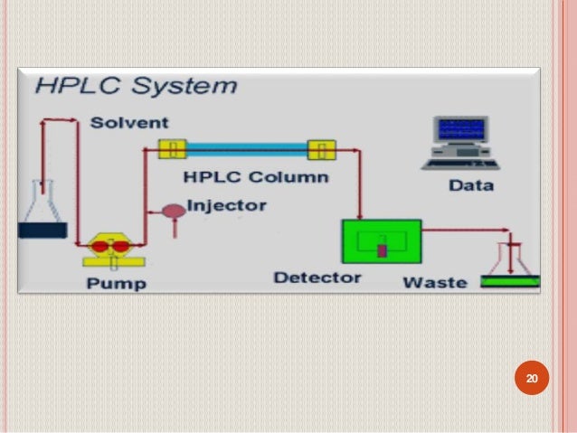
Schematic of the internal layout of an Agilent Cary 5000 UV-Vis-NIR spectrophotometer, showing the main components. Note that this is a.36 pages Block Diagram of a Mass Spectrometer Ion Source Detector "High" Vacuum Sample In Computer Tandem Mass Spectrometry or MS/MS m/z. S Q H L F A P I S E K 200 400 600 800 1000 1200 0 20 40 60 80 100 Relative Abudance m/z. S Q H L F A P I S E K 147 276 363 477 574 645 792 905 1042 1170 216 353 467 614 685 782 895 982 Block Diagram and Explanation A basic block diagram of an HPLC is shown in Figure 1. Figure 1: Block Diagram of an HPLC ... also need to know what wavelengths in the UV-VIS spectrum you want to monitor. A diode array detector has a range of 210-400 nm and for samples, the default program setting are fine. The
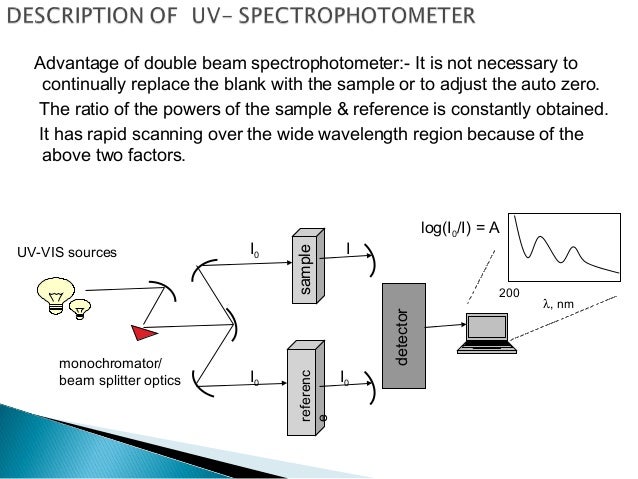
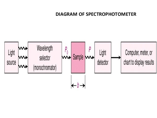
Uv vis block diagram. UV spectroscopy is an important tool in analytical chemistry. The other name of UV (Ultra-Violet) spectroscopy is Electronic spectroscopy as it involves the promotion of the electrons from the ground state to the higher energy or excited state. In this article I will explain the basic principle, working and all the applications of UV spectroscopy. Chapter 1: UV-Visible & Fluorescence Spectroscopy 4 Figure 1-3: An example UV-Vis spectrum, showing a λmax at 591.1 nm. 1.4 Fluorescence Spectroscopy Fluorescence is a complementary technique to UV-Vis absorption. It occurs in the same wavelength range, but results from an excited state emitting a photon of a lower energy than it absorbed. UV-vis spectroscopy is a cost-effective, simple, versatile, non-destructive, analytical technique suitable for a large spectrum of organic compounds and some inorganic species. As a function of wavelength, UV-vis spectrophotometers measure the absorption or transmission of light that passes through a medium. In order to classify and measure the ... UV/Vis spectrophotometer is used in the quantitative determination of concentrations of the absorber in the solutions of transition metal ions and highly conjugated organic compounds. The UV-Vis Spectrophotometer (Varian Cary 100) (Figure 1) was established in the Core Lab in the Core Lab (201D) in 2007 with the DRDO grant of the Institute.
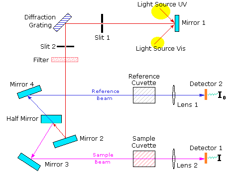
Download scientific diagram | Block Diagram of UV-Visible Spectrometer. from publication: Effect of Doping on Nano Cadmium-Selenide (CdSe) -Assessment Through UV-VIS Spectroscopy | Cadmium ... The principle of Ultra Violet (UV) Spectrophotometer. Ankur Choudhary. Oct 12, 2017 · 3 min read. Ultraviolet-visible spectroscopy is considered an important tool in analytical chemistry. In fact ... UV-Vis: Qualitative analysis Internuclear distances and force constants corresponding to the excited electronic states differ from those corresponding to ground electronic states! 250 nm = 40000 cm-1 = 1.2 x 10¹⁵ Hz 400 nm = 25000 cm-1 = 7.5 x 10¹⁴ Hz 750 nm = 13333 cm-1 = 4 x 10¹⁴ Hz UV Vis 21 Nov 2021 — UV spectroscopy is a type of absorption spectroscopy in which light of the ultra-violet region (200-400 nm) is absorbed by the molecule ...
Ultraviolet-visible (UV-Vis) spectroscopy is one of the most popular analytical techniques because it is very versatile and able to detect nearly every molecule. With UV-Vis spectroscopy, the UV-Vis light is passed through a sample and the transmittance of light by a sample is measured. UV spectroscopy or UV-visible spectrophotometry (UV-Vis or UV/Vis) refers to absorption spectroscopy or reflectance spectroscopy in part of the ultraviolet and the full, adjacent visible regions of the electromagnetic spectrum.This means it uses light in the visible and adjacent ranges. The absorption or reflectance in the visible range directly affects the perceived color of the chemicals ... Introduction: UV-Visible spectroscopy is a mature and well-established analytical technique used extensively in many industry sectors including Environmental Analysis, Pharmaceutical Testing, Food and Beverage Production etc. Spectroscopy is the measurement and interpretation of electromagnetic radiation absorbed or emitted when the molecules or atoms or ions of a sample moves from one energy ... 1.2 UV-1700 Outline UV-1700 PharmaSpec - a spectrophotometer for ultr aviolet and visible region that is the first in its class to amply clear the 1nm resolution barrier to conform to wavelength resolution regulations specified in the European Pharmacopoeia - is a sister instrument to the highly rated UV-1600. The UV-1700 has the following ...
spectroscopy
A Schematic Diagram Of Typical Single Beam Uv Absorbance Monitor Scientific. 6 A Dual Beam Uv Vis Spectrophotometer 8 Scientific Diagram. Parison Between Single Beam And Double Atomic Absorption Spectrometer Systems. Spectrophotometry In 2021 Tools For Lied Food Science Felixinstruments.

A rocky beach on the Island of Vis Croatia
UV-Visible spectroscopy is widely used in the field of analytical chemistry, especially during the quantitative analysis of a specific analyte. For example, the quantitative analysis of transition metal ions can be achieved with the help of UV-Visible spectroscopy. Furthermore, the quantitative analysis of conjugated organic compounds can also ...
10.3: UV/Vis and IR Spectroscopy - Chemistry LibreTexts
Block diagram of a single-beam atomic absorption spectrometer. Radiation from a line source is focused on ... UV/Vis Spectra for Atoms When a beam of UV or Vis radiation passes through a medium containing atoms, only a ... energy level diagram for sodium's occupied and unoccupied valence shell atomic orbitals.

The UV-Visible spectrometer sketch diagram | Download ...
Have a look at this schematic diagram of a double-beam UV-Vis. spectrophotometer;. Instruments for measuring the absorption of U.V. or visible radiation are ...

Block diagram of the CCD spectrometer | Download ...
Below is a diagram of the 508 PV™ Microscope Spectrophotometer integrated with a microscope so that the system can measure UV-visible-NIR spectra in both reflectance and transmission of microscopic samples. Additionally, ...

Block diagram of the UV-LED exposure system. | Download ...
Physikalisch-chemisches Praktikum I UV/VIS Spectroscopy { 2017 cent peaks (or troughs) in the time-frozen electromagnetic wave, and is given in meters, centimetres or nanometres (10 9 meters). Visible wavelengths cover a range from approx-imately 400 to 750 nm. The frequency is the number of wave cycles that travel past

Inked 2
UV-Visible Spectroscopy A diagram of the components of a typical spectrometer are shown in the following diagram. The functioning of this instrument is relatively straightforward. A beam of light from a visible and/or UV light source (colored red) is separated into its component wavelengths by a prism or diffraction grating.
Optical system for detection of lignan using UV visible ...
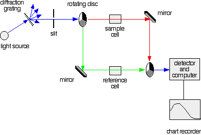
This page describes a double beam UV-visible absorption spectrometer in enough detail to meet the requirements of UK-based chemistry courses for 16 - 18 year olds. If you pass white light through a coloured substance, some of the light gets absorbed. A solution containing hydrated copper(II) ions ...

Block Diagram of UV-Visible Spectrometer. | Download ...
The diagram below shows a simple UV/visible absorption spectrum for buta-1,3-diene—a molecule which will be detailed later. Absorbance (on the vertical axis) is just a measure of the amount of light absorbed. The higher the value, the more particular the wavelength is being absorbed. Absorption maxima diagram
Block diagram of NO and N2O sensor | Download Scientific ...
Ultraviolet-visible (UV-Vis) spectrophotometry is a technique used to measure light absorbance across the ultraviolet and visible ranges of the electromagnetic spectrum. When incident light strikes matter it can either be absorbed, reflected, or transmitted. The absorbance of radiation in the UV-Vis range causes atomic excitation, which refers ...

Schematic Diagram Of A Single Beam Uv Vis ...
In Ultravoilet/Visible (UV-Vis) spectroscopy, electromagnetic radiation is emitted within the ... Schematic of our single beam diode array spectrometer.

Schematic Diagram Uv Vis Spectrophotometer - SHELVESCRIBE
A diagram of the components of a typical spectrometer are shown in the following diagram. The functioning of this instrument is relatively straightforward. A ...

Difference Between Single Beam And Double Uv Spectrometer ...
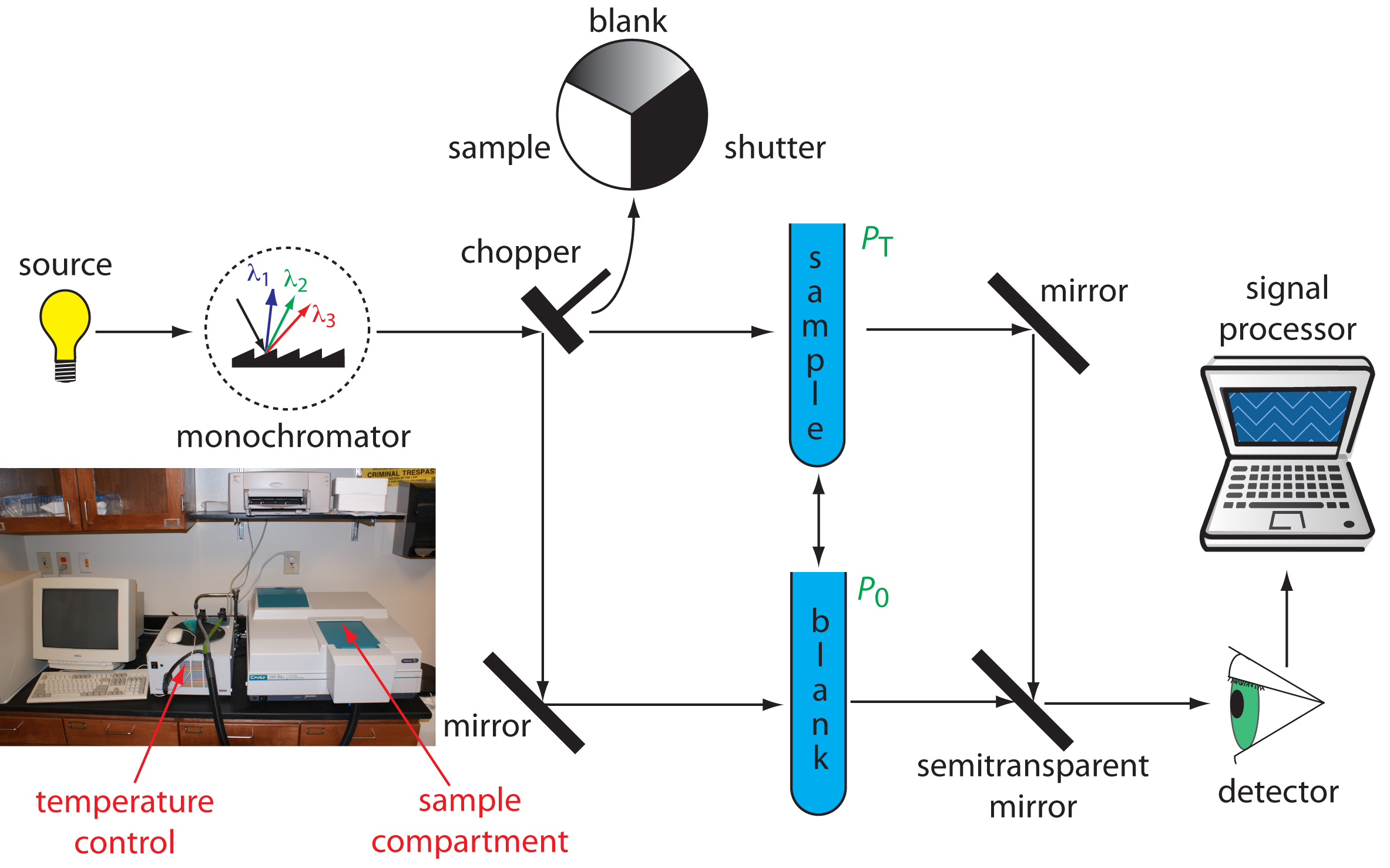
indicate absorbance or transmittance. The block diagram (Fig 2) below illustrates the relationship between these parts. Fig 2 In your spectrophotometer, light from the lamp is focused on the entrance slit of the monochromator where the collimating mirror directs the beam onto the grating.

Schematic Diagram Uv Vis Spectrophotometer - SHELVESCRIBE
The NIR region of UV/Vis/NIR instruments uses two different types of solid-state detectors. A silicon photodiode utilizes the internal photoelectric effect, the phenomenon whereby the electrical properties of the detector itself change when light strikes it. ... That bounces it onto the detector. It is following the red path in the diagram at ...

Sophisticated Analytical Instrument Facility
Fig.-block diagram of instrumentation of UV-spectrophotometer 22. amplifier Read out Fig.- block diagrammatic representation of UV-spectrophotometer 23. It is important that the power of the radiation source does not change abruptly over its wavelength range.
Ultra violet (UV) spectroscopy, introduction,principle ...
straightforward. We will not do UV-Vis becauase it is not very useful for structure identification. It is an extremely important tool for quantitating substances and is used widely. 1. Molecular interaction with electromagnetic radiation. (13.1-2) Molecules have electromagnetic fields derived from their electrons and nuclei. We
Spectrophotometry
Block Diagram and Explanation A basic block diagram of an HPLC is shown in Figure 1. Figure 1: Block Diagram of an HPLC ... also need to know what wavelengths in the UV-VIS spectrum you want to monitor. A diode array detector has a range of 210-400 nm and for samples, the default program setting are fine. The
Single Beam Spectrophotometer - New Images Beam
Block Diagram of a Mass Spectrometer Ion Source Detector "High" Vacuum Sample In Computer Tandem Mass Spectrometry or MS/MS m/z. S Q H L F A P I S E K 200 400 600 800 1000 1200 0 20 40 60 80 100 Relative Abudance m/z. S Q H L F A P I S E K 147 276 363 477 574 645 792 905 1042 1170 216 353 467 614 685 782 895 982

Working Of Double Beam Spectroscopy - The Best Picture Of Beam
Schematic of the internal layout of an Agilent Cary 5000 UV-Vis-NIR spectrophotometer, showing the main components. Note that this is a.36 pages

Schematic Diagram Of Fluorescence Spectroscopy - Wiring ...

Difference Between Single Beam and Double Beam ...

Schematic Diagram Of A Single Beam Uv Vis ...

Schematic diagram of a double beam thermal lens ...

TIDA-00155 DLP Near-Infrared Spectrometer for Optical ...

ORGANIC SPECTROSCOPY INTERNATIONAL: UV-Visible Spectroscopy

UV-VISIBLE SPECTROPHOTOMETER - Applied Analytical Systems

Schematic Diagram Of Double Beam Uv Vis Spectrophotometer ...

Double-beam spectrophotometers

Halsey's concert in London

2 : Schematic representation of a typical fluorescence ...

Difference Single Beam And Double Spectrophotometer - New ...
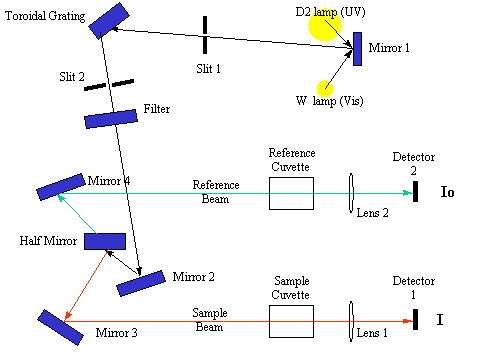
A Double Beam Absorption Spectrometer - Chemistry LibreTexts

Block Diagram of UV-Visible Spectrometer. | Download ...

spectroscopy - What is the role of a monochromator in AAS ...
Schematic Diagram Uv Vis Spectrophotometer - SHELVESCRIBE
Hplc
Schematic Diagram Of Double Beam Uv Vis Spectrophotometer ...

Schematic diagram of typical FTIR spectrophotometer ...
0 Response to "38 uv vis block diagram"
Post a Comment