38 rat dissection diagram labeled
Rat Dissection - GCSE/A Level Biology - YouTube In this video, I talk you through how to do a dissection of a rat to highlight the major mammalian organ systems. It shows the excretory system, digestive sy... PDF Rat External Anatomy - Springfield Public Schools The rat's body is divided into six anatomical regions: cranial region - head cervical region - neck pectoral region - area where front legs attach thoracic region - chest area abdomen - belly pelvic region - area where the back legs attach 1. Note the hairy coat that covers the rat and the sensory hairs (whiskers) located on the rat's face ...
A high-resolution anatomical rat atlas - PMC But until now, these rat atlases have looked only at regional anatomy (Toga et al. 1995; Bard et al. 1998), or are of low resolution such as the computer tomography atlas (Montgomery et al. 2001). In this study, we have developed a high-resolution Sprague-Dawley (SD) rat atlas that covers the anatomy of the whole rat.
Rat dissection diagram labeled
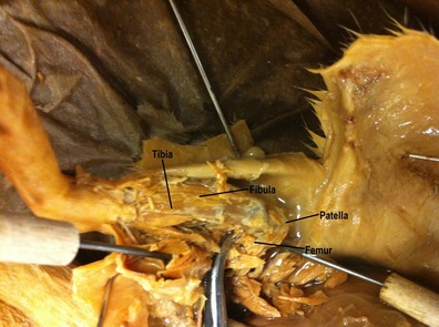
PDF Rat brain pictures - Western University Rat brain pictures Dorsal aspect of brain and rostral two Ventral aspect of the brain, and junction of segments of spinal cord. medulla with spinal cord. Dissection: Medial view of the right cerebrum. Hippocampus is visible after removing brain stem and most of the right thalamus. Dissection: Dorsal view of brain stem after PDF Including pregnant female - VWR International External jugular Internal jugular Lateral thoracic Vertebral Anterior vena cava Intercostal Posterior vena cava Renal Right gonadal Common iliac External iliac Investigation: Rat Dissection - The Biology Corner Students start with the external anatomy, noting features of the rat such as whiskers and the nictitating membrane of the eye. Students carefully skin the rat to reveal the major muscle groups and expose the hip joint and review the names of bones, diagrams of the rat skeleton and muscles are included.
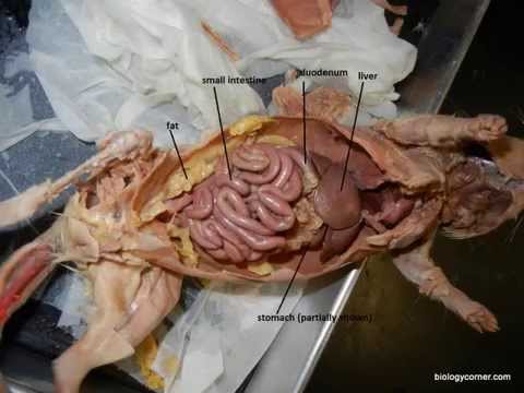
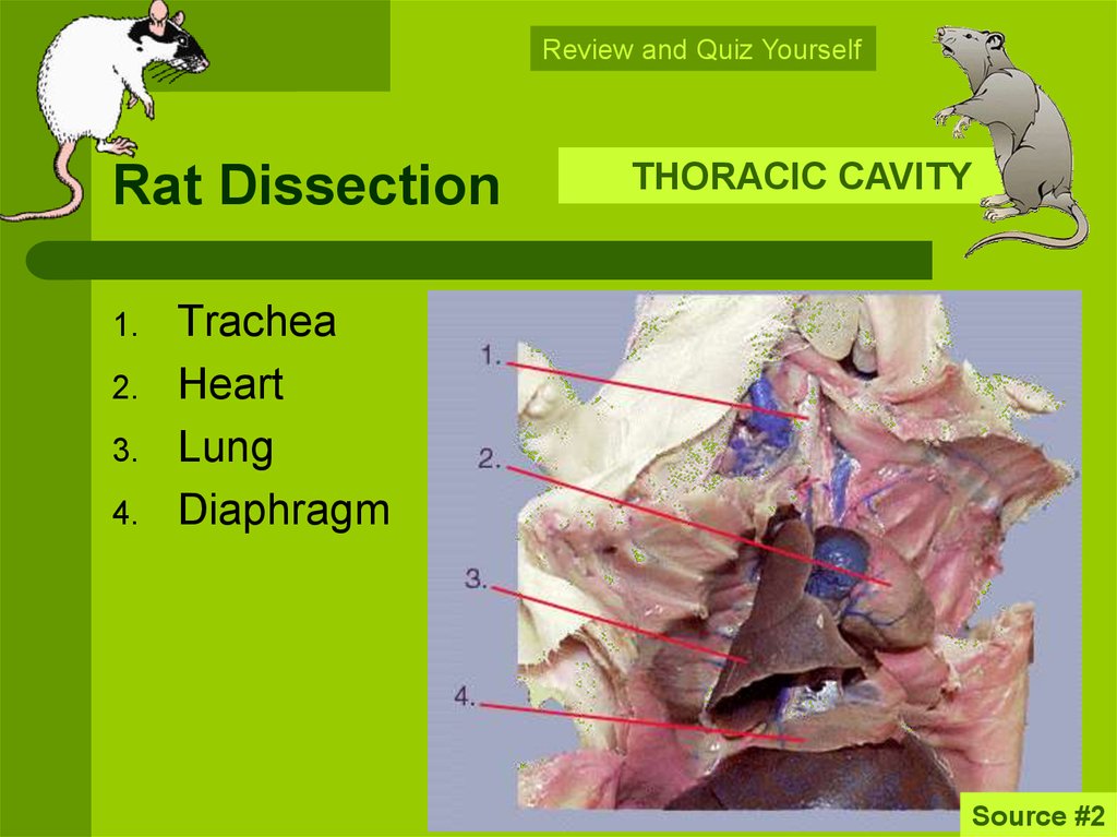
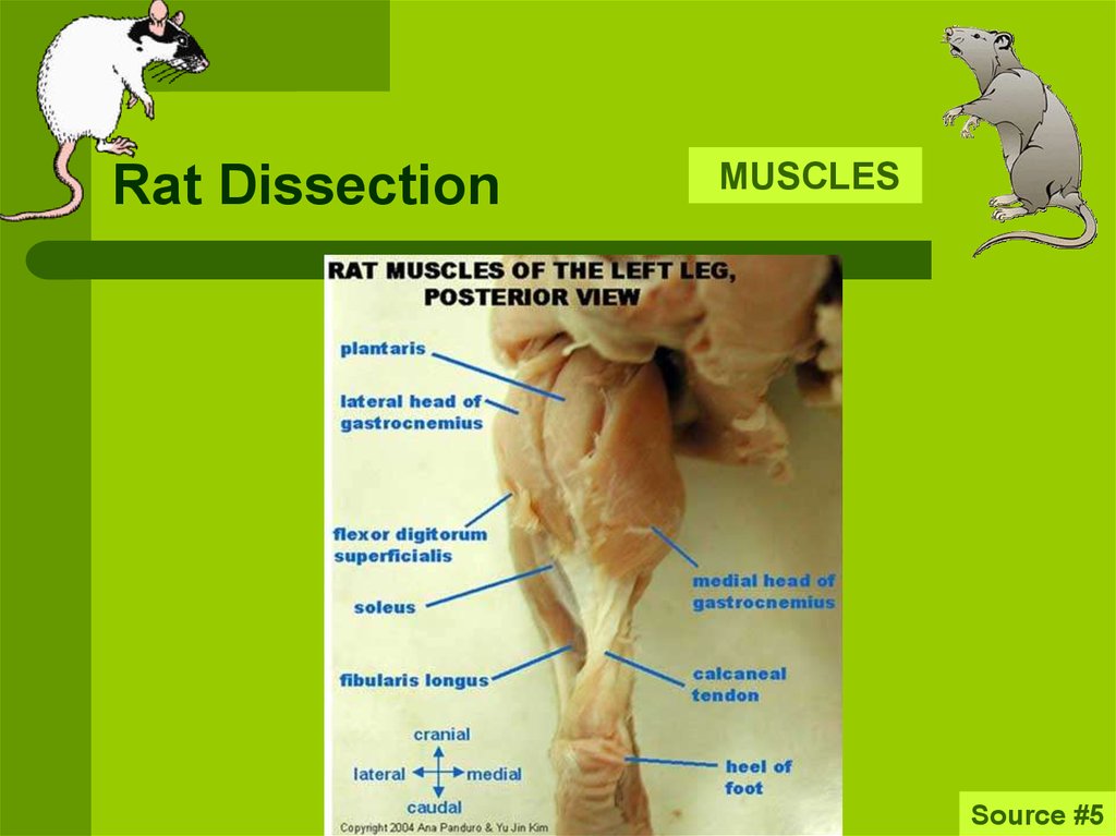
Rat dissection diagram labeled. University of Tennessee at Martin PDF Rat Dissection Guide The diagram below illustrates the muscles of the ventral surface of the rat. Be able to identify those listed. Use the photographs on the following pages and your lab atlas to assist you. Head and Throat muscles Digastric - this V shaped muscle follows the lower jaw line. It functions to open the mouth. Rat Dissection (complete) - Google Docs 1.Note the hairy coat that covers the rat and the sensory hairs (whiskers) located on the rat's face, called vibrissae. 2. The mouth has a large cleft in the upper lip which exposes large front... DOCX RAT DISSECTION - Eagle Mountain-Saginaw Independent School District Keep the tip of your dissection tool pointed upwards. Note: when you cut through the thoracic cavity, you will encounter bone. 3. Once the incisions have been made, pin both skin flaps to the side of the rat. 4. Locate the trachea. The trachea is a tube that extends from the neck to the chest. It is white and lined with cartilage.
Dissection guide to the rat - SlideShare Dissection guide to the rat 1. Dissection Guide to the Rat The Norway rat (Rattus norvegicus) Family:Muridae 2. Anatomical Terms Cranial-toward the head Caudal-toward the tail Dorsal-toward the backbone Ventral-toward the belly side Lateral-toward the side Medial-toward the middle Proximal-closer to the the base of a structure Distal-farther away from a structure PDF Dissection of the Rat - Alvin Independent School District Introduction to Dissection of the Rat In this laboratory exercise, the anatomy of the rat will be examined in some detail. You will get to know and love your preserved rat over the course of this dissection. The classification of the Rat (Rattus norvegicus) *Find the missing classification levels Kingdom: Phylum: Subphylum: Class: Order: Rat Dissection (complete) - Google Docs Headings you add to the document will appear here. Dissection of the Rat. Rat External Anatomy. The Muscular and Skeletal System of the Rat. Structures of the Head and Neck. Checkpoint: Rat Dissection - The Thoracic Organs. The Abdominal Organs. Checkpoint: Test Your Knowledge. Rat Dissection Manual - aurumscience.com Anatomical terms gives students practice using the most common anatomical terms used in rat dissections. This include differentiating between left and right (from the specimen's perspective), cranial, caudal, dorsal, and lateral. The Muscular System helps students identify all of the major surface muscles of the rat.
DOCX RAT DISSECTION - Pearland High School Keep the tip of your dissection tool pointed upwards. Note: when you cut through the thoracic cavity, you will encounter bone. 3. Once the incisions have been made, pin both skin flaps to the side of the rat. 4. Locate the trachea. The trachea is a tube that extends from the neck to the chest. It is white and lined with cartilage. PDF Rat External Anatomy Free Pdf 7th, 2022Frog Dissection External Anatomy Answer KeyFrog Dissection Lab Report - FLIPPED OUT SCIENCE In Regards Of The External Anatomy Of A Leopard Frog: A. It Is Easy To Tell The Sex Of The Animal. B. The Cloaca Is At The Anterior End Of The Animal. C. The Feet Of The Hind Limbs Have 5 Toes. D. All Of The Above. Mar 5th, 2022. BIO370-Rat Dissection Labeled dissections of male & female Brown Rats (Rattus norvegicus) Return to Unlabeled Rat Dissection This page last updated 8 August 2007 by Udo M. Savalli ( ) Dissection of Rat (With Diagram) | Zoology - Biology Discussion Dissection: Put the specimen on its back on a dissecting tray. Fix it with pins passing through the limbs. Lift the skin of the abdomen with a pair of forceps and make a small cut at about the middle of the abdomen. Starting from the cut give an incision extending up to the snout anteriorly and the genital opening posteriorly.
Rat Anatomy - YouTube
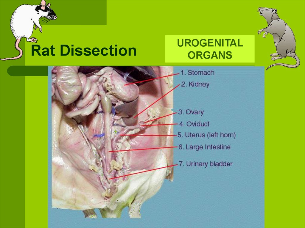
Anatomy - Rat Guide Anatomy Anatomy-Diagram Male Reproductive System. 501 Bella . March 27, 2006 at 15:54 (last edited September 15, 2020 at 13:56) ... The Rat Guide and its affiliates accept no responsibility for misuse or misunderstanding of its information. This guide in whole or part, exists solely for the purpose of recognizing and understanding the care and ...

Rat Anatomy | The Biology Corner
PDF Rat Dissection Lab - Mrs Carnahan's Pre-AP Biology Terms to know for dissection: Dorsal - the back or upper surface Ventral - the belly or lower surface Lateral - the side Anterior - the front or head end Posterior - the hind or tail end Medial - toward the midline of the animal Proximal - closer to the midline of the body Distal - farther from the midline of the body Grading: Your grade for this laboratory experiment will consist of four parts.

Dissection of a Rat
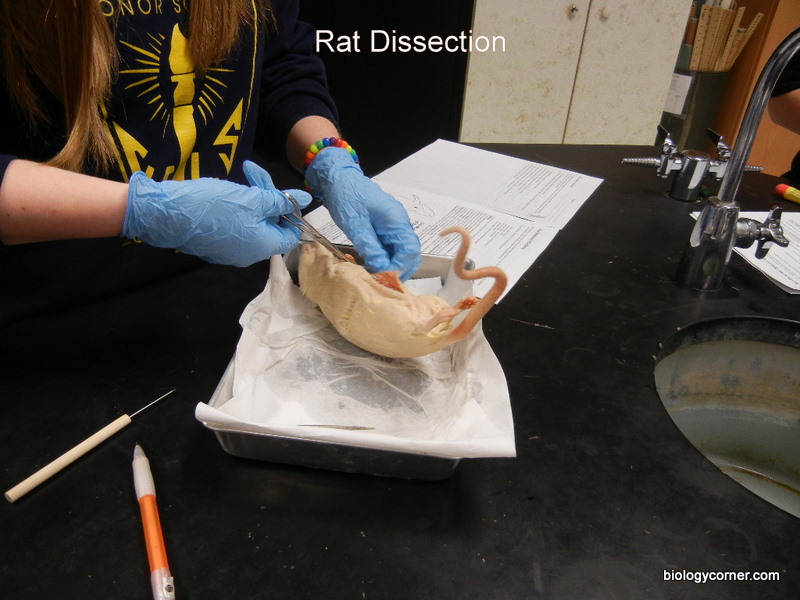
Virtual Rat Dissection Step by Step - The Biology Corner Rat External Anatomy. Step 1: In the biology lab, you will be working with specimens that have been preserved in chemicals and you will be working with sharp instruments. Before you start, obtain safety goggles, and nitrile gloves. Nitrile gloves come in different sizes, most women will wear a medium and most men will wear a large.
Rat Dissection - online presentation
Rat Anatomy - Rat Guide Tap or move your mouse over the rat diagram below to view the organ names. *Tip: on smaller devices, you may want to flip your device to landscape if you can't see the full width of the image. Medical Illustration by Chris McKee for the Rat Guide. Additional descriptions will be added by the Rat Guide Team.

Rat dissection guide pdf - dobraemerytura.org
Rat Dissection Practice | Biology Quiz - Quizizz Which body region is indicated by #5? Identify the organ/structure that is marked with a red star. Identify the organ/structure that is marked with a red star. Identify the organ/structure that is marked with a red star. Identify the organ/structure that is marked with a red star.
.PNG)
Digestion in Ruminants & Rodents - Presentation Health and Disease
rat anatomy labeled A Color Atlas Of Sectional Anatomy Of The Rat - Cosmo Bio Co.,Ltd. . anatomy. Rat Dissection - Online Presentation en.ppt-online.org. rat dissection ppt organs intended slides following. Rat Dissection-- Thoracic Cavity & Circulatory System - YouTube . rat dissection cavity thoracic system circulatory. Alan ...

Biology 11: Fetal Rat Dissection
Rat Dissection Anatomy Quiz - PurposeGames.com This is an online quiz called Rat Dissection Anatomy. There is a printable worksheet available for download here so you can take the quiz with pen and paper. Your Skills & Rank. Total Points. 0. Get started! Today's Rank--0. Today 's Points. One of us! Game Points. 10. You need to get 100% to score the 10 points available.
Rat Dissection - online presentation
Labeled Rat - Rat Dissection - Weebly Rat Dissection. Rat Dissection. Home External Features Muscular System Oral Cavity Abdominal Cavity Cervical Cavity Thoracic Cavity Urogenital Page Nervous System Contact About Untitled External pictures of the rat Powered by Create your own unique website with ...

My Labs!: Rat Dissection - 9/10/2012
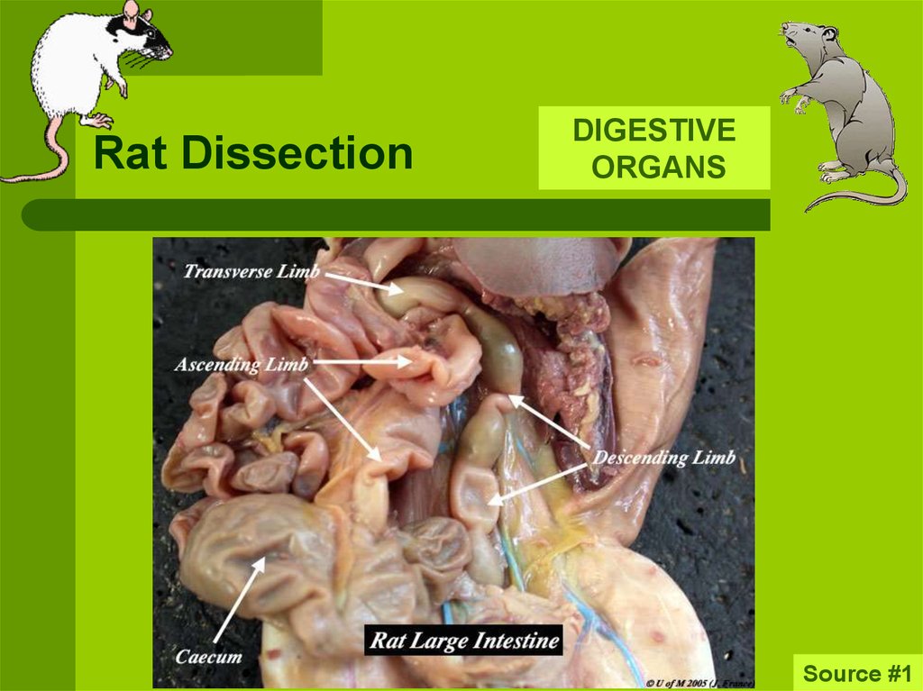
Rat Dissection Flashcards | Quizlet Anatomy rat dissection quiz- labeling parts Tags related to this set Anatomy And Physiology Human Anatomy And Physiology 1 External Iliac Artery Femoral Artery Caudal Vena Cava Great Vessels Of The Heart Terms in this set (113) heart lung trachea diaphragm liver stomach spleen pancreas small intestine large intestine cecum (see-kum) rectum kidney
Anatomy & Physiology Lecture Notes - Rat dissection
Anatomy - RAT DISSECTION Diagram | Quizlet front of the body, towards the head Posterior back of body, towards the tail Dorsal toward the back or spine Ventral toward the belly Nipple projection from the breast into which the lactiferous ducts open Urethra tube leading from the urinary bladder to the outside of the body; opening of the urinary system Anus
Rat Dissection - Biology 11 Honours - Animalia Labs
Investigation: Rat Dissection - The Biology Corner Students start with the external anatomy, noting features of the rat such as whiskers and the nictitating membrane of the eye. Students carefully skin the rat to reveal the major muscle groups and expose the hip joint and review the names of bones, diagrams of the rat skeleton and muscles are included.
Rat Dissection - online presentation
PDF Including pregnant female - VWR International External jugular Internal jugular Lateral thoracic Vertebral Anterior vena cava Intercostal Posterior vena cava Renal Right gonadal Common iliac External iliac
rat_dissection_title.JPG
PDF Rat brain pictures - Western University Rat brain pictures Dorsal aspect of brain and rostral two Ventral aspect of the brain, and junction of segments of spinal cord. medulla with spinal cord. Dissection: Medial view of the right cerebrum. Hippocampus is visible after removing brain stem and most of the right thalamus. Dissection: Dorsal view of brain stem after
Rat Dissection - online presentation

Label the White Rat Muscles
0 Response to "38 rat dissection diagram labeled"
Post a Comment