42 cow eye dissection labeled diagram
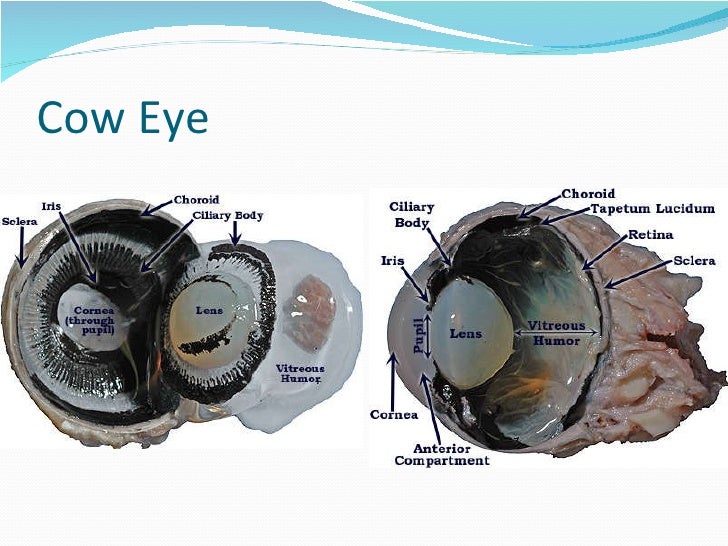
Cow Eye Diagrams - aw-drivein List of Cow Eye Diagrams. 1. Anatomy Eyes Pig Eyes. 2. Eye Dissection Labeling. 3. Virtual Eye Dissection Eye Parts Eyes Eye 4. Teacher Dissection Page Labeling Ideas Eyes ... Eye Dissection Diagram Labeled Eye Eyes. 18. Eye Anatomy Physiology. 19. Dissection Detailed Eye Dissection Video Part 2 20. Dissecting Cows Eyeball ... Cow eye - dissection and label - SlideShare 1. Cow eye - Dissection and Label By Leslie Young Section 63. 2. Cow eye shown with labeled cornea. The cornea is the transparent front part of the eye that covers the iris, pupil, and anterior chamber. The cornea, with the anterior chamber and lens, refracts light, with the cornea accounting for approximately two-thirds of the eye's total ...
[Solved] How to label a cow eye dissection? | Course Hero In a cow eye dissection, the first step is to remove the cow's eye from its socket. This can be done by cutting around the circumference of the eye with a sharp knife. Once the eye is removed, it can be placed on a dissecting tray. The next step is to designate the various components of the eye. The cornea is the eye's transparent, outermost layer.

Cow eye dissection labeled diagram
Cow Eye Dissection Guide - Google Slides Cow Eye. Use the point of a scissors or a scalpel to make an incision through the layers of the eye capsule (similar to figure 1); there are three layers from the exterior: sclera, whitish/grey, continuous with the transparent cornea, choroid, thin dark black layer and the retina, thin greyish/pink layer. Use a scissors to dissect the entire ... Cow's Eye Dissection - Eye diagram - Exploratorium Learn how to dissect a cow's eye in your classroom. This resource includes: a step-by-step, hints and tips, a cow eye primer, and a glossary of terms. Cow's Eye Dissection - Eye diagram WMU Psychology Department: Lisa Baker | Cow eyes, Dissection, How to ... Eyeball Anatomy. Parts Of An Eye. Eye Definition. The first line of protection of the eyes is provided by the lids, which prevent access of foreign bodies and assist in the lubrication of the corneal surface. Lid closure and opening are accomplished by the orbicularis oculi and levator palpebri muscles; the orbicularis oculi operates on both ...
Cow eye dissection labeled diagram. Cow Eye Dissection - The Biology Corner The cow eye is a fantastic specimen for students of all ages to dissect. The structures are clear, dissection easy to accomplish and usually kids enjoy the lab. The lab guide for students outlines the procedure for the dissection and you can view the eye gallery to see photographs of the dissection. PDF Cow Eye Dissection - UW SCIENCE EXPLORERS How it works: The cow's cornea has many layers to make it thick and strong. When the cow is grazing, blades of grass may poke the cow's eye, but the cornea protects the inner eye. 9)Look at the back half of the eyeball. On the inside back half of the eyeball, you can see some blood vessels that are part of a thin fleshy film. Cow Eye Diagram Labeled - All About Cow Photos Anatomy cow eye diagram quizlet eye diagram cliparts co cow eye dissection anatomy cow eye dissection anatomy. ... and function of the eyes eye disorders merck manuals how do we see light ask a biologist human eye diagram quizlet eye anatomy cow s eye dissection diagram. Related. Related Posts. Why Does My Cow Moo At Night . October 6, 2021 ... eye diagram labeled - anatomyedu99.z21.web.core.windows.net brain function structure functions diagram lobes macmillan labelled showing. Cow Eye Dissection - YouTube . cow eye dissection. Label The Muscles Of The Eye - PurposeGames . purposegames. 3d Eye Model 32 Pcs Assembled Human Anatomy Model New 3D Structure Of . auge. Photoreceptor Cell ...
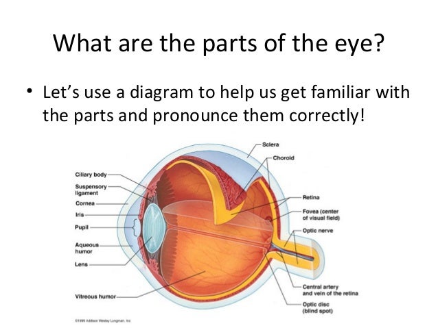
Cow Eye Dissection & Anatomy Project | HST Learning Center On the posterior side of the eye, nestled in the fat and muscle tissue, there is a noticeably round protuberance that feels stiffer than the surrounding tissue. This is the optic nerve, and it sends the images collected in the eye to the brain. Cow Eye Dissection: Internal Anatomy. 1. Place the cow's eye on a dissecting tray. The eye most ... Cow Eye Dissection Diagram | Quizlet A ring of muscle tissue that forms the colored portion of the eye around the pupil and controls the size of the pupil opening. The transparent structure behind the pupil that changes shape to help focus images on the retina. Muscles that help change the shape of the lens. Located in the back of the eye, contains the rods and cones. PDF Cow Eye Dissection Lab - Home Science Tools Cow Eye Dissection 2/6 Cow Eye Observation: External Anatomy Look carefully at the preserved cow eye. The most noticeable part of the eye is the large mass of gray tissue that surrounds the pos-terior (back) of the eye and is attached to the sclera. The second most noticeable part of the eye is the cornea, located in the ante- Cow Eye Dissection | Carolina.com Students explore the external and internal anatomy, learning how structures work together to create images from incoming light. A preserved cow eye dissection can be carried out in 1-2 class periods and only requires basic dissecting instruments. Explore the internal and external anatomy of the cow eye using the procedural steps below.
PDF Cow Eye Dissection: Examining Structure and Function the eye. 4. Using scissors or a scalpel, carefully cut the eye in half. Separate the front from the back of the eye. a. Describe the sclera, the external part of the eye that you cut through. What can you infer about its functions? Procedure Cornea Optic nerve Fat Muscle Sclera Cornea Procedure continued on the next page. Cow Eye Dissection: PDF Dissection 101: Cow Eye eye to the brain, using the following. (optic nerve, iris, pupil, sclera, cones, rods, cornea, retina, lens and vitreous humor) Use a labeled drawing if it is PDF Cow Eye Dissection Guide - Central Bucks School District DISSECTION OF THE COW EYE Please make sure to wear gloves and safety glasses when you are dissecting, and make sure to clean up thoroughly after the lab. Also, the cow eyes can be rather slippery, so use caution when handling and cutting them. You will need a scalpel and forceps. 1. First, identify the most external structures of the eye. Cow Eye Dissection - The Biology Corner 1. Examine the outside of the eye. You should be able to find the sclera, or the whites of the eye. This tough, outer covering of the eyeball has fat and muscle attached to it. 2. Locate the covering over the front of the eye, the cornea. When the cow was alive, the cornea was clear. In your cow's eye, the cornea may be cloudy or blue in color.
33 besten Animal a&p Bilder auf Pinterest | Anatomie, Pferdepflege und ...
bones structures of cow dissection cow eye. Cow Leg Bones Diagram / Human Skeleton Labeled Diagram . Human Skeleton brittnid-boxing.blogspot.com. skeleton skeletal. Lab 5 . lab patella mandible wtamu lab5 mammalogy edu. Hawktunics.html campus.murraystate.edu. vitreous humor eye avian retina tunics contains. Hot Water And Old Bones

Cow Eye Dissection Parts Labeled - All About Cow Photos
Cow Eye Dissection Labeled Diagram - All About Cow Photos Cow s eye dissection diagram neur 320 art and vision cow eye dissection lab cow s eye dissection dissecting a lens eye picture 2694249. ... Cow eye diagram label wiring diagrams folder lab 9 2 eye dissection physics matters preserved cow eyes ward s science solved retina biology 30 15 eye dissection name ion diagram of cow wiring diagrams show.
Cow Eye Labeled Diagram - ClipArt Best
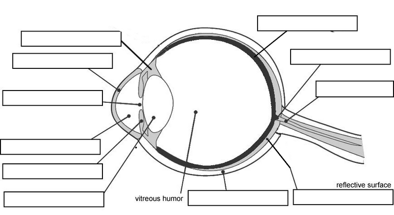
Lab 10—Labeled Cow Eye The Cow Eye (Labeled) Return to: Lab 10 Page BIO 137 Main Page Be sure to practice using the unlabeled images. Coronal section, anterior view (lens and vitreous humor displaced) Sagittal section: This page created and maintained by Udo M. Savalli. Last updated April 18, 2006.

WMU Psychology Department: Lisa Baker | Cow eyes, Anatomy bones, Anatomy
Cow Eye Dissection & Parts of the Eye Diagram | Quizlet A ring of muscle tissue that forms the colored portion of the eye; controls the size of the pupil opening. Located in the back of the eye, contains the rods and cones. The transparent structure behind the pupil that changes shape to help focus images on the retina. Location of the retina where there are no rods or cones.
Cow Eye Dissection
cow anatomy diagram for kids Parts Of A Turtle: Useful Turtle Anatomy With Pictures • 7ESL 7esl.com. turtle parts body animal anatomy english vocabulary morphology useful 7esl. Cow Eye Dissection Worksheet | Cow Eyes, Dissection, Frog Dissection . cow eye dissection worksheet eyes anatomy diagram tell three. Transparent Skeletal System Png - Skeletal ...

Eyes Coloring Worksheet | cow s eye dissection tutorial cow s eye ...
Cow Eye Dissection Teaching Resources | Teachers Pay Teachers This is a comprehensive dissection guide of the cow eye, designed for a high school or early college Biology or Anatomy & Physiology class. The guide includes step-by-step instructions and labeled diagrams that will lead students through the external anatomy of the eye, followed by dissection of the internal structures.

Cow's Eye Dissection - Eye diagram | Veterinaria, Salud, Along the way
cow internal anatomy Cow's eye dissection. Eye diagram cow eyes anatomy labeled exploratorium human dissection edu chart learning cliparts biology physiology med museum science muscles animal. Planarian system digestive platyhelminthes hydra structure internal phylum cavity gastrovascular simple freshwater felina mouth cnidaria

Sheep Eye Dissection - YouTube
leg anatomy diagram Dissected Cow Eye Clearly Showing How Eye Can Be Understood And How . cow lens anatomy eye dissected dissection labeled human eyes camera vet sagittal section understood clearly showing muscle open. Anatomy Tibia And Fibula . tibia fibula. Knee Joint Cross Section - Medical Art Library medicalartlibrary.com
Sheep eye dissection
WMU Psychology Department: Lisa Baker | Cow eyes, Dissection, How to ... Eyeball Anatomy. Parts Of An Eye. Eye Definition. The first line of protection of the eyes is provided by the lids, which prevent access of foreign bodies and assist in the lubrication of the corneal surface. Lid closure and opening are accomplished by the orbicularis oculi and levator palpebri muscles; the orbicularis oculi operates on both ...
Eye anatomy
Cow's Eye Dissection - Eye diagram - Exploratorium Learn how to dissect a cow's eye in your classroom. This resource includes: a step-by-step, hints and tips, a cow eye primer, and a glossary of terms. Cow's Eye Dissection - Eye diagram

Parts and Functions - Dissecting A COW EYE
Cow Eye Dissection Guide - Google Slides Cow Eye. Use the point of a scissors or a scalpel to make an incision through the layers of the eye capsule (similar to figure 1); there are three layers from the exterior: sclera, whitish/grey, continuous with the transparent cornea, choroid, thin dark black layer and the retina, thin greyish/pink layer. Use a scissors to dissect the entire ...

Cow Eye Dissection Diagram Labeled | Cow Eye | Biology 100: Anatomy ...

1000+ images about Biology 100: Anatomy & Phys. on Pinterest | Anatomy ...
0 Response to "42 cow eye dissection labeled diagram"
Post a Comment