39 which of the following structures is labeled d in the diagram?
FINAL EXAM: Human Anatomy & Medical Terminology - Quizlet Which of the following statements about diagnostic and therapeutic procedures is correct? Select one: A. An indwelling catheter is one that remains in the body for any prolonged period of time. ... Which of the structures labeled in diagram below prevents the backflow of blood into the left ventricle? Select one: A. 1 B. 5 C. 4 D. 2. 1. NCBI Conserved Domain Database (CDD) Help 3-dimensional structures and conserved core motifs: NCBI Conserved Domain Curators have re-evaluated and modified multiple sequence alignments imported from outside sources, and made them agree with what we can infer from three-dimensional structure and three-dimensional structure superposition. Curated alignments contain aligned blocks ...
Identify the labeled structures on the following diagram of translation. Short Answer Question: Identify the labeled structures on the following diagram of translation. Part A is the _____ Part B is the _____ Part C is the _____ Maharashtra State Board HSC Science (General) 12th Board Exam. Question Papers 255. Textbook Solutions 14289. MCQ Online ...

Which of the following structures is labeled d in the diagram?
Ternary Phase Diagram - an overview | ScienceDirect Topics WebTernary phase diagrams are used to represent all possible mixtures of three solvents [1]; they are described in Chapter 3.Here, we shall indicate how they should be used to minimize the solvent consumption. Figure 2.1 (top) shows the methanol–chloroform–water ternary phase diagram with the tie-lines in the biphasic domain. Five particular compositions are … Which best describes the structure labeled x in the diagram katsxlove. The structure labeled X in the diagram is a membrane protein. Need a bit more clarification? Get a high-quality answer with step-by-step explanations from a professional in just minutes instead! Get a verified answer. Advertisement. Anatomy 102 Chapter 12 study guide Diagram - Quizlet Which of the following structures is labeled B in the diagram? a) axon terminal b) trigger zone c) cell body d) peripheral process e) dendrites.
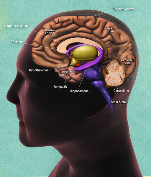
Which of the following structures is labeled d in the diagram?. What is a Network? | Webopedia Web31.08.1996 · A network is defined as a group of two or more computer systems linked together. There are many types of computer networks, including the following: Local-area networks (LANs): The computers are geographically close together (that is, in the same building). Wide-area networks (WANs): The computers are farther apart and are … Labeled Diagrams of the Human Brain You'll Want to Copy Now The height of the human brain is about 3.6 inches and it weighs about 4 to 5 lbs at birth and 3 lbs in adults. The total surface area of the cerebral cortex is about 2,500 cm2 and when stretched, it will cover the area of a night table. The brain is composed of 77 to 78% water and 10 to 12% lipids. It contains 8% proteins 1% carbohydrates, 2% ... Locate the following structures and label them in the - Course Hero Label the following structures in the image above. • Cochlea • Vestibule • Semicircular canals • Oval window 5. What part (s) of the inner ear do the nerves connect to? 6. Where do these nerves project in the brain? (For each structure you choose, be sure to select the book icon for more information.) 7. Locate the round window. Cell Biology - Wiki - Scioly.org WebVor 1 Tag · DNA (deoxyribonucleic acid) is one of two long molecules known as nucleic acids, which code for the genetic information found within cells.It is made up of molecules known as nucleotides, which are made up of a nitrogenous base, a phosphate group, and a deoxyribose sugar.The phosphates of one nucleotide bonds to the carbon of another, …
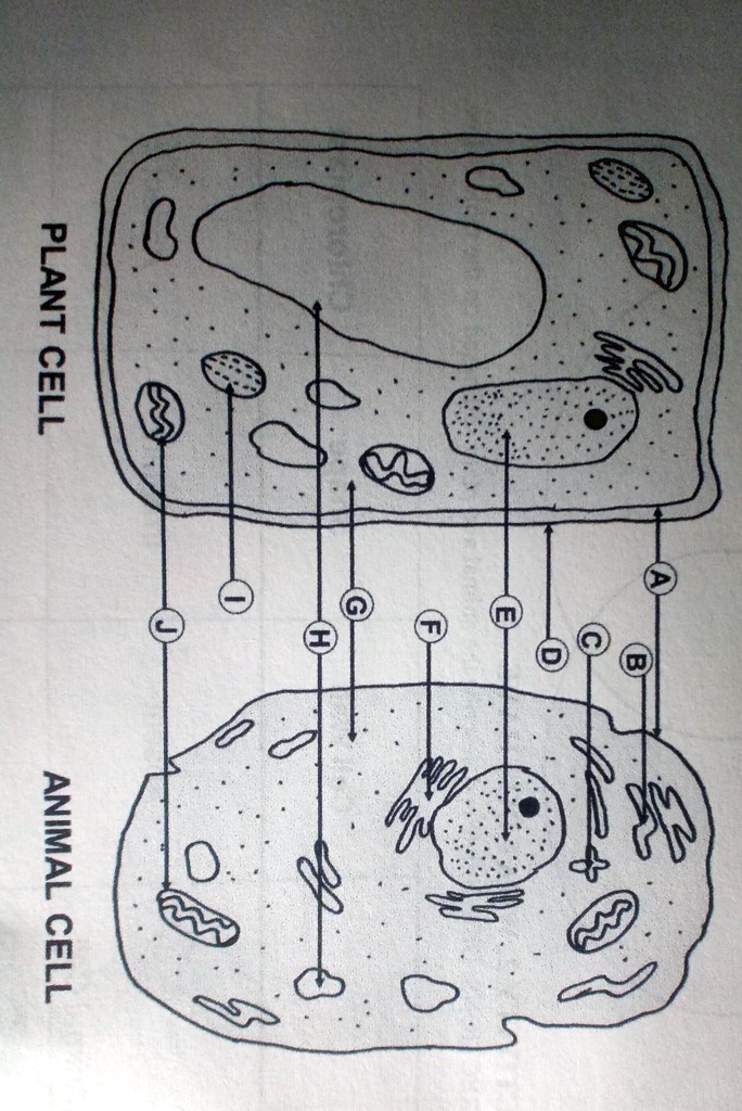
Structure of Cell: Definition, Types, Diagram, Functions - Embibe Some of these structures are (1) Cell Wall (2) Mitochondria (3) Chloroplast (4) Cell Membrane and (5) Nucleus . Q3. What is the structure of a human cell? Ans: A cell structure is composed of many components which are present inside the cell. The basic structure of a cell consists of three essential parts that are cell membrane (plasma membrane ... Plant and Animal Cell: Labeled Diagram, Structure, Function - Embibe 3. It contains a network of a thread-like structure called chromatin fibres. 4. There is a dark structure inside the nucleus, called the nucleolus, which is without membrane and rich in RNA. 5. The nucleus controls metabolic activities taking place in the cell. 6. Nucleolus controls gene expression. Mitochondria: 1. Cell: Structure and Functions (With Diagram) - Biology Discussion The nuclear membrane is a double layered structure surrounding the nucleus containing many nuclear pores. These pores allow different materials to move in and out of nucleus. The pores have octagonal 'doors' made of protein which open and close on either side depending on specific signals. Lipids - Michigan State University The structure of vitamin D can be described as a steroid in which ring B is cut open and the remaining three rings remain unchanged. The precursors of vitamins A and D have been identified as the tetraterpene beta-carotene and the steroid ergosterol, respectively. Structures for these will be displayed by clicking on the vitamin diagram above.
Tree (graph theory) - Wikipedia WebDefinitions Tree. A tree is an undirected graph G that satisfies any of the following equivalent conditions: . G is connected and acyclic (contains no cycles).; G is acyclic, and a simple cycle is formed if any edge is added to G.; G is connected, but would become disconnected if any single edge is removed from G.; G is connected and the 3-vertex … HUMAN A & p TEST 5 Flashcards - Quizlet Which of the following structures is labeled D in the diagram? Lumbar enlargement. Image: Which of the following structures is labeled D in the diagram? ER Diagram (ERD) - Definition & Overview | Lucidchart WebMake sure all your entities and relationships are labeled. You can translate relational tables and ER diagrams back and forth, if that helps you achieve your goal. Make sure the ER diagram supports all the data you need to store. There may be different valid approaches to an ER diagram. A Labeled Diagram of the Human Heart You Really Need to See The human heart resembles the shape of an upside-down pear, weighing between 7-15 ounces, and is little larger than the size of the fist. It is enclosed in a bag-like structure called the pericardium, and is located between the lungs, that is in the middle of the chest, behind and slightly to the left of the sternum or breast bone.
human digestive system | Description, Parts, & Functions ...
In the diagram a tendon is formed by the merging of the following ... 38) In the diagram, a tendon is formed by the merging of the following structures a) C, D, and H b) D and E c) H and C d) C and D e) All of these choices are correct. Answer: a a ) C , D , and H 39) Which of the following structures are made of dense regular connective tissue? a) Fb) A c) I d) Both F and I e) All of these choices.

The toposiomerase IIIalpha-RMI1-RMI2 complex orients human ...
Tree (graph theory) - Wikipedia A labeled tree is a tree in which each vertex is given a unique label. The vertices of a labeled tree on n vertices are typically given the labels 1, 2, …, n. A recursive tree is a labeled rooted tree where the vertex labels respect the tree order (i.e., if u < v for two vertices u and v, then the label of u is smaller than the label of v).

10.2 Skeletal Muscle – Anatomy & Physiology
Which of the following structures is labeled a in the - Course Hero a) axon terminal b) trigger zone c) cell body d) peripheral process e) dendrites Answer: e. e ) dendrites. 58) Which of the following structures is labeled B in the diagram? a) axon terminalb) trigger zone c) cell bodyd) peripheral process e) dendrites Answer: d. d ) peripheral process.
Trivia Quiz Questions On Ear Model - ProProfs Quiz
Plant Cell- Definition, Structure, Parts, Functions, Labeled Diagram Functions of the plant cell (plasma) membrane. In-plant cells the cell membrane separated the cytoplasm from the cell wall. It has a selective permeability hence it regulates the contents that move in and out of the cell. It also protects the cell from external damage and provides support and stability to the cell.
The Digestive System Flashcards - Easy Notecards
quiz 13 &14 a&p lecture Flashcards - Quizlet Which of the following structures is labeled D in the diagram? Image: Lumbar enlargement.

Quia - AP Chapter 6 - Cells (basic)
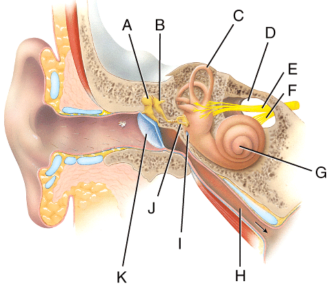
Human Ear Diagram - Bodytomy Auditory Ossicles: The three small bones in the middle ear, called malleus, stapes, and incus, are connected. These bones together are called the auditory ossicles, and their purpose is to let the sound that strikes the eardrum, further into the inner ear.

Skin 1: the structure and functions of the skin | Nursing Times
A&P 2 Final Diagrams Flashcards | Quizlet b) special senses neuron c) motor neuron d) association neuron e) dendrites Which of the following structures is labeled A in the diagram? a) axon terminal b) trigger zone c) cell body d) peripheral process e) dendrites c) C Which of the structures in this diagram would be included in a ganglion? a) A b) B c) C d) D e) E a) A

Lumbar Spine Anatomy
Plant Cell and Animal Cell Diagram Quiz - Biology Multiple Choice Quizzes 1. Which of the following is a justification for considering diagram 1 as a plant cell? Presence of mitochondrion Presence of vacoule Presence of cell wall Presence of plasma membrane 2. The structure labeled A is present in all prokaryotic and eukaryotic cells. The structure is Ribosomes Cell wall Cell membrane Endoplasmic reticulum 3.

Homologous Structure Vector Illustration Biological Species ...
Chapter 13 Wiley Flashcards - Quizlet Which of the following structures is labeled D in the diagram? Cervical enlargement. Lumbar enlargement. Obturator nerve. Conus medullaris. Cauda equina.

4.1 Chemical Energy and ATP - ppt download
Identify the labeled structures. A: B: C: D: E: - Brainly.com Get a high-quality answer with step-by-step explanations from a professional in just minutes instead! Get a verified answer. Advertisement.

Renal cortex - Wikipedia
Chapter 16: Structural Design, 2020 FBC - Building, 7 th edition WebIn lieu of the basic load combinations specified in Section 1605.3.1, structures and portions thereof shall be permitted to be designed for the most critical effects resulting from the following combinations.When using these alternative allowable stress load combinations that include wind or seismic loads, allowable stresses are permitted to be increased or …
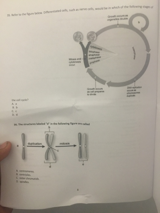
Solved Refer to the figure below. Differentiated cells, such ...
Labeled Diagram of the Human Lungs - Bodytomy Given below is a labeled diagram of the human lungs followed by a brief account of the different parts of the lungs and their functions. Each lung is enclosed inside a sac called pleura, which is a double-membrane structure formed by a smooth membrane called serous membrane. The outer membrane of this structure is called parietal pleura and is ...
THE SKELETAL SYSTEM
Lipids - Michigan State University WebThe structure of vitamin D can be described as a steroid in which ring B is cut open and the remaining three rings remain unchanged. The precursors of vitamins A and D have been identified as the tetraterpene beta-carotene and the steroid ergosterol, respectively. Structures for these will be displayed by clicking on the vitamin diagram above.
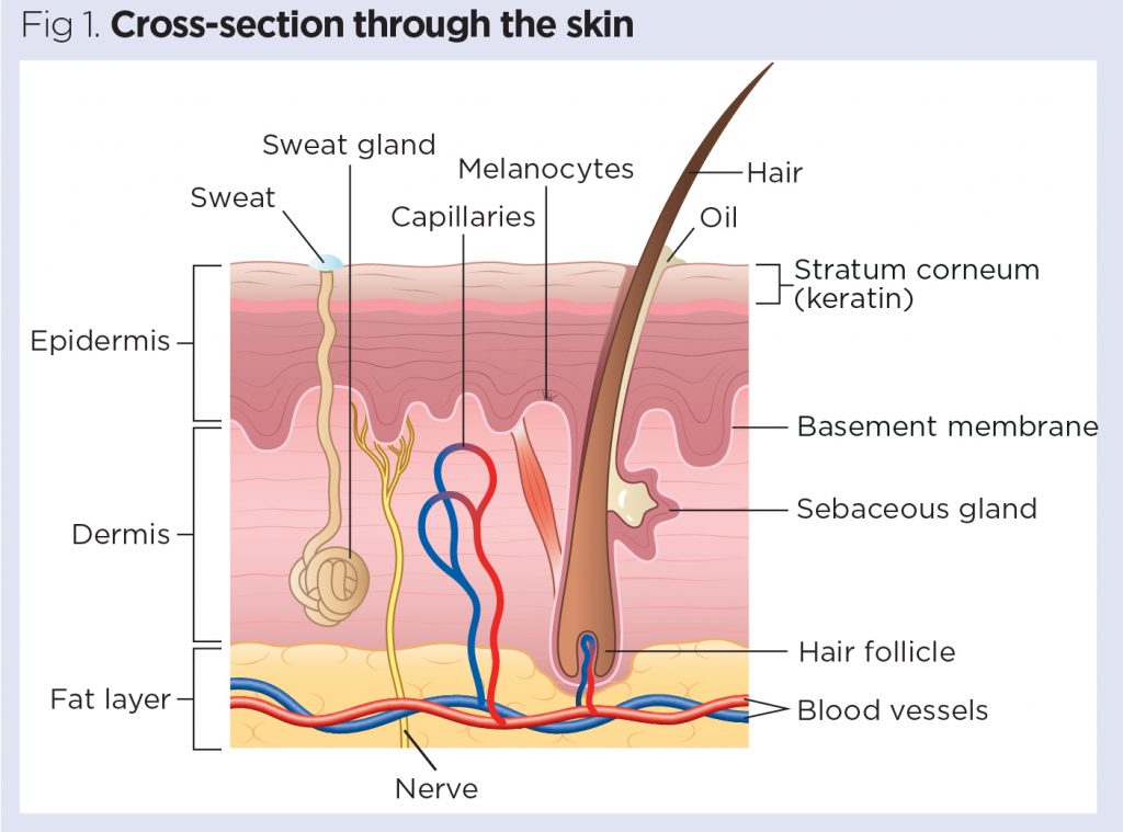
Skin 1: the structure and functions of the skin | Nursing Times
Anatomy and Physiology Unit 9 Quiz (Nervous Tissue, Spine ... Which of the following structures is labeled C in the diagram? ... D E. E. Image: Which of the labeled cells in the diagram is a neuroglial cell that is.

What Is a Neuron? Diagrams, Types, Function, and More
Labeled Sarcomere Diagram Dodge Durango Wiring Diagram. Start studying UNIT 5: Label the parts of the Sarcomere. Learn vocabulary, terms, and more with flashcards, games, and other study tools. Draw and label a diagram to show the structure of a sarcomere, including Z lines, actin filaments, myosin filaments with heads, and the resultant light and dark bands.
Anencephaly: MedlinePlus Genetics
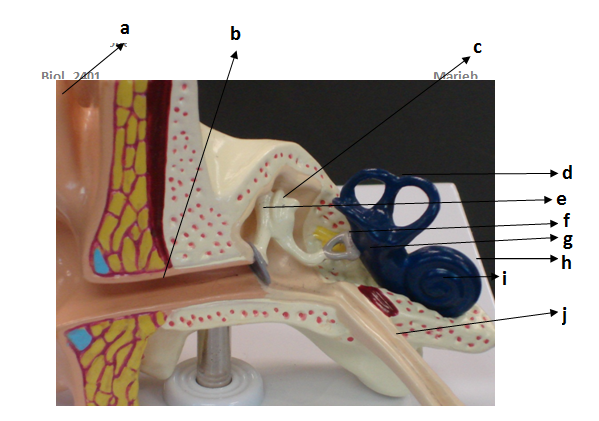
Solved 1. Locate the following structures and label them in - Chegg Locate the following structures and label them in the diagram above: a. Tympanum b. Cochlea c. Incus d. Stapes e. Malleus 2. For the structures listed above, list them in order, from outermost to innermost. 3. For the structures listed a-d above, list the role each of them has in hearing. This problem has been solved! See the answer

1. Give the name and functions of the structure labeled A on ...
ER Diagram (ERD) - Definition & Overview | Lucidchart An Entity Relationship (ER) Diagram is a type of flowchart that illustrates how “entities” such as people, objects or concepts relate to each other within a system. ER Diagrams are most often used to design or debug relational databases in the fields of software engineering, business information systems, education and research.
Male Reproductive System: Structure & Function
Structure of Bacterial Cell (With Diagram) - Biology Discussion Capsule: It is an outer covering of thin jelly-like material (0.2 μm in width) that surrounds the cell wall. Only some bacterial species possess capsule. Capsule is usually made of polysaccharide (e.g. pneumococcus), occasionally polypeptide (e.g. anthrax bacilli) and hyaluronic acid (e.g. streptococcus).

Chapter 13 Wiley Flashcards | Quizlet
The Structure of an Atom Explained With a Labeled Diagram Basic Diagram of an Atom. Most of an atom is just empty space and consists of a positively charged nucleus of protons and neutrons surrounded by a cloud of negatively charged electrons. The center of an atom is the nucleus and one or more electrons surrounding the nucleus. When one says an atom is electrically neutral, it means that the number ...
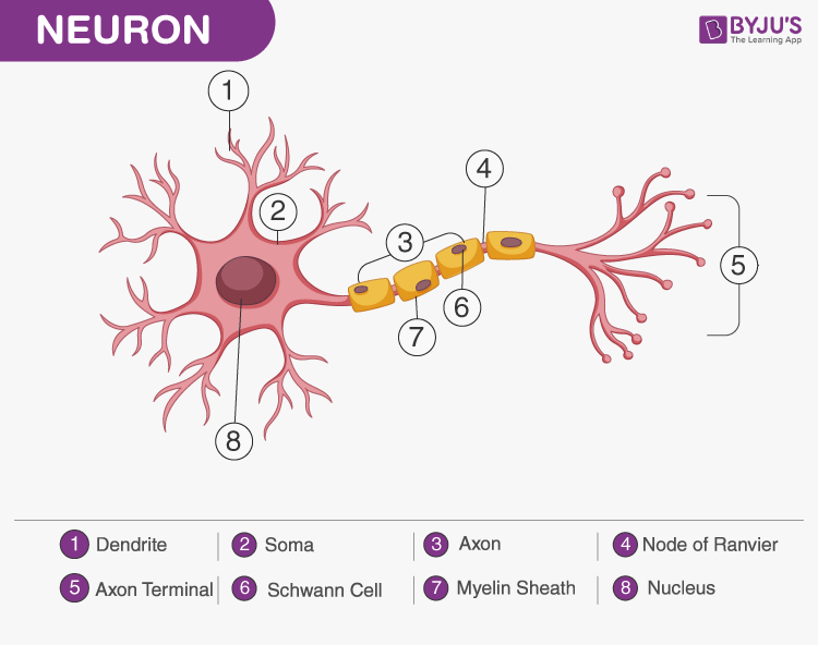
A Labelled Diagram Of Neuron with Detailed Explanations
Ternary Phase Diagram - an overview | ScienceDirect Topics A point on the diagram represents a composition that is specified in terms of mole fraction or weight fraction. The point, (0.3, 0.4, 0.3) is at the center of the small triangle in the diagram and is located by following the red diagonal 60° line at red 0.3 and the horizontal line at blue 0.4 or any combination of two of the coordinates (A, B, C).
SOLVED: '_______1. Identify the structure labelled D, which ...
Dopamine - Wikipedia WebDopamine (DA, a contraction of 3,4-dihydroxyphenethylamine) is a neuromodulatory molecule that plays several important roles in cells. It is an organic chemical of the catecholamine and phenethylamine families. Dopamine constitutes about 80% of the catecholamine content in the brain. It is an amine synthesized by removing a carboxyl …

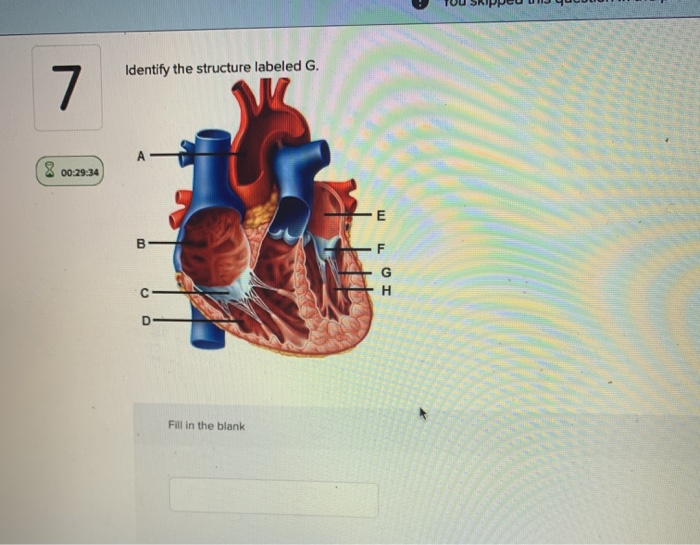
Heart Anatomy: Labeled Diagram, Structures, Blood Flow ...
Cell Biology - Wiki - Scioly.org Sep 11, 2022 · Tertiary structures form when the R groups of the amino acids interact, often incorporating multiple secondary structures as a part of the final protein shape. Finally, quaternary structures form when multiple protein molecules known as subunits come together to form a final protein. Lipids

Basic Histology of the Eye and Accessory Structures - EyeWiki
Which of the following structures is labeled D in the diagram ... [Solved] Which of the following structures is labeled D in the diagram? A)Lumbar enlargement B)Cervical enlargement C)Conus medullaris D)Cauda equina ...
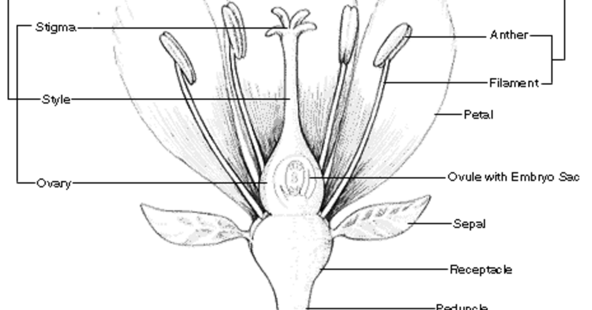
Parts of a Flower: An Illustrated Guide | AMNH
NCBI Conserved Domain Database (CDD) Help - National Center … WebThis document includes help for the Conserved Domain Database (CDD), the CD-Search Tool, and Batch CD-Search.These resources can be used to help elucidate protein function. Note that conserved domain data continue to evolve as research progresses. Comments about the data are welcome and can be sent to info@ncbi.nlm.nih.gov. The "How To" …

Hepatitis viruses comparison | Alila Medical Images
What is a Network? | Webopedia Aug 31, 1996 · Image: Network Topology diagram (v.) To connect two or more computers together with the ability to communicate with each other. Network topologies. Network topology describes how a network is arranged physically and logically. This description includes how links and nodes are connected in relation to each other.
Anatomy and Physiology Quiz 2 Name: A) lymphocytes B) neurons ...
Chapter 12 Anatomy Test Flashcards - Quizlet D. Image: Which type of circuit is involved in solving mathematical problems? ... Which of the following structures is labeled D in the diagram?

Digestive organs: Diagram, stomach, intestines, and more
What is the structure labeled with the X in the diagram above A Leaf B ... 42) What is the structure labeled X in the diagram above of a cross section of a tree trunk? A) Phloem B) Cambium. C) Cork. D) Sapwood. E) Heartwood. Sapwood is all the light colored wood between the heartwood (Z) and cambium (Y). Phloem and cork are outside the cambium.Y Z. D ) Sapwood .
A&P Lecture Exam 3 Flashcards - Easy Notecards
Solved Which of the following structures is labeled D in the - Chegg Epi mater Question: Which of the following structures is labeled D in the diagram? a. Lumbar enlargement b. Cervical enlargement c. Conus medullaris d. Cauda equina e. Obturator nerve Which of the three spinal meninges is the most superficial? a. Arachnoid mater b. Dura mater c. Meninx mater d. Pia mater e. Epi mater This problem has been solved!

The stepwise 3-D atlas reconstruction procedure. (A) A ...
Chapter 13 Wiley Flashcards | Quizlet Which of the following structures is labeled D in the diagram? Cervical enlargement Lumbar enlargement Obturator nerve Conus medullaris Cauda equina 1 Only During childbirth, anesthesia is administered into the epidural space of the spinal column between which of the following vertebrae? 1. L4 and L5 2. T3 and T4 3. S4 and S5 4. C3 and C4 1 only
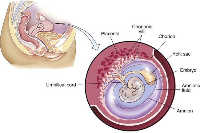
Pregnancy, Childbirth, and the Puerperium | Basicmedical Key
Chapter 13 Flashcards | Quizlet Which of the following structures is labeled B in the diagram> cervical enlargement. Which of the following structures is labeled D in the diagram? lumbar enlargement. Which of the following structures is labeled G in the diagram? cauda equina. The structure labeled A in the diagram belongs to which group of spinal nerves?

What is the structure labeled E in the diagram below ...
Definition and Examples of Parallel Structure - ThoughtCo Web24.06.2020 · "The use of parallel structures," says Ann Raimes in Keys for Writers, "helps produce cohesion and coherence in a text." In traditional grammar, the failure to express such items in similar grammatical form is called faulty parallelism.
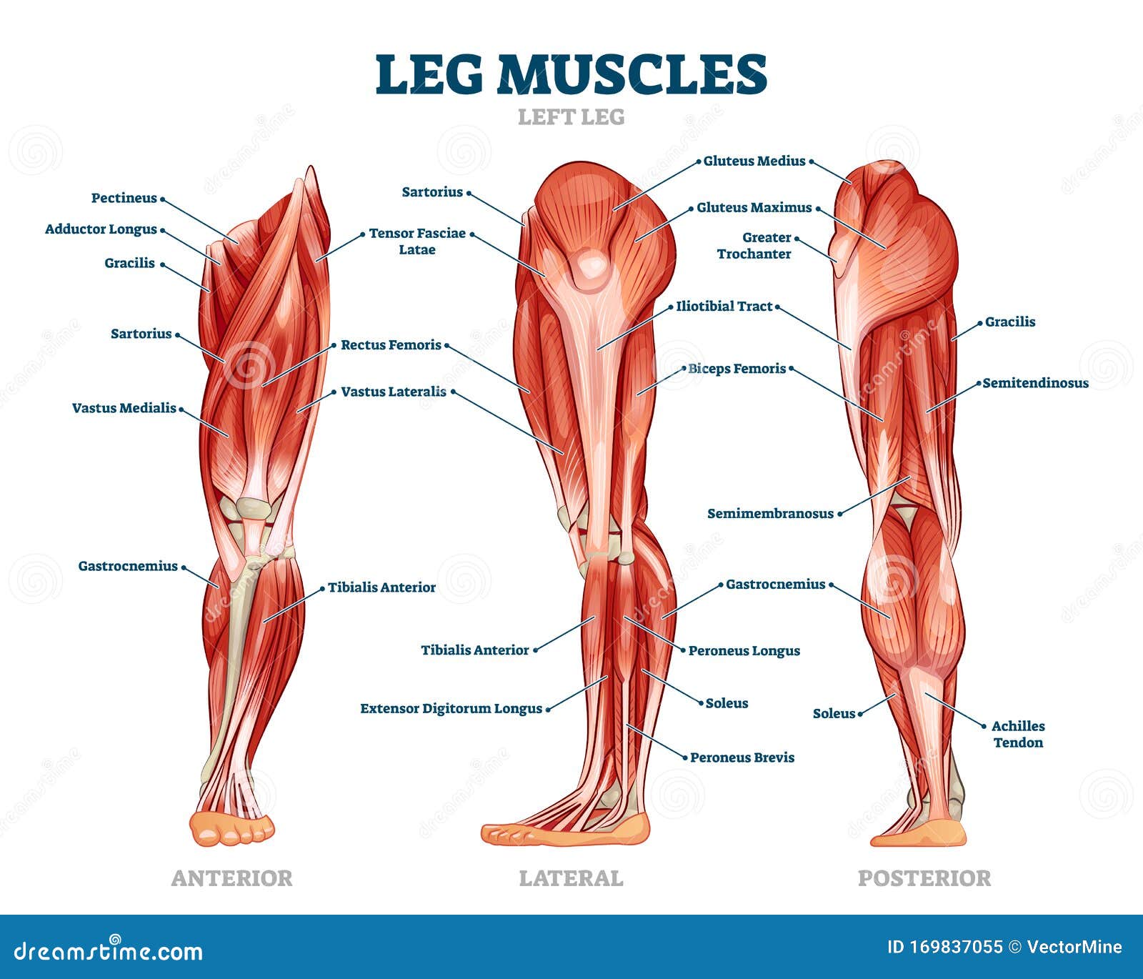
Leg Muscle Anatomical Structure, Labeled Front, Side and Back ...
Unit 4 Picture Questions Flashcards | Quizlet Which of the following structures is labeled D in the diagram? A) Conus medullaris B) Cauda equina C) Cervical enlargement D) Obturator nerve E) Lumbar enlargement C) coccygeal nerves The structure labeled I in the diagram belongs to which group of spinal nerves? A) cervical nerves B) lumbar nerves C) coccygeal nerves D) thoracic nerves
Solved * You received no credit for ti Identify the | Chegg.com
Anatomy 102 Chapter 12 study guide Diagram - Quizlet Which of the following structures is labeled B in the diagram? a) axon terminal b) trigger zone c) cell body d) peripheral process e) dendrites.
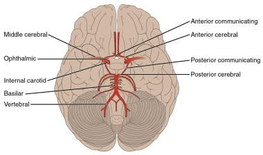
Circle of Willis Anatomy: Overview, Gross Anatomy, Natural ...
Which best describes the structure labeled x in the diagram katsxlove. The structure labeled X in the diagram is a membrane protein. Need a bit more clarification? Get a high-quality answer with step-by-step explanations from a professional in just minutes instead! Get a verified answer. Advertisement.
:max_bytes(150000):strip_icc()/FemalereproductivesystemwithimagediagramKinwunGetty-5b7c291b95d14f229aba86d8d7f0f229.jpg)
Female Anatomy: Labeled Diagrams of the Reproductive System
Ternary Phase Diagram - an overview | ScienceDirect Topics WebTernary phase diagrams are used to represent all possible mixtures of three solvents [1]; they are described in Chapter 3.Here, we shall indicate how they should be used to minimize the solvent consumption. Figure 2.1 (top) shows the methanol–chloroform–water ternary phase diagram with the tie-lines in the biphasic domain. Five particular compositions are …
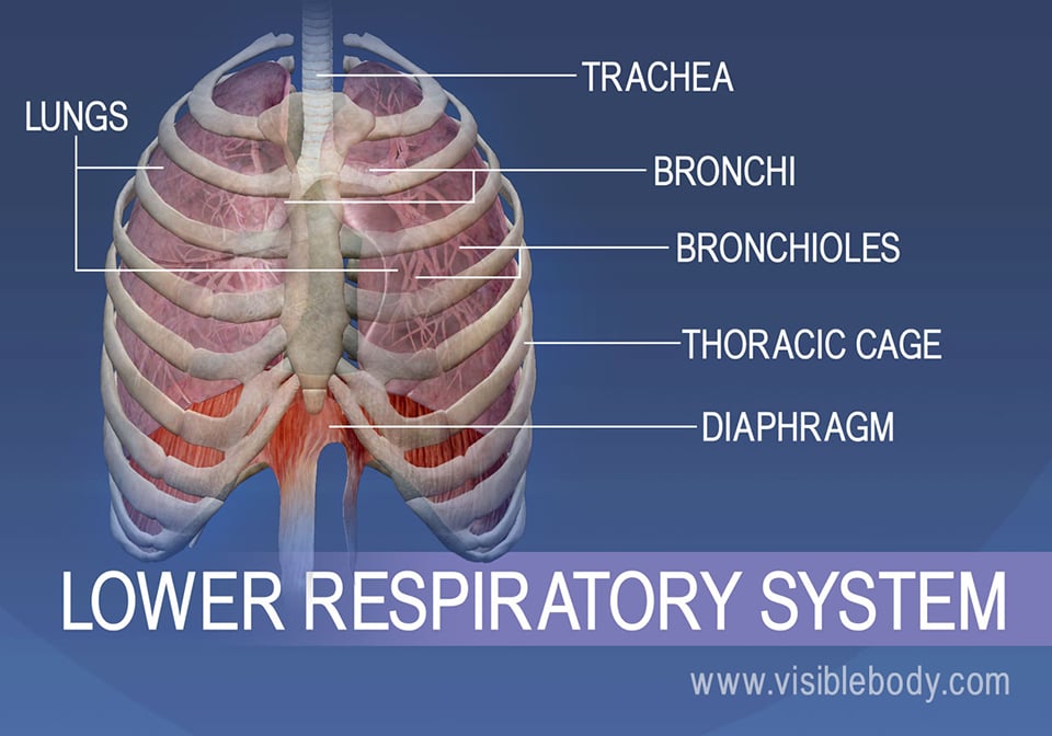
Lower Respiratory System | Respiratory Anatomy
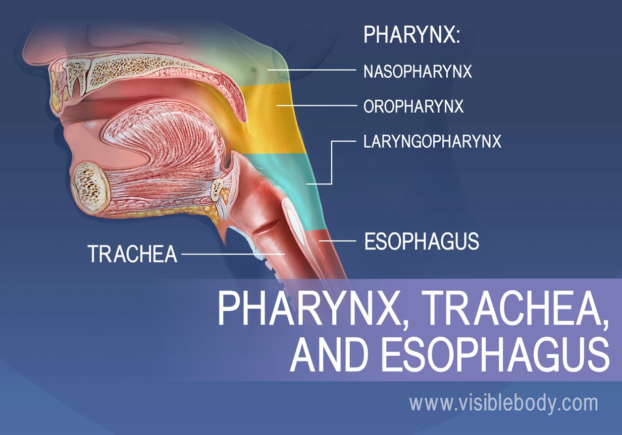
Upper Respiratory System | Respiratory Anatomy
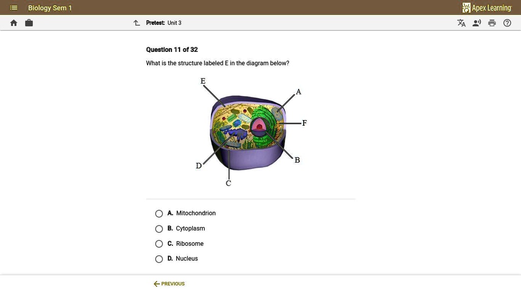
SOLVED: 'What is the structure labeled E in the diagram below ...
/FemaleUrinarySystemKocakayaaliGetty-0e3fdba3f5f34026873c3cfb02baecc9.jpg)
Female Anatomy: Labeled Diagrams of the Reproductive System
0 Response to "39 which of the following structures is labeled d in the diagram?"
Post a Comment