41 in the diagram, where is the left auricle of the left atrium?
Auricle and Atrium|Function|Difference|Applied Anatomy Describing the structure: Auricle is ear like conical muscular appendage to the heart. It is located in the anterior wall of upper heart chamber i.e atrium It is an appendage to Atrium which served by increasing blood holding capacity and blood pumping capacity. Left Auricle - Anatomy Pictures and Information - Innerbody The left auricle, also known as the left atrial appendage (LAA), is a flap of heart wall on the anterior surface of the left atrium of the heart. Often confused with the left atrium, it is one of the most prominent structural features of the left atrium and plays an important role in the pumping of blood within the heart.
Keyword: "atrium" | ClipArt ETC - FCIT 1, The trachea or windpipe; 2 and 3, right and left common carotid arteries; 4 and 5, right and left… Heart and Lungs The heart and lungs. 1, right ventricle; 3, right auricle (atrium); 6, 7, pulmonary artery; 9, aorta;…

In the diagram, where is the left auricle of the left atrium?
auricle of left atrium Heart hypoplastic left syndrome aortic stenosis congenital hlhs ventricle critical genes defect discovered cause rare ever chamber obgynkey 17 Heart. 15 Pics about 17 Heart : Difference Between Auricle and Atrium | Compare the Difference Between, The heart seen from the anterior surface with opened heart chambers and also Right Atrium. Major arteries, veins and nerves of the body: Anatomy | Kenhub 15.8.2022 · Major arteries By definition, an artery is a vessel that conducts blood from the heart to the periphery. All arteries carry oxygenated blood–except for the pulmonary artery.The largest artery in the body is the aorta and it is divided into four parts: ascending aorta, aortic arch, thoracic aorta, and abdominal aorta.. After receiving blood directly from the left ventricle of the … The main structural distinction between the left atrium and the right ... In reality, it is the right atrium that contains the pectinate muscles. The remaining options either correspond to both atrium, or neither. The auricle is another word for ear. This is a cavity present on both the right and left atrium which allows them to extend their blood carrying capacity in situations of need.
In the diagram, where is the left auricle of the left atrium?. Diagram of Blood Flow Through the Heart - Bodytomy The right and left side or chambers of the heart work in tandem with each other. Blood from all over the body will reach the heart through pulmonary veins; inferior vena cava and the superior vena cava. The deoxygenated blood will first enter the right atrium and simultaneously the left atrium receives oxygenated blood from the lungs. Left atrial enlargement | Radiology Reference Article 31.7.2022 · Clinical presentation. An enlarged left atrium can have many clinical implications, such as: Ortner syndrome: left recurrent laryngeal nerve palsy secondary to compression from enlarged left atrium; dysphagia megalatriensis: compression of esophagus between the enlarged left atrium and vertebral bodies; atrial fibrillation: via a multiple wavelet mechanism Left Atrial Enlargement: Causes, Symptoms, and Treatment - Healthline taking medications, such as beta-blockers, calcium channel blockers, alpha-beta-blockers, and diuretics. eating a heart-healthy diet. limiting salt. being physically active and maintaining a ... Left Atrial Anatomy Revisited | Circulation: Arrhythmia and ... The left atrium has a distinctive atrial appendage and an atrial body that comprises component parts that blend into one another. The patterns of general myocardial arrangement in the left atrial wall and the presence of interatrial muscle bundles may provide some anatomic background to atrial and interatrial conduction. Understanding the ...
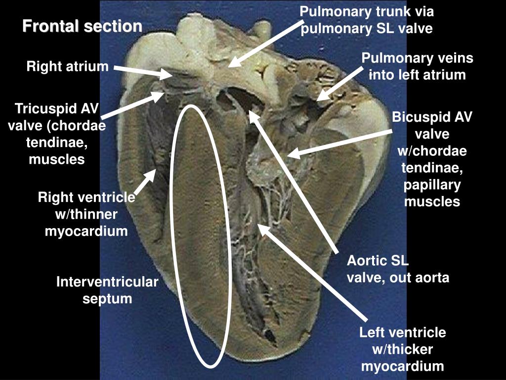
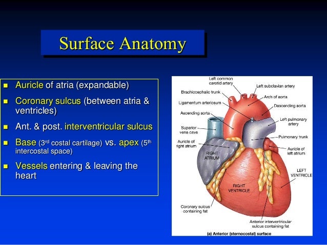
Solved Correctly label the following external anatomy of the - Chegg Expert Answer. THE LABELLED DIAGRAM OF THE GIVEN IMAGE IS ATTACHED BELOW: RIGHT ATRIUM RECEIVE DEOXYGENATED B …. View the full answer. Transcribed image text: Correctly label the following external anatomy of the anterior heart Left atrium Right ventricle Right auricle Left ventricle Coronary sulcus Right atrium. Human Heart – Diagram and Anatomy of the Heart - Innerbody 30.7.2020 · The heart contains 4 chambers: the right atrium, left atrium, right ventricle, and left ventricle. The atria are smaller than the ventricles and have thinner, less muscular walls than the ventricles. The atria act as receiving chambers for blood, so they are connected to the veins that carry blood to the heart. Learn Insta - Life Processes Class 10 Important Questions and Answers ... 3.8.2020 · Left Atrium – Receiving oxygenated blood from lungs. Right Atrium – Receiving deoxygenated blood from the body as well as walls of the heart. Question 17. (a) Draw a diagram of an excretory unit of human kidney and label the following : Bowman’s capsule, Glomerulus, collecting duct, Renal artery. Right and left atrium | Acland's Video Atlas of Human Anatomy The left atrium, like the right one, has a blind pouch, the left auricle or atrial appendage, which projects upwards and forwards. In diastole, the blood that's in the left atrium passes forwards into the left ventricle through the left atrio-ventricular valve, or mitral valve, which is here. To see inside the left atrium we'll remove this part ...
Carnivore Anatomy Lab 12 Introduction - University of Minnesota pulmonary trunk (splits into right & left pulmonary aa.) ligamentum arteriosum (fetal ductus arteriosus; connection to the aorta) Note: pulmonary vv. from the lungs enter the left atrium: left atrium left auricle left atrioventricular orifice left atrioventricular valve (human mitral/bicuspid valve) (parietal & septal cusps) left ventricle A&P Chpt 20 Flashcards | Chegg.com In the diagram, where is the left auricle of the left atrium? a) C b) F c) G d) H e) I. Answer: c. In the diagram, which labeled structure prevents blood flow from the right ventricle back into the right atrium? ... left atrium, left ventricle, right ventricle, pulmonary trunk, pulmonary veins, aorta Right and left atrium Diagram | Quizlet Left atrium - inflow portion receives blood from the pulmonary vein - its internal surface is smooth and it is derived from the pulmonary veins themselves Left atrium - outflow portion located anteriorly and includes the left auricle. Lined by pectinate muscles and is derived from the embryonic atrium Cardiac Cycle: Physiology, Diagram, Phases and Sample Questions Cardiac Cycle is the time taken by the one systole followed by immediate diastole of the heart. In common terms one heartbeat of a person is known as one cardiac cycle. It is the chain of events that occurs inside the human heart when the heart beats. The human heart is a muscular organ that pumps blood through a set of connections between ...
8 a Transthoracic two-dimensional echocardiography (see also Video 1 ...
Sheep Heart Dissection Lab for High School Science | HST The left side of their heart is on their left, but since you are facing them, it is on your right. 1. Identify the right and left sides of the heart. Look closely and on one side you will see a diagonal line of blood vessels that divide the heart. The half that includes all of the apex (pointed end) of the heart is the left side.
PPT - Sheep Heart Dissection PowerPoint Presentation, free download ...
Heart - Wikipedia The human heart is situated in the mediastinum, at the level of thoracic vertebrae T5-T8.A double-membraned sac called the pericardium surrounds the heart and attaches to the mediastinum. The back surface of the heart lies near the vertebral column, and the front surface sits behind the sternum and rib cartilages. The upper part of the heart is the attachment point for several large …

3Lab_Heart_Anatomy Flashcards | Quizlet
Left Atrium Function, Definition & Anatomy | Body Maps - Healthline The left atrium is one of the four chambers of the heart, located on the left posterior side. Its primary roles are to act as a holding chamber for blood returning from the lungs and to act as a...

Heart Structures Flashcards | Easy Notecards
The Heart: Diagram and anatomy of the human heart The left atrioventricular valve (also known as the "Mitral" or "bicuspid") is located between the left auricle and the left ventricle. The pulmonary semi-lunar valve is located at the point where the right ventricle meets the pulmonary artery. The Aortic Semi-Lunar Valve is located at the point where the aorta emerges from the left ventricle.
Heart Anatomy - Right Atrium - 3D Anatomy Tutorial - YouTube
Quiz 16 heart Flashcards | Quizlet In the diagram, where is the left auricle of left atrium? B. In the diagram, where is the ascending aorta? E and I. ... In the diagram, where does the blood pass from the right atrium into the right ventricle? A. In the diagram, where are the semilunar valves? B and D.
Heart by dr. armaan singh
Difference Between Atrium and Auricle - Pediaa.Com Auricle refers to an ear-shaped pouch in the atrium of the heart. Each auricle is attached to the anterior surface of each atrium. Thereby, the two auricles are called the left auricle and the right auricle. Auricle is a wrinkled structure. It resembles a dog's ear. The major purpose of the auricle is to increase the capacity of each atrium.

Human Heart Muscle Structure Image & Photo | Bigstock
ATRIUM DIAGRAM Diagram | Quizlet Start studying ATRIUM DIAGRAM. Learn vocabulary, terms, and more with flashcards, games, and other study tools. Home. ... left auricle... left superior pulmonary vein... valve of fossa ovale... left atrium... interatrial septum... inferior vena cava... left AV valve...
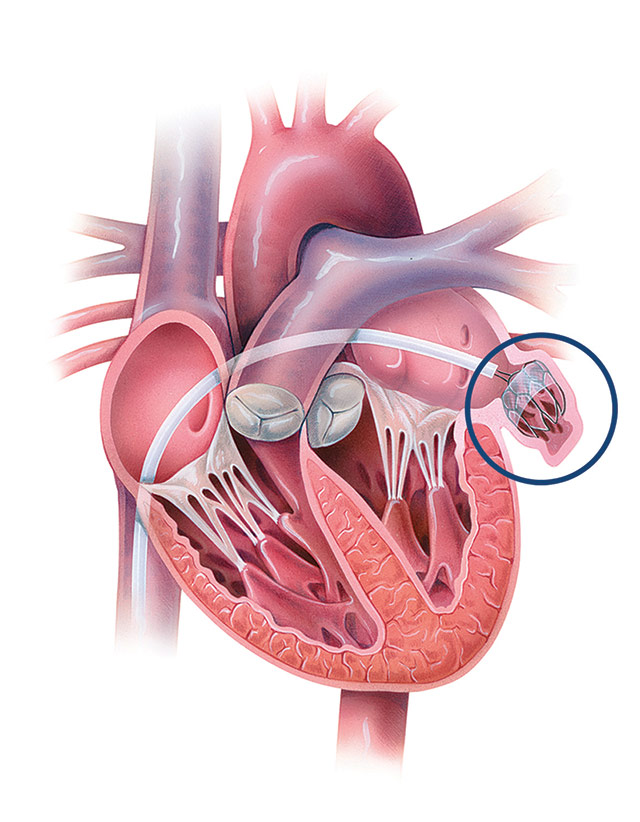
Left Atrial Appendage Closure Procedures | Johns Hopkins Medicine
Heart (right and left atrium): Anatomy and function | Kenhub In the anatomical position, the left atrium is located between the 5th to 8th thoracic vertebrae if the individual is supine (lying flat) or the 6th to 9th vertebrae in someone who is standing erect. Also posteriorly related to the left atrium are the descending aorta, esophagus, and the previously described pulmonary veins.
0 Response to "41 in the diagram, where is the left auricle of the left atrium?"
Post a Comment