38 spinal cord cross section diagram labeled
Spinal cord: Anatomy, functions, and injuries - Medical News Today This article looks at the spinal cord's function and anatomy and includes an interactive diagram. ... Looking at the spinal cord cross-section, the top wings of the gray matter "butterfly ... Spinal Cord Quiz: Cross-Sectional Anatomy | GetBodySmart Spinal Cord - Cross-Sectional Anatomy. Start Quiz. Want to learn faster? Look no further than these interactive, exam-style anatomy quizzes. Learn anatomy faster and remember everything you learn. Start Now. Related Articles. Parts of the Brain Quiz. Test your knowledge with the parts of the brain and their functions in a fun and interactive ...
Spinal Cord Diagram with Detailed Illustrations and Clear Labels - BYJUS The spinal cord is one of the most important structures in the human body. It is the most important structure for any vertebrate. Anatomically, the spinal cord is made up of nervous tissue and is integrated into the spinal column of the backbone. Main Article: Spinal Cord - Anatomy, Structure, Function, and Spinal Cord Nerves; Also Read:
Spinal cord cross section diagram labeled
Spinal Cord Cross Section Labeling Quiz - PurposeGames.com Cross section of the spinal cord and the structures involved . Cross section of the spinal cord and the structures involved. English en. Login. Login Register Free Help; Start; Explore. Games; ... Anatomy. Science. spinal. nerves. Spine. Nervous System. Spinal Cord. CNS. Human Body. Games by same creator. Simple Muscle Structure 9p Shape Quiz. › en › libraryBrainstem: Definition, anatomy, parts, function | Kenhub Aug 15, 2022 · Cross section of the medulla at the level of the hypoglossal nerve (overview diagram) The spinal trigeminal tract and nucleus are located in the lateral aspect of the medulla at this level. Both structures are found dorsal to the lateral reticular nucleus, which is medial to the nucleus ambiguus and posterior accessory olivary nucleus. Spinal Cord Tissue Exploration Of The Human Spinal Cord . nervous neurons spinal labeled cord system brain neuron nerve cell diagram cells human magnification microscope under motor section cross axon > Spinal Ganglion 1 HPO bsmt3.fortunecity.ws. hpo spinal ganglion lpo. Use Of A Self-Delivering Anti-CCL3 FANA Oligonucleotide As An www ...
Spinal cord cross section diagram labeled. Duke Neurosciences - Lab 2: Spinal Cord & Brainstem: Surface and ... A cross-section through the spinal cord is illustrated schematically in Figure 2.6 and 3.4. The gray matter forms the interior of the spinal cord; it is surrounded on all sides by the white matter. The white matter is subdivided into dorsal (or posterior), lateral, and ventral (or anterior) columns. spinal nerve cross section - Microsoft Spinal cord cross-section, detailed anatomy. - Nervous System. 35 Pictures about Spinal cord cross-section, detailed anatomy. - Nervous System : the spinal cord cross section - Google Search | Spinal nerves anatomy, 13.2 Ganglia and Nerves - Anatomy & Physiology and also Solved: Identify the features indicated in the spinal cord cros. Solved Label the following parts of a spinal cord on the - Chegg label the following parts of a spinal cord on the cross- section diagram. a white matter b. grey matter c. donat root ganglion d. nerve fibers e interneuron t synapse g sensory neuron h. motor neuron k reflex act label the following parts of a reflex act on the diagram of a boy stepping on a tack and jerking his leg away. a. sensory neuron b. … en.wikipedia.org › wiki › Posterior_thoracic_nucleusPosterior thoracic nucleus - Wikipedia Anatomy. It occupies the medial part of the base of the posterior grey column and appears on the transverse section as a well-defined oval area.. It begins caudally at the level of the second or third lumbar nerve, and reaches its maximum size opposite the twelfth thoracic nerve.
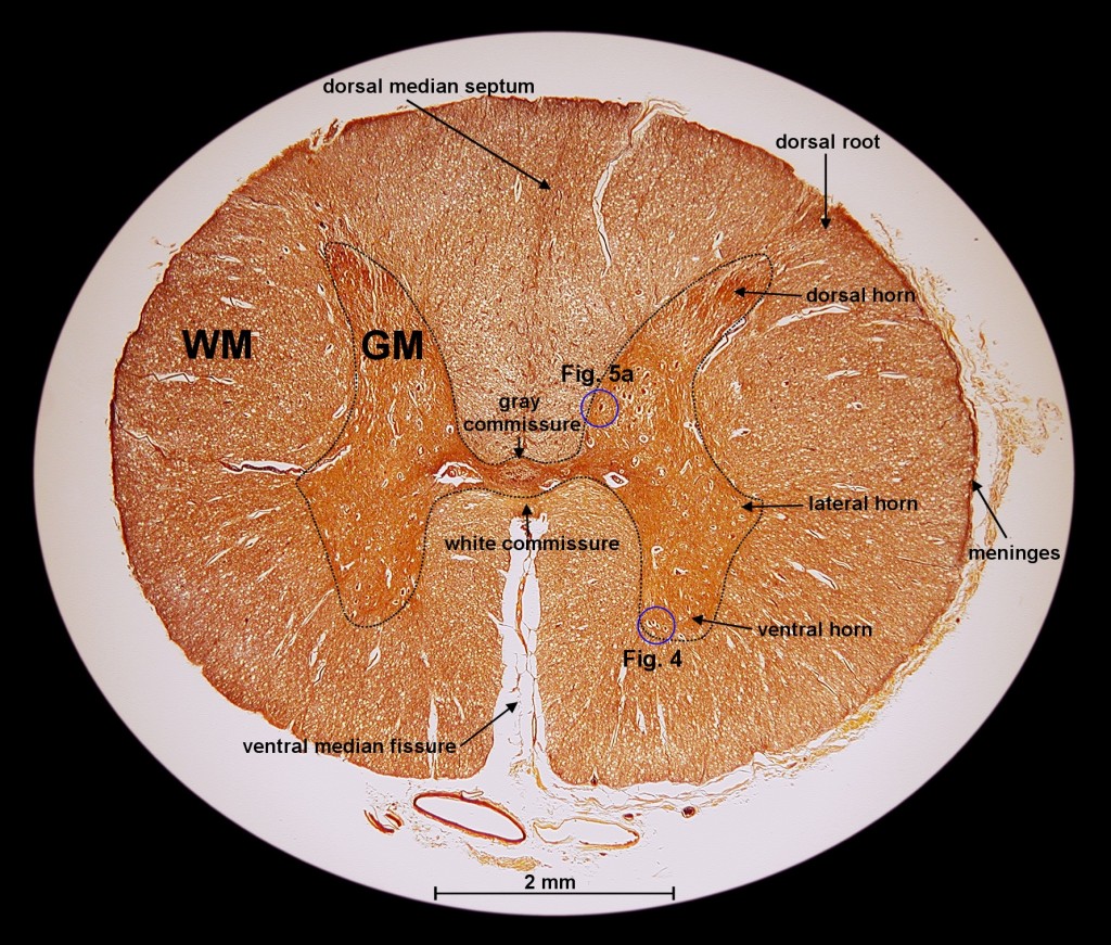
openstax.org › books › anatomy-and-physiology1.6 Anatomical Terminology - Anatomy and Physiology - OpenStax Apr 25, 2016 · A section is a two-dimensional surface of a three-dimensional structure that has been cut. Modern medical imaging devices enable clinicians to obtain “virtual sections” of living bodies. We call these scans. Body sections and scans can be correctly interpreted, however, only if the viewer understands the plane along which the section was made. Spinal cord - austincc.edu Spinal Cord 40X. Cross sections of the spinal cord are so large that you will not be able to see the whole thing on the microscope--you will have to move back and forth or use a dissecting microscope. This section is from a slide that includes both the spinal cord and a vertebra. You are looking at the spinal cord in anatomical position, with the anterior or ventral part at the bottom and the posterior (post) or dorsal part at the top. Single-cell RNA sequencing reveals time- and sex-specific ... - Nature 11.02.2022 · A 5 mm section of the spinal cord ... The Venn diagram was obtained by comparing top 1000 highly expressed genes of each dataset. Cross-species analysis of single-cell transcriptomic data. To ... Neck Pain: Revision 2017: Clinical Practice Guidelines Linked to … Congenital narrowing of the spinal canal may also increase the risk for developing spinal canal stenosis later in life. 106 Magnetic resonance imaging (MRI) is useful in determining the diagnosis of myelopathy. 114 Clinical tests used in the diagnostic process for cervical myelopathy generally have low sensitivity; therefore, they should not be used when screening for and diagnosing this ...
Spinal Cord: Diagram, Anatomy, Structure and Function - Collegedunia A cross-section of the spinal cord reveals the following key areas: Gray matter is the dark, butterfly-shaped region of the spinal cord composed of nerve cell bodies. White matter: The white matter in the spinal cord surrounds the grey matter and contains cells coated in myelin, which speeds up nerve transmission. Gray matter nerve cells are not as heavily coated with myelin. PDF Scanned Document - Bronx High School of Science SPINAL CORD AND REFLEX ACT Cross Section of Spinal Cord Label the following parts of a spinal cord on the cross-section diagram. Name a, b. c. d. h, white matter grey matter dorsal root ganglion nerve fibers interneuron synapse sensory neuron motor neuron (à)çpjnal cord newon. neuwn Posterior thoracic nucleus - Wikipedia The posterior thoracic nucleus, (Clarke's column, column of Clarke, dorsal nucleus, nucleus dorsalis of Clarke) is a group of interneurons found in the medial part of lamina VII, also known as the intermediate zone, of the spinal cord.It is mainly located from the cervical vertebra C7 to lumbar L3–L4 levels and is an important structure for proprioception of the lower limb. Spinal cord cross section (Gray's illustration) - Radiopaedia Some of Gray's illustrations of the spinal cord.
Exploration of the Human Spinal Cord
Spinal Cord Diagram: Detailed Illustration - Collegedunia The spinal cord transmits sensory outputs from the brain to various parts of the body. The spinal cord induces flexible movements. The spinal cord acts as the main coordinating center of all the reflex actions of our body. The white matter of the spinal cord consists of myelin which performs the function of electrical insulation.
Spinal Cord Cross Section Explained (with Videos) | New Health Advisor
Brainstem: Definition, anatomy, parts, function | Kenhub 15.08.2022 · Brainstem tectum, tegmentum and basal area (diagram) The tectum is the roof of the cavity while the tegmentum forms the ventral covering.The central cavity of the neural tube becomes the aqueduct of Sylvius, the fourth ventricle, and the central canal of the spinal cord.Therefore the tectum is the area dorsal to the aqueduct of Sylvius (in the midbrain) and …
Transverse Sections of Spinal Cord | ClipArt ETC
Spinal cord: Anatomy, structure, tracts and function | Kenhub Like the vertebral column, the spinal cord is divided into segments: cervical, thoracic, lumbar, sacral, and coccygeal. Each segment of the spinal cord provides several pairs of spinal nerves, which exit from vertebral canal through the intervertebral foramina. There are 8 pairs of cervical, 12 thoracic, 5 lumbar, 5 sacral, and 1 coccygeal pair of spinal nerves (a total of 31 pairs).

Schwann Cell Anatomy - Human Anatomy - GUWS Medical
The spinal cord | Human Anatomy and Physiology Lab (BSB 141 ... The spinal cord in cross-section has a central region of darker gray matter and the rest is lighter white matter. The gray matter is made up of neuroglia cells and neuron cell bodies. The white matter is made up of neuron axons, mostly but not all myelinated. The dorsal horns are the thinner projections of dark matter that jut out from the rest ...
0 Response to "38 spinal cord cross section diagram labeled"
Post a Comment