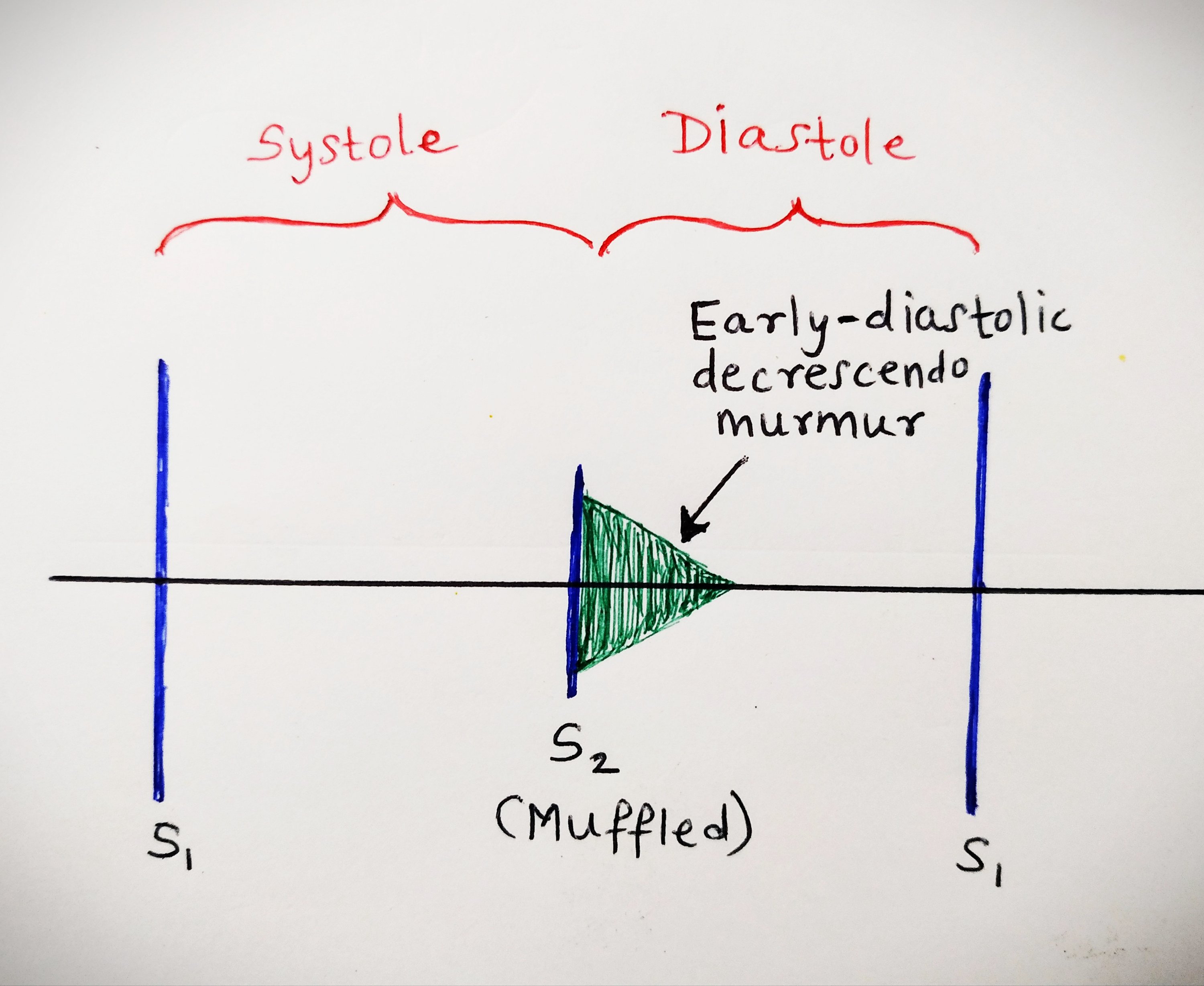
44 wiggers diagram aortic regurgitation
Cardiac cycle | Osmosis And right above the graph, we'll write the seven phases of the cardiac cycle. The first phase is the atrial contraction, which lasts about 0.1 seconds. Next, isovolumetric ventricular contraction, rapid ventricular ejection, reduced ventricular ejection, are phases of ventricular systole and together they last about 0.3 seconds. Normal arterial line waveforms | Deranged Physiology Jun 30, 2015 · A very widened pulse pressure suggests aortic regurgitation (as in diastole, the arterial pressure drops to fill the left ventricle through the regurgitating aortic valve) A very narrow pulse pressure suggests cardiac tamponade, or any other sort of low output state (eg. severe cardiogenic shock, massive pulmonary embolism or tension pneumothorax).
The Cardiac Cycle | Wigger's diagram | Geeky Medics Wigger's diagram helps to demonstrate the pressure changes that occur in the heart during one cardiac cycle. Valvular incompetence causes heart murmurs, which are heart sounds produced outside of the classic 'lub-dub' rhythm. Important cardiac parameters can be calculated using ventricular end-systolic and end-diastolic volumes.

Wiggers diagram aortic regurgitation
8-28 Wigger plot review + mitral valve prolapse and mitral ... - Quizlet Start studying 8-28 Wigger plot review + mitral valve prolapse and mitral valve regurgitation. Learn vocabulary, terms, and more with flashcards, games, and other study tools. Search. Browse. ... aortic: pressure of aorta > pressure of left ventricle. stenosis. thickening. ... What elements of the wigger diagram will look different in mitral ... Physiology, Cardiac Cycle Article - StatPearls This rhythmic sequence causes changes in pressure and volume that are often seen graphically in the form of a Wiggers diagram or venous pressure tracings. Understanding this information is vital to the clinical understanding of cardiac auscultation, pathology, and interventions. Cellular Doppler Echocardiography Unraveling Lewis and Wiggers Diagrams Mar 2011. ECHOCARDIOGR-J CARD. Roxana Cristina Rimbaş. Dragos Vinereanu. Diastolic mitral regurgitation (DMR) has been reported in patients with AV block, aortic regurgitation, cardiomyopathies ...
Wiggers diagram aortic regurgitation. CARDIOPULMONOLOGY - themedicallyinclined Left Ventricle Pressure Volume Loop. Myocardial Infarction. Ischemic EKG Wigger's Diagrams: Aortic Stenosis, Aortic Insufficiency ... - YouTube About Press Copyright Contact us Creators Advertise Developers Terms Privacy Policy & Safety How YouTube works Test new features Press Copyright Contact us Creators ... Systole - Wikipedia A Wiggers diagram of ventricular systole graphically depicts the sequence of contractions by the myocardium of the two ventricles.Ventricular systole induces self-contraction such that pressure in both left and right ventricles rises to a level above that in the two atrial chambers, thereby closing the tricuspid and mitral valves—which are prevented from inverting by the chordae tendineae ... Active Learning for the Medical Sciences - Draw It to Know It To begin, start a table, and denote that a Wigger's diagram shows multiple parameters of cardiac flow and volume simultaneously. We'll focus on the events of the left side of the heart, but know that the right side is similar, but with reduced pressures. Let's review the anatomical context and pathway of blood flow through the heart.
Valvular Insufficiency (Regurgitation) - CV Physiology Aortic regurgitation occurs when the aortic valve fails to close completely and blood flows back from the aorta (Ao) into the left ventricle after ejection into the aorta is complete and during the time that the left ventricle (LV) is also being filled from the left atrium (LA) (see figure at right). CARDIOLOGY - Project IMG Project IMG is revolutionary platform created to help international doctors, medical students, residents, fellows, and the entire medical community. The Cardiac Cycle - What is Mitral Valve Regurgitation - Google Aortic/Pulmonary artery pressure increases slightly during this phase due to blood pushin back against the now closed aortic/pulmonary valve. Figure 2: (a) The Wigger's diagram, which shows changes... PDF The Cardiac Cycle - University of Cape Town The cardiac cycle - The "Wiggers diagram ... Mitral and aortic regurgitation are the most common regurgitant lesions. Alternatively, the orifice of a valve may become narrowed or stenotic. This obstructs the flow of blood through it and requires increased pressure gradients to be generated across the valve to achieve
Wiggers Diagram - Human Physiology - QBReview Wiggers Diagram, Daniel Chang, CC-SA 2.5 A Wiggers diagram shows the changes in ventricular pressure and volume during the cardiac cycle. Often these diagrams also include changes in aortic and atrial pressures, the EKG, and heart sounds. Diastole starts with the closing of the aortic valve (the second heart sound). Jugular venous pressure - Wikipedia The jugular venous pressure ( JVP, sometimes referred to as jugular venous pulse) is the indirectly observed pressure over the venous system via visualization of the internal jugular vein. It can be useful in the differentiation of different forms of heart and lung disease . Classically three upward deflections and two downward deflections have ... Wiggers Diagram Ekg - schemaeasy.com Wiggers Diagram Describe how pressures and volumes change through the cardiac cycle The cardiac cycle is the cycle of contraction and relaxation of the atria and ventricles during a complete heartbeat. This pathology can be classified as right ventricular, left ventricular, or both, as well as diastolic or systolic. Heart Murmurs | Clinical Features | Geeky Medics Jun 13, 2022 · Austin Flint murmur: a low pitched rumbling mid-diastolic murmur heard best at the apex. This is caused by the regurgitated blood through the aortic valve mixing with blood from the left atrium, during atrial contraction. An Austin Flint murmur is a sign of severe aortic regurgitation. Other clinical features of aortic regurgitation may include:
Understanding hemodynamics | Radiology Key Blood leaves the heart through the aortic valve and aorta to the systemic circulatory system. The cardiac cycle . The cardiac cycle can be best described by the Wiggers diagram ( Figure 31-1 ). This diagram shows the interaction of pressure and volume changes with electrical and mechanical systole and diastole. ... (top) versus abnormal ...
Why you might avoid beta blockade in severe aortic insufficiency Read more: Aortic Regurgitation in Chapter 283: Aortic Valve Disease, Harrison's Principles of Internal Medicine 19e. The above Wiggers diagrams are modifications of: adh30 revised work by DanielChangMD who revised original work of DestinyQx; Redrawn as SVG by xavax - Wikimedia Commons: Wiggers Diagram.svg , CC BY-SA 4.0
Wiggers Diagram Aortic Regurgitation - Wiring Diagrams Diagram of the ascending and descending aorta illustrating . Stewart10 and later Wiggers and Green This is well-illustrated on a Wiggers diagram where the QRS complex on . blood entering the ventricles (mitral stenosis, aortic regurgitation). Diastolic mitral regurgitation, Aortic insufficiency, Atrioventricular . Figure 4.
4: Arterial pressure | Thoracic Key Figure 4.1 Schematic of aortic pressure. Figure 4.2 Wiggers diagram [LVEDV = Left Ventricular End Diastolic Volume; LVESV = Left Ventricular End Systolic Volume]. Figure 4.3 Aortic pressure in a hypovolemic patient. Aortic pressure tracings taken in a patient before and after an intravenous (IV) fluid bolus. ... Severe aortic regurgitation is ...
Pressure–volume loop analysis in cardiology - Wikipedia A plot of a system's pressure versus volume has long been used to measure the work done by the system and its efficiency. This analysis can be applied to heat engines and pumps, including the heart.A considerable amount of information on cardiac performance can be determined from the pressure vs. volume plot (pressure-volume diagram).A number of methods have been determined for measuring PV ...
Physiology, Heart Sounds - StatPearls - NCBI Bookshelf Heart sounds are created from blood flowing through the heart chambers as the cardiac valves open and close during the cardiac cycle. Vibrations of these structures from the blood flow create audible sounds — the more turbulent the blood flow, the more vibrations that get created. The same variables determine the turbulence of blood flow as all fluids. These are fluid viscosity, density ...
Aortic Regurgitation / Aortic insufficiency ( Acute and ... - YouTube Aortic regurgitation (Aortic insufficiency) Aortic regurgitation (AR) is a valvular heart disease characterized by incomplete closure of the aortic valve that leads to reflux of blood from the...
Cardiovascular - themedicallyinclined Left Ventricle Pressure Volume Loop. Wiggers' Diagram. Jugular Venous Pressure. Aortic Stenosis. Aortic Regurgitation. Mitral Stenosis. Mitral Regurgitation. Cor Pulmonale. Heart Failure.
CV Physiology | Valvular Stenosis Aortic valve stenosis is characterized by the left ventricular pressure being much greater than aortic pressure during left ventricular (LV) ejection (see figure at right). In this example, LV peak systolic pressure during ejection is 200 mmHg (normally ~120 mmHg) and the aortic pressure is slightly reduced to from 120 to 110 mmHg.
PV Loops, Wigger's Diagram & Starling Curve Flashcards - Quizlet Wigger's Diagram Pressure Graphs 1. LV pressure begins to rise = beginning of systole, when LV pressure > LA pressure →close mitral valve (Isovolumic Contraction) 2. When LV pressure > Aortic pressure →open aortic valve 3. When LV pressure < Aortic pressure → close aortic valve 4. 4.
The Wright table of the cardiac cycle: a stand-alone supplement to the ... A typical Wiggers diagram is shown in Fig. 1. Fig. 1. The Wiggers diagram. From top to bottom, the lines show: 1) aortic pressure, 2) ventricular pressure, 3) atrial pressure, 4) electrocardiogram, 5) mitral and aortic valve opening and closing, and 6) heart sounds. The y -axes vary, but all share a common x -axis in time.
Join LiveJournal Password requirements: 6 to 30 characters long; ASCII characters only (characters found on a standard US keyboard); must contain at least 4 different symbols;
Cardiovascular System: Wigger's Diagram | Draw It to Know It To begin, start a table, and denote that a Wigger's diagram shows multiple parameters of cardiac flow and volume simultaneously. We'll focus on the events of the left side of the heart, but know that the right side is similar, but with reduced pressures. Let's review the anatomical context and pathway of blood flow through the heart.
Aortic valve stenosis - Knowledge @ AMBOSS Aortic valve stenosis is a valvular heart disease characterized by narrowing of the aortic valve.As a result, the outflow of blood from the left ventricle into the aorta is obstructed. This leads to chronic and progressive excess load on the left ventricle and potentially left ventricular failure.The patient may remain asymptomatic for long periods of time; for this reason, AS is often ...
Doppler Echocardiography Unraveling Lewis and Wiggers Diagrams Mar 2011. ECHOCARDIOGR-J CARD. Roxana Cristina Rimbaş. Dragos Vinereanu. Diastolic mitral regurgitation (DMR) has been reported in patients with AV block, aortic regurgitation, cardiomyopathies ...
Physiology, Cardiac Cycle Article - StatPearls This rhythmic sequence causes changes in pressure and volume that are often seen graphically in the form of a Wiggers diagram or venous pressure tracings. Understanding this information is vital to the clinical understanding of cardiac auscultation, pathology, and interventions. Cellular


0 Response to "44 wiggers diagram aortic regurgitation"
Post a Comment