43 sea urchin anatomy diagram
Distinct regulatory states control the elongation of individual ... The sea urchin larval skeletogenesis provides an excellent platform to tackle this question. In the early stages of sea urchin skeletogenesis, skeletogenic genes are uniformly expressed in the skeletogenic lineage. Yet, during skeletal elongation, skeletogenic genes are expressed in distinct spatial sub-domains. Embryology and Development of the Musculoskeletal System First, it is a relatively slow process. The divisions occur approximately 12 to 24 hours apart. Second, there is a unique orientation of the cells with relation to one another. The first cleavage is a meridional division, but, in the second division, one pair of cells divides meridionally and the other divides equatorially ( Fig. 1.1 ).
Overview, Characteristics, & Examples - Study.com For example, as shown in the echinodermata diagram below, the sea stars often seen on the ocean floor or along the beach possess five arms that extend from a central body containing the mouth and...

Sea urchin anatomy diagram
Use of Echinoderm Embryos to Study the Basic Mechanisms of ... In recent years, it has been shown that the ECM of the sea urchin embryo is a very complex structure, consisting of a number of layers organising during different developmental steps. Using specific antibodies, different storage compartments of the ECM components, like granules and vesicles, have been identified in the unfertilised egg cytoplasm. Architecture and evolution of the cis-regulatory system of the ... - eLife The embryonic skeleton of euechinoid sea urchins, the best studied taxon, is formed by a specialized population of skeletogenic cells known as primary mesenchyme cells (PMCs). These cells are the progeny of the large micromeres (LMs), four cells that arise near the vegetal pole during early cleavage. depts.washington.edu › vurchin › indexVirtual Urchin - Urchin Anatomy - University of Washington Urchin Anatomy. Title Anatomy Quiz . Explore the Internal and External Anatomy of the Sea Urchin. Click on the urchin to see inside. Click again for outside. Start ...
Sea urchin anatomy diagram. Sea otter - Wikipedia The sea otter (Enhydra lutris) is a marine mammal native to the coasts of the northern and eastern North Pacific Ocean.Adult sea otters typically weigh between 14 and 45 kg (30 and 100 lb), making them the heaviest members of the weasel family, but among the smallest marine mammals.Unlike most marine mammals, the sea otter's primary form of insulation is an exceptionally thick coat of fur, the ... Cell Organelles (Plant, Animal)- Structure, Functions, Diagrams Centrioles are the tubular structure that measures about 0.2µm × 0.5µm which is open on both sides until it carries a flagellum or cilium. The wall of the centriole is made up of nine groups of microfilament which is arranged in a circle. Each group is a triplet formed by the A, B, and C tubules. Transcript: Kindergarten Designated ELD w/ Science Teacher: Sea otters need to survive in the ocean ecosystem and, if that is their favorite food, they need to have something on their anatomy so that they can eat that. So, what do sea otters have that help them eat sea urchins? Student 1: Um, sharp teeth! Teacher: Mahava, you think they have sharp teeth? Free Worksheets For Apologia Zoology 2: Swimming Creatures Diagram the anatomy with this printable. Purple Sea Urchin Printable - Purple sea urchin printable diagram anatomy sheet. Sand Dollar Printable - Sand dollars are beautiful sea animals. Learn about what they eat and their predators with this informational sheet. Sea Cucumbers Printable - Printable diagram and labeling sheet of a sea cucumber.
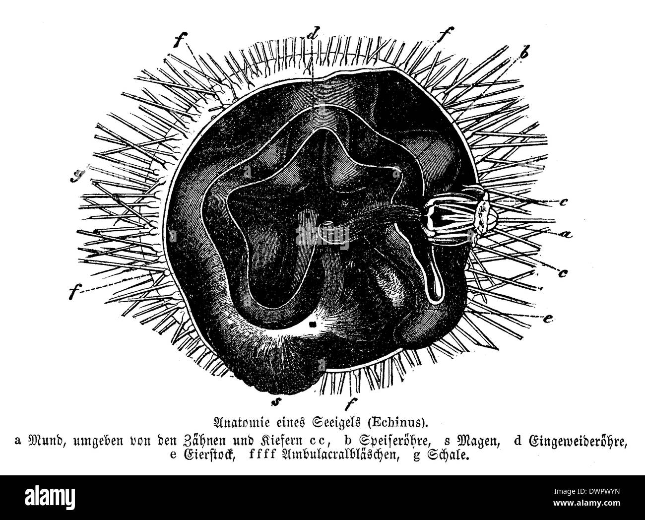
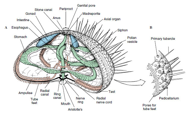
Mitochondria- Definition, Properties, Structure, Functions 140,000 to 150,000 in eggs of sea urchin 300,000 in oocytes of amphibians. It is found that only 500 to 1600 mitochondria are present in the liver cells of the rat. As compared to the animal cell, the number of mitochondria is less in green plants. Architecture and evolution of the cis-regulatory system of the ... The gene regulatory network (GRN) that underlies echinoderm skeletogenesis is a prominent model of GRN architecture and evolution. KirrelL is an essential downstream effector gene in this network ... Spine Healing Therapy - Vertigo Treatment This mouth, often called Aristotle's Lantern, is claw-like with 5 teeth-like plates. Urchins use these teeth to pull, tear and rip off algae from the rocks. These teeth continue to grow throughout the sea urchin's life. The spines also have the ability to transport food to the mouth, serving as both protection and as a feeding-filter for the ... Do Sea urchins have eyes? Or are they plants? - Quora The spherical, hard shells of sea urchins are round and spiny, ranging in diameter from 3 to 10 cm (1 to 4 in). Sea urchins move slowly, crawling with tube feet, and also propel themselves with their spines. Although algae are the primary diet, sea urchins also eat slow-moving ( sessile) animals.
Animal Kingdom: NEET MCQ Questions [150+ Solved] - medicoholic Solve the below free NEET mock test for a better understanding of the various topics. 150+ important MCQs (multiple choice questions) are given in this NEET question bank. Animal Kingdom is the fourth chapter in the unit 'Diversity in the Living World' of class XI or class 11th Biology NCERT. Do note that NEET (which is conducted by NTA) is ... anatomydiagram123.z21.web.core.windows.net › seasea urchin anatomy diagram sea urchin anatomy diagram Sea Star – "OCEAN TREASURES" Memorial Library. 9 Images about Sea Star – "OCEAN TREASURES" Memorial Library : Sea Urchin - Echinus melo - Digestive Systems In Different Phylums, Strongylocentrotus spp., sea urchin: model echinoderm, facts, life and also The Echinoid Directory - Natural History Museum. Sea Urchin Fish Facts | Echinoidea - AZ Animals Sea urchins are animals that are typically small, spiny and round. They live in all the earth's oceans, at depths ranging from the tide line to 15,000 feet. Because they cannot swim, they live on the sea floor. Their main defense against more agile predators like eels and otters is their hard, spiny test, or shell. 3 Sea Urchin Facts Arthropod - Wikipedia Arthropods form the phylum Arthropoda. They are distinguished by their jointed limbs and cuticle made of chitin, often mineralised with calcium carbonate. The arthropod body plan consists of segments, each with a pair of appendages. Arthropods are bilaterally symmetrical and their body possesses an external skeleton.
Sea urchin - Wikipedia
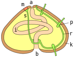
Lab 4 Report S2022 (1) - LAB 4 REPORT ANIMAL DIVERSITY NAME: ID #: CRN ... (e) Observe the fins under the stereomicroscope. Name a characteristic feature. (Hint 3 look at the bony parts of the fins). EXERCISE 4: Anatomy of a Crayfish. On the drawings provided, label the external structures of a crayfish listed in the Protocol.. On the diagram of the sea urchin label the external oral structures listed in the Protocol.. On the diagram of the sea urchin label the ...
Sea urchin - Wikipedia
Acrosome: Reaction, Function & Definition - Study.com This vesicle contains soluble proteolytic enzymes (depicted in the diagram as pink contents) and inner membrane proteins, such as bindin (depicted as cyan-colored dots). The nucleus (depicted in...
Purple Sea Urchin Anatomy
› figure › The-anatomy-of-theThe anatomy of the sea urchin Strongylocentrotus ... Download scientific diagram | The anatomy of the sea urchin Strongylocentrotus droebachiensis. from publication: A Guide to the Sea Urchin Reproductive Cycle and Staging Sea Urchin Gonad Samples ...
External Anatomy of the sea urchin
tiger muscular system - zirpp.org po box 310 phoenix md 21131; ano ang kahalagahan ng oracle bone; how to jump off ladder wwe 2k20. is ghalta male or female; join class action lawsuit against vaccine mandate

Sea Urchin Anatomy by Abiogenisis on DeviantArt
Biology - WCCC Library - Warren County Community College Library ... Digital Anatomist Interactive Atlases This website provides 2-D and 3-D images from cadaver sections, MRI scans, and computer reconstructions of the human body. Users can click on images to view different angles of the organs. Embryology Tutorial This website includes Amphibian and Sea Urchin Embryology tutorials.
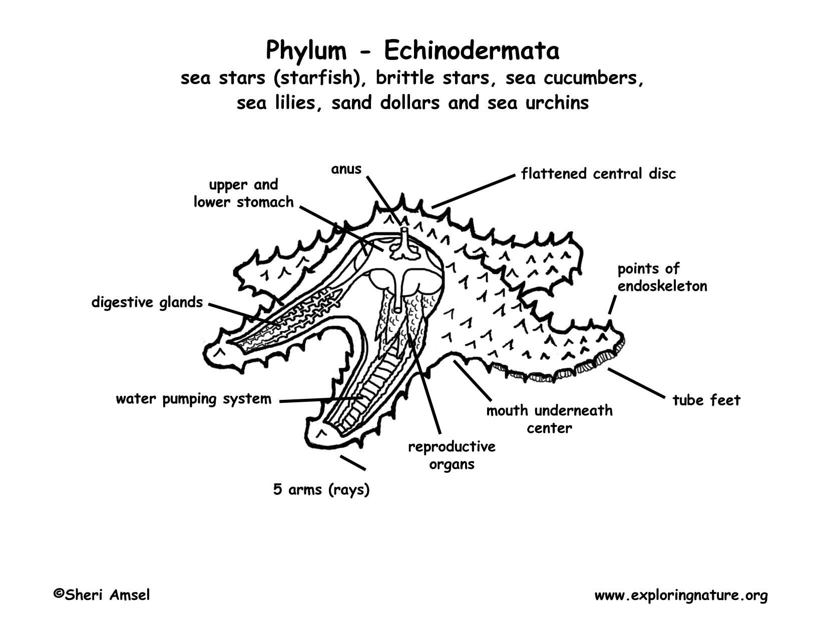
Phylum - Echinodermata (starfish, sea urchins, sand dollars, etc.)
A Reaction-Diffusion Model of Human Brain Development Cortical folding exhibits both reproducibility and variability in the geometry and topology of its patterns. These two properties are obviously the result of the brain development that goes through local cellular and molecular interactions which have ...
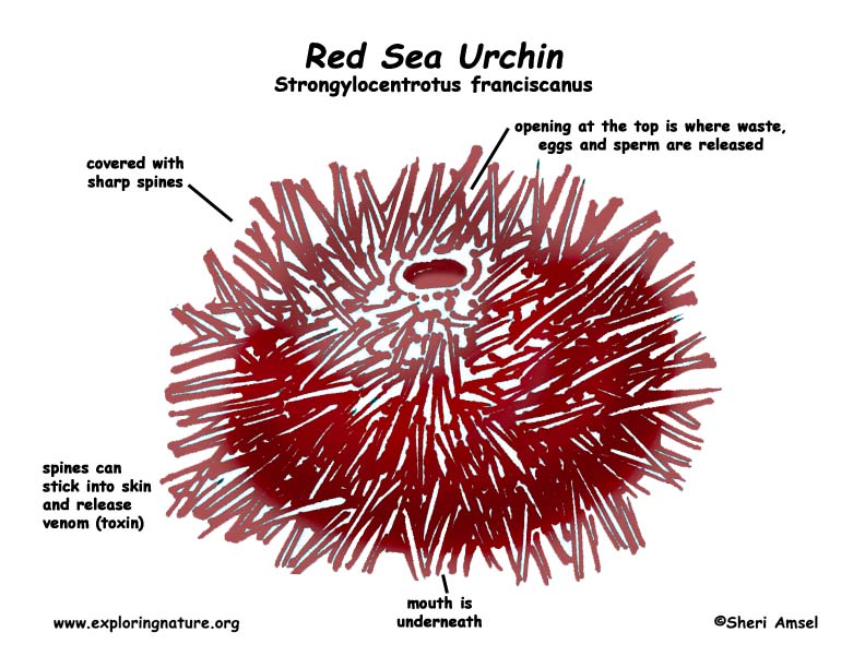
Sea Urchin (Red)
quizlet.com › 291538152 › sea-urchin-anatomy-diagramSea Urchin Anatomy Diagram | Quizlet Start studying Sea Urchin Anatomy. Learn vocabulary, terms, and more with flashcards, games, and other study tools.
Class Echinoidea | Echinoderms | The Diversity of Animal Life
Download PDF, ePub, Kindle eBooks - Blogger Male Anatomy Diagram - Internal Male Anatomy Diagram Quizlet - Aug 22, 2015 · sea urchin anatomy one look at a sea urchin and you can see why they would be called sea hedgehogs. Male Anatomy Diagram - Internal Male Anatomy Diagram Q… [DOWNLOAD] "Liebe" by Richard David Precht # eBook PDF Kindle ePub Free
Worms | From the Archives
elodea leaf cells characteristics of life - design Fitness Open the PDF File titled "Characteristics of Life Worksheet" 3. The gamete producing gametophyte is the persistent plant. Sea Urchins (Lytechinus pictus) Video: Sea urchin cell division. The irst kind, called monoecious, can develop both … response to stimuli. characteristics of life.

Port Phillip Bay Taxonomy Toolkit
humananatomychart.z21.web.core.windows.net › seasea urchin anatomy diagram Diagram Sea Star Internal Anatomy we have 9 Pictures about Diagram Sea Star Internal Anatomy like Sea Urchin Anatomy by Abiogenisis on DeviantArt, sea creatures Archives - Padi Scuba Diving in Protaras Cyprus and also Class Echinoidea | Echinoderms | The Diversity of Animal Life. Read more: Diagram Sea Star Internal Anatomy amazeas.blogspot.com

Diagram Sea Star Internal Anatomy
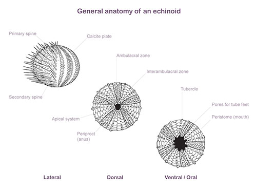
› structures › structureStructure of Sea Urchin (Echinus): With Diagram | Zoology In this article we will discuss about the structure of Sea Urchin (Echinus) with the help of a diagram. 1. It is commonly known as “sea urchin” and is formed in shallow water in both rocky and sandy place in sea. 2. Body is Sub-globular and convex or dome-shaped above and flattened below. The aboral and oral surfaces are distinct.
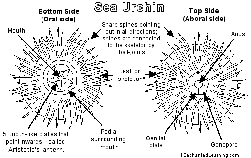
Sea Urchin- Enchanted Learning Software
2022 Science Art Contest Winners | Science Says - UC Davis These ocean friends (the angular unicorn snail, purple sea urchin, shag-rug sea slug, striped shore crab, and bat star) each have a tool box of various adaptations and strategies they use to brave the fluctuating temperatures, solar radiation, and changing water levels. When I was an undergraduate student, I took courses in Bodega Marine ...

sea creatures Archives - Padi Scuba Diving in Protaras Cyprus
My Quest to make art for the living room wall - EMULSIVE Urchin Print. I liked the shot of the bottom of the sea urchin, so I put the negative in the carrier (currently just 2 pieces of cardboard with a 6×6 cm hole in) and did a test strip. I only have two lenses for my enlarger right now: a 50mm and a 150mm. The 50mm won't cover a negative that big, so my only option is the 150mm.
How to Catch Sea Urchin - The Scuba Doctor
Species-Specific Proteins in the Oviducts of Snail Sibling Species ... The anatomy of the adult reproductive system (pallial oviduct) is the only reliable feature used for species identification in females of these species. ... Block diagram, representing a comparison of the domain structure of TRP and SRCR-OP from L. obtusata and L. fabalis. ... Bindin, the protein of the sea urchin's sperm cells surface, ...
-internal-anatomy-a-novel-1741-7007-6-33-4.jpg/220px-Systematic-comparison-and-reconstruction-of-sea-urchin-(Echinoidea)-internal-anatomy-a-novel-1741-7007-6-33-4.jpg)
Wikisource:WikiProject Open Access/Programmatic import from PubMed ...
Sea Urchin Illustration Antique Reproduction Print. Kitchen | Etsy UK Kitchen Wall Art, Butterfly Diagram Print, Species Chart, Vintage Decor. #I1 Ad by studio75wallart Ad from shop studio75wallart studio75wallart From shop studio75wallart
0 Response to "43 sea urchin anatomy diagram"
Post a Comment