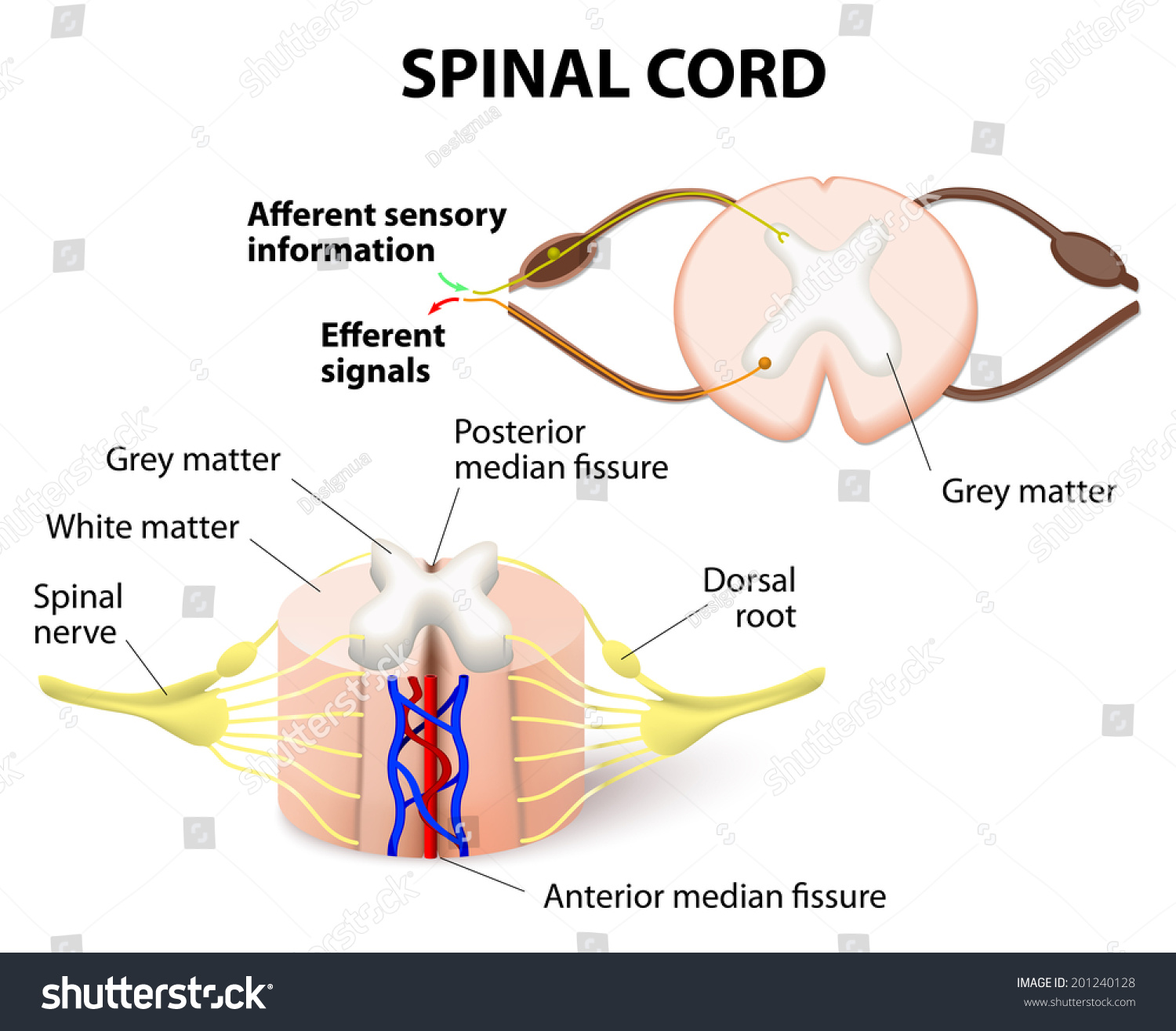
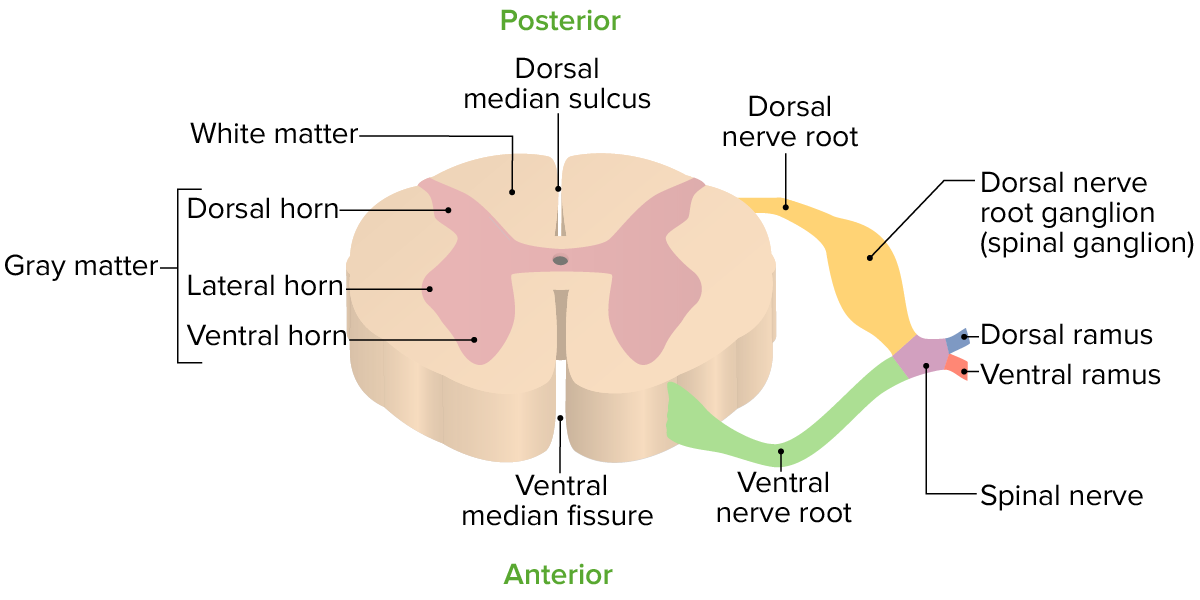
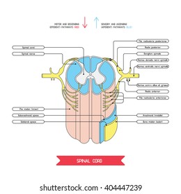
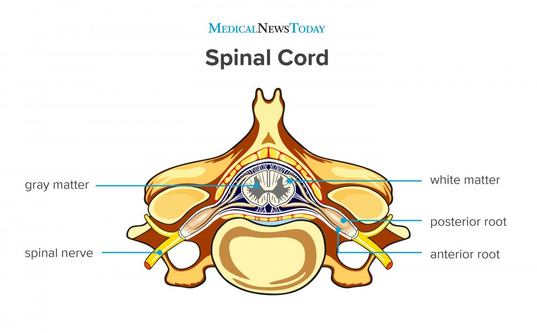
38 spinal cord cross section diagram
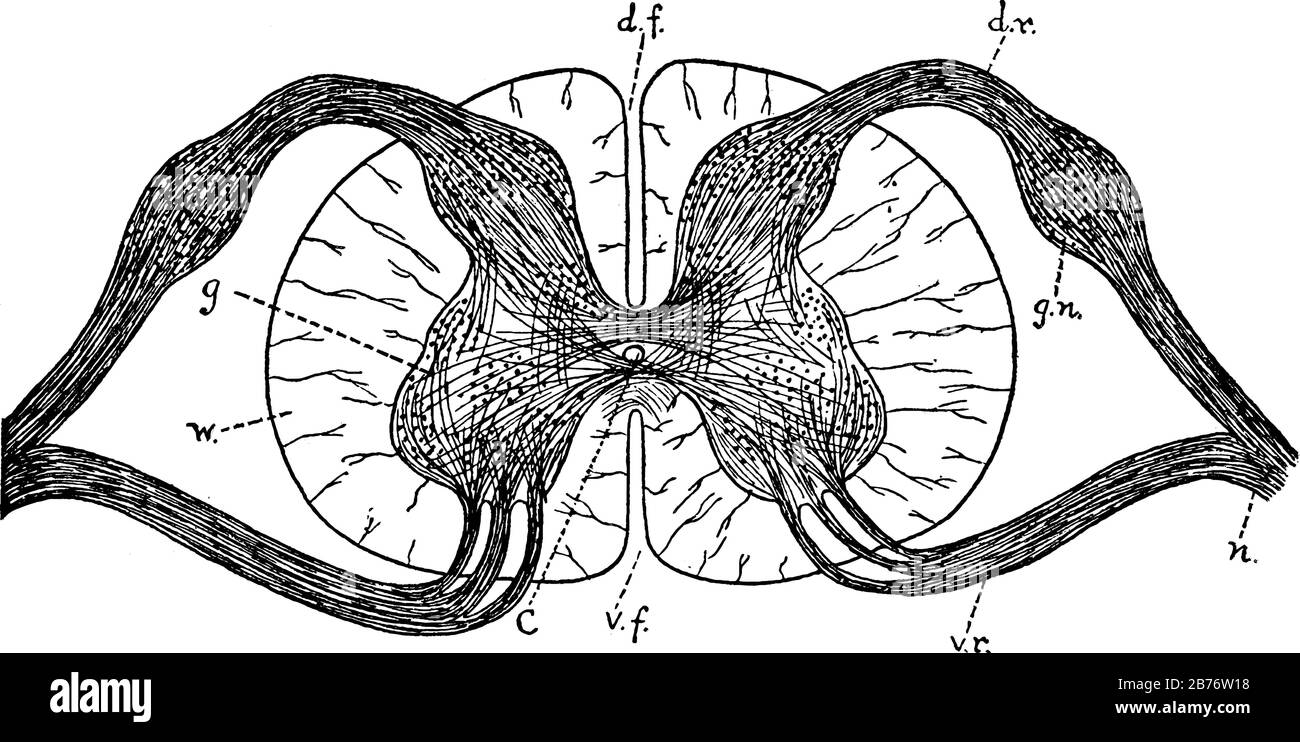
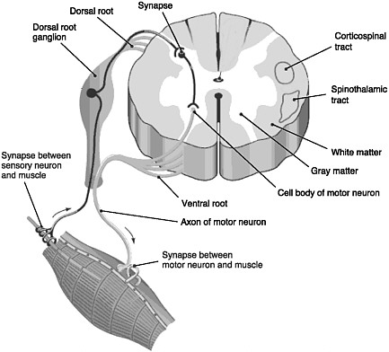
Solved 2. Figure 13-7 is an outline of a cross section ... Figure 13.7. Cross Section of Spinal Cord 12 १ 9 13 3. What is the function of each of the following; Question: 2. Figure 13-7 is an outline of a cross section through the spinal cord. Complete the diagram by drawing the roots and the beginning of the spinal nerve. Add to the diagram a simple reflex arc and label its components. 6 8 १ 1 R ... Anatomy of the Spinal Cord (Section 2, Chapter 3 ... Each spinal nerve is composed of nerve fibers that are related to the region of the muscles and skin that develops from one body somite (segment). A spinal segment is defined by dorsal roots entering and ventral roots exiting the cord, (i.e., a spinal cord section that gives rise to one spinal nerve is considered as a segment.) (Figure 3.4).
Cross Section Spinal Cord Diagram Labeled - spinal cord ... Here are a number of highest rated Cross Section Spinal Cord Diagram Labeled pictures upon internet. We identified it from obedient source. Its submitted by government in the best field. We admit this kind of Cross Section Spinal Cord Diagram Labeled graphic could possibly be the most trending topic behind we share it in google plus or facebook.

Spinal cord cross section diagram
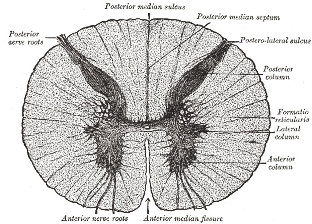
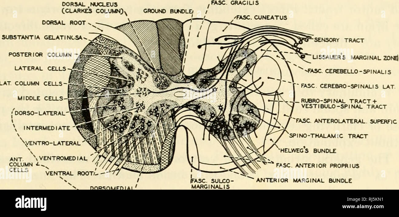
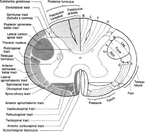
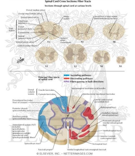
Learn the spinal cord with diagrams and quizzes | Kenhub Take a look at the spinal cord cross section diagram below. Here you can see the white and gray matter of the spinal cord and the associated structures such as funiculi, lamina and tracts. Thinking of the information you learned in the video, spend some time linking the location of the labeled structures with what you know about their function. Cross-sections of the Spinal Cord - SmartDraw Cross-sections of the Spinal Cord. Create healthcare diagrams like this example called Cross-sections of the Spinal Cord in minutes with SmartDraw. SmartDraw includes 1000s of professional healthcare and anatomy chart templates that you can modify and make your own. Spinal Cord Cross Section Photos and Premium High Res ... Browse 881 spinal cord cross section stock photos and images available, or search for spinal cord nerve or autonomic nervous system to find more great stock photos and pictures. female anatomy diagram - spinal cord cross section stock illustrations. pregnancy - spinal cord cross section stock illustrations.
Spinal cord cross section diagram. Spinal Cord Cross Section Stock Photos, Pictures & Royalty ... Browse 2,471 spinal cord cross section stock photos and images available, or search for spinal cord nerve or spinal cord injury to find more great stock photos and pictures. section of the human vertebral column and cross-section of spinal cord. section of the human vertebral column and cross-section of spinal cord. Refer to the following diagram which shows a cross section ... 2011. 19. Refer to the following diagram, which shows a cross-section of the spinal cord of a mammal: Sensory neurons transmit impulses from the receptor, and motor neurons transmit impulses to the effector. Normally when receptor X is stimulated, muscles Y and Z contract. Which one of the following circumstances will cause only muscle Z ... Spinal cord schematic diagram with all sections - cervical ... Spinal cord schematic diagram with all sections - cervical spine, thoracic spine, lumber spine, sacrum, coccyx. and diagram of vertebra. ... vector illustration medical scheme with human head and neck cross section. Vector illustration of cervical vertebrae. Medical scheme with close-up skull and isolated C1 atlas, C2 axis, C3, C4, C5, C6 and ... Body Cavities and Membranes: Labeled Diagram, Definitions Oct 24, 2021 · We can use the circular cross-section below as a reference. The cross-section illustrates as if we are looking down at the spinal cord, and it shows the layers of the spinal cavity discussed above. The spinal cord shown in red is in the center of the spinal cavity. The spinal cavity is enclosed by the vertebral column shown in green.
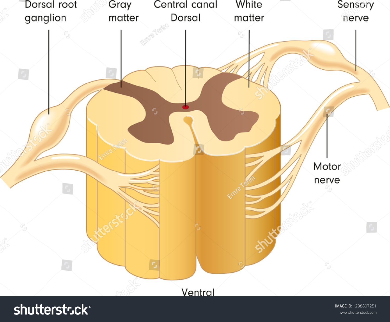
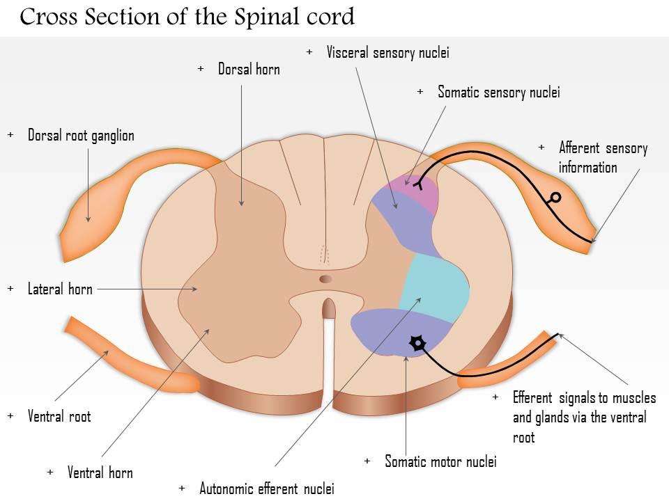
Spinal Cord - Anatomy, Structure, Function, & Diagram Cross-section of spinal cord displays grey matter shaped like a butterfly surrounded by a white matter. Grey matter consists of the central canal at the centre and is filled with a fluid called CSF (Cerebrospinal fluid). It consists of horns (four projections) and forms the core mainly containing neurons and cells of the CNS. Spinal cord- Cross section labeled w/ functions Diagram ... Start studying Spinal cord- Cross section labeled w/ functions. Learn vocabulary, terms, and more with flashcards, games, and other study tools. Spinal cord - Wikipedia Diagrams of the spinal cord. Cross-section through the spinal cord at the mid-thoracic level. Cross-sections of the spinal cord at varying levels. Cervical vertebra A portion of the spinal cord, showing its right lateral surface. The dura is opened and arranged to show the nerve roots. The diagram given below depicts the cross section of the ... The diagram given below depicts the cross section of the spinal cord Study the same and then answer the Questions that follow: (i) Name the process that is being depicted. (ii) Name the parts labelled 2, 5, and 6. (iii) Name the cells in contact with the part labelled '1'. (iv) What is the function of the parts labelled 3, 4 and 7 ?
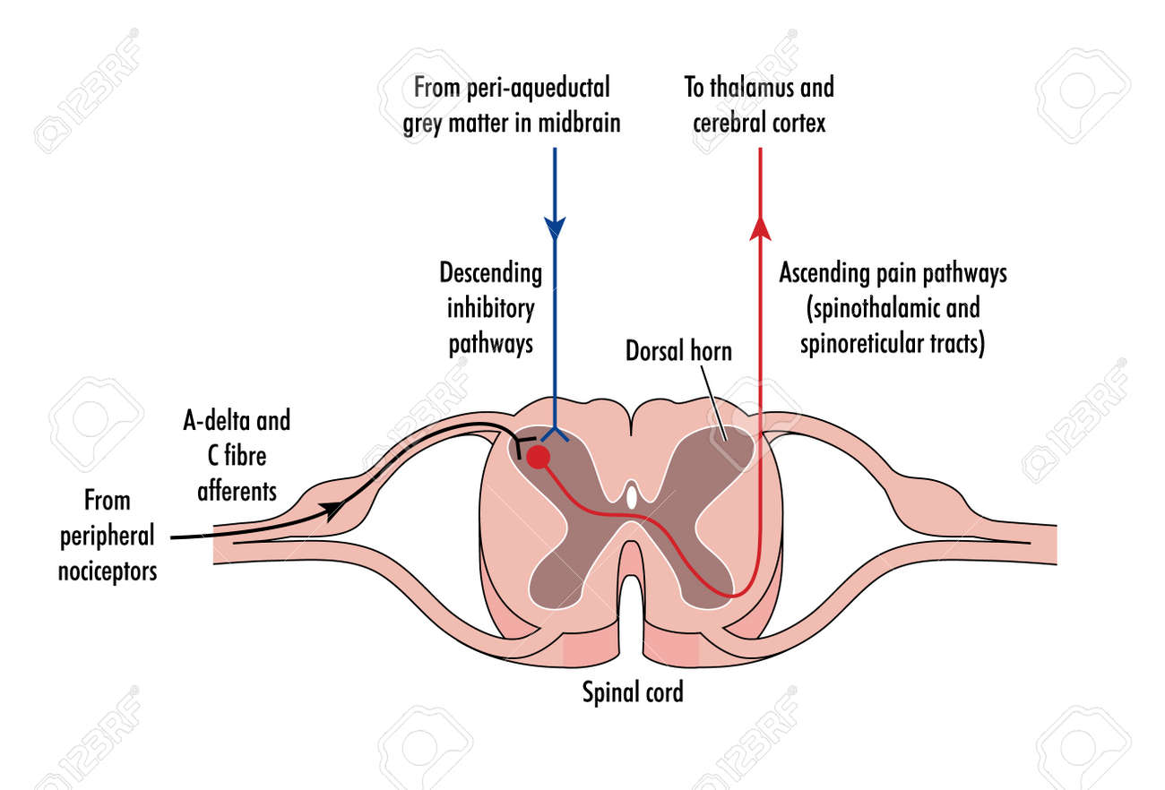
Spinal Cord Cross Section Explained (with Videos) | New ... Spinal Cord Cross Section Looking at a cross section of the spinal cord, you would see gray matter shaped like a butterfly surrounded by white matter. The gray matter is the core and ends up to be four projections that are known as horns. At the back are two dorsal horns and away from the back are two ventral horns. PDF Anatomy and Physiology of the Spinal Cord A complete spinal cord injury means that there is a total blockage of signals from the brain to your sacral nerves. An incomplete spinal cord injury means there is some preservation of nerves from the brain to the lowest part of the spinal cord, the sacral level. The amount of movement and feeling that Spinal cord: Anatomy, structure, tracts and function | Kenhub Spinal cord (cross section) The gray matter is the butterfly-shaped central part of the spinal cord and is comprised of neuronal cell bodies. It shows anterior, lateral, and posterior horns. White matter surrounds the gray matter and is made of axons. It contains pathways that connect the brain with the rest of the body. Anatomy of the spinal cord - eAnatomy The 10 spinal laminae of the spinal cord are shown on a second diagram about the grey matter of the spinal cord. Then, two axial sections of the spinal cord and adjacent structures allow the organisation of a spinal nerve to be displayed with its various branches (sensitive posterior root, anterior motor root, meningeal branch, muscular ...
Spinal cord crosssectional views upper diagram - Spinal Cord Last Updated on Sun, 27 Dec 2020 | Spinal Cord The upper diagram is a cross-section through the spinal cord at the C8 level, the eighth cervical segmental level of the spinal cord (not the vertebral level, see Figure 1). The gray matter is said to be arranged in the shape of a butterfly (or somewhat like the letter H).
Solved Spinal Nerves and Nerve Plexuses. Label the ... Spinal Nerves and Nerve Plexuses. Label the following spinal cord cross section diagram. Note the direction and origin/end of the axons in order to label them correctly. Question: Spinal Nerves and Nerve Plexuses. Label the following spinal cord cross section diagram. Note the direction and origin/end of the axons in order to label them correctly.
Brain, spinal cord and peripheral nervous system anatomy | Kenhub Feb 22, 2022 · The central mass of the spinal cord is a butterfly-shaped grey matter which contains neuronal cell bodies. Master spinal cord anatomy with our study materials. Spinal cord: Cross section Explore study unit
Spinal cord - austincc.edu Spinal Cord 40X Cross sections of the spinal cord are so large that you will not be able to see the whole thing on the microscope--you will have to move back and forth or use a dissecting microscope. This section is from a slide that includes both the spinal cord and a vertebra.
PDF Scanned Document - Bronx High School of Science SPINAL CORD AND REFLEX ACT Cross Section of Spinal Cord Label the following parts of a spinal cord on the cross-section diagram. Name a, b. c.
Duke Neurosciences - Lab 2: Spinal Cord & Brainstem ... A cross-section through the spinal cord is illustrated schematically in Figure 2.6 and 3.4. The gray matter forms the interior of the spinal cord; it is surrounded on all sides by the white matter. The white matter is subdivided into dorsal (or posterior), lateral, and ventral (or anterior) columns.
Spinal Cord Quiz: Cross-Sectional Anatomy - GetBodySmart Spinal Cord - Cross-Sectional Anatomy. Start Quiz. Want to learn faster? Look no further than these interactive, exam-style anatomy quizzes. Learn anatomy faster and remember everything you learn. Start Now. Related Articles. Parts of the Brain Quiz. Test your knowledge with the parts of the brain and their functions in a fun and interactive ...
Cross-Sectional Anatomy - The Central Nervous system Cross-sectional anatomy of the spinal cord The spinal cord appears to be somewhat flat with two grooves that mark its surface. The two grooves are named as follows: the ventral (anterior) median fissure and the more shallow dorsal (posterior) median sulcus.
Spinal Cord Anatomy - Parts and Spinal Cord Functions A spinal needle is inserted between two vertebrae at level L3/L4 or L4/L5, where there is no risk of accidental injury to the spinal cord (which ends at L1 to L2). Cross-Sectional Anatomy of Spinal Cord. The spinal cord, like the brain, consists of two kinds of nervous tissue called gray and white matter.
Home Page: Archives of Physical Medicine and Rehabilitation Apr 04, 2019 · The 40th Episode of the Archives of Physical Medicine’s RehabCast features Tiago Jesus and Christina Papadimitriou on the growth of the person centered rehabilitation model in practice - it’s about putting the person, not the patient, at the center of what we do, and doing it collaboratively.
Nervous System Worksheet Answers - WikiEducator Jan 14, 2008 · 8. The diagram below shows a section of a dog’s brain. Add the labels in the list below and, if you like, colour in the diagram as suggested. Cerebellum - blue; Spinal cord - green; Medulla oblongata - orange; Hypothalamus - purple; Pituitary gland - red; Cerebral hemispheres – yellow. 9. Match the descriptions below with the terms in the list.
PDF Transverse Sections of the Spinal Cord - Elsevier.com Transverse Sections of the Spinal Cord 23 The spinal cord is perhaps the most simply arranged part of the CNS. Its basic structure, indicated in a schematic drawing of the eighth cervical segment (Figure 2-1), is the same at every level—a butterfl y-shaped core of gray matter surrounded by white matter. An often indistinct central
Brainstem - Wikipedia The brainstem (or brain stem) is the posterior stalk-like part of the brain that connects the cerebrum with the spinal cord. In the human brain the brainstem is composed of the midbrain, the pons, and the medulla oblongata.
Spinal Cord Cross Section Labeling - Printable About this Worksheet. This is a free printable worksheet in PDF format and holds a printable version of the quiz Spinal Cord Cross Section Labeling.By printing out this quiz and taking it with pen and paper creates for a good variation to only playing it online.
Spinal Nerves - Veterian Key Jul 18, 2016 · Because the caudal part of the spinal cord (S-1 caudally) and the nerves that leave it resemble a horse’s tail, this part of the spinal cord (the conus medullaris), with the spinal roots coming from it, is called the cauda equina (see Chapter 16). The cauda equina is therefore a part of the peripheral nervous system.
spinal cord cross sections Diagram | Quizlet Start studying spinal cord cross sections. Learn vocabulary, terms, and more with flashcards, games, and other study tools.
Spinal cord: Anatomy, functions, and injuries Looking at the spinal cord cross-section, the top wings of the gray matter "butterfly" reach toward the spinal bones. The bottom wings are toward the front of the body and its internal organs.
Spinal Cord Cross Section Photos and Premium High Res ... Browse 881 spinal cord cross section stock photos and images available, or search for spinal cord nerve or autonomic nervous system to find more great stock photos and pictures. female anatomy diagram - spinal cord cross section stock illustrations. pregnancy - spinal cord cross section stock illustrations.
Cross-sections of the Spinal Cord - SmartDraw Cross-sections of the Spinal Cord. Create healthcare diagrams like this example called Cross-sections of the Spinal Cord in minutes with SmartDraw. SmartDraw includes 1000s of professional healthcare and anatomy chart templates that you can modify and make your own.
Learn the spinal cord with diagrams and quizzes | Kenhub Take a look at the spinal cord cross section diagram below. Here you can see the white and gray matter of the spinal cord and the associated structures such as funiculi, lamina and tracts. Thinking of the information you learned in the video, spend some time linking the location of the labeled structures with what you know about their function.





:background_color(FFFFFF):format(jpeg)/images/library/11473/spinal-membranes-and-nerve-roots_english.jpg)








0 Response to "38 spinal cord cross section diagram"
Post a Comment