38 diagram of eye muscles
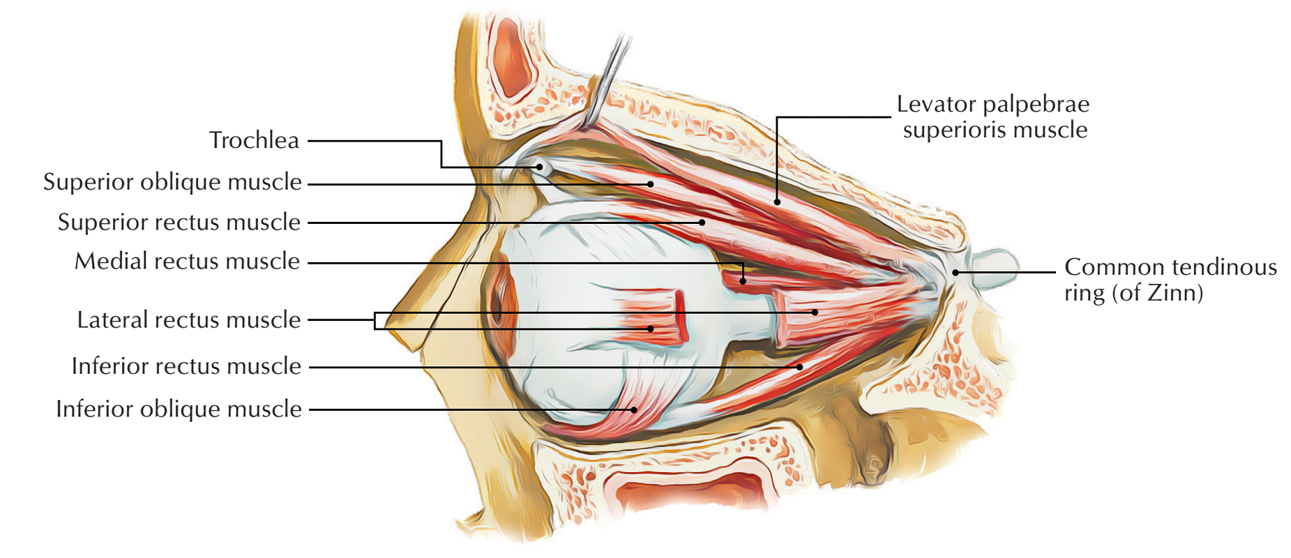
Eye anatomy: Muscles, arteries, nerves and lacrimal gland ... Intrinsic ocular muscles which are within the eyeball itself and control how the eyes accommodate Six extraocular muscles move the eye: superior rectus, inferior rectus, medial rectus, lateral rectus, superior oblique and inferior oblique muscles; and one other, levator palpebrae superioris, opens the eyelid. Structure and Functions of Human Eye with labelled Diagram Human Eye Diagram: Contrary to popular belief, the eyes are not perfectly spherical; instead, ... From the muscles and tissues to nerves and blood vessels, every part of the human eye is responsible for a certain action. Furthermore, contrary to popular belief, the eye is not perfectly spherical; instead, it is two separate segments fused ...
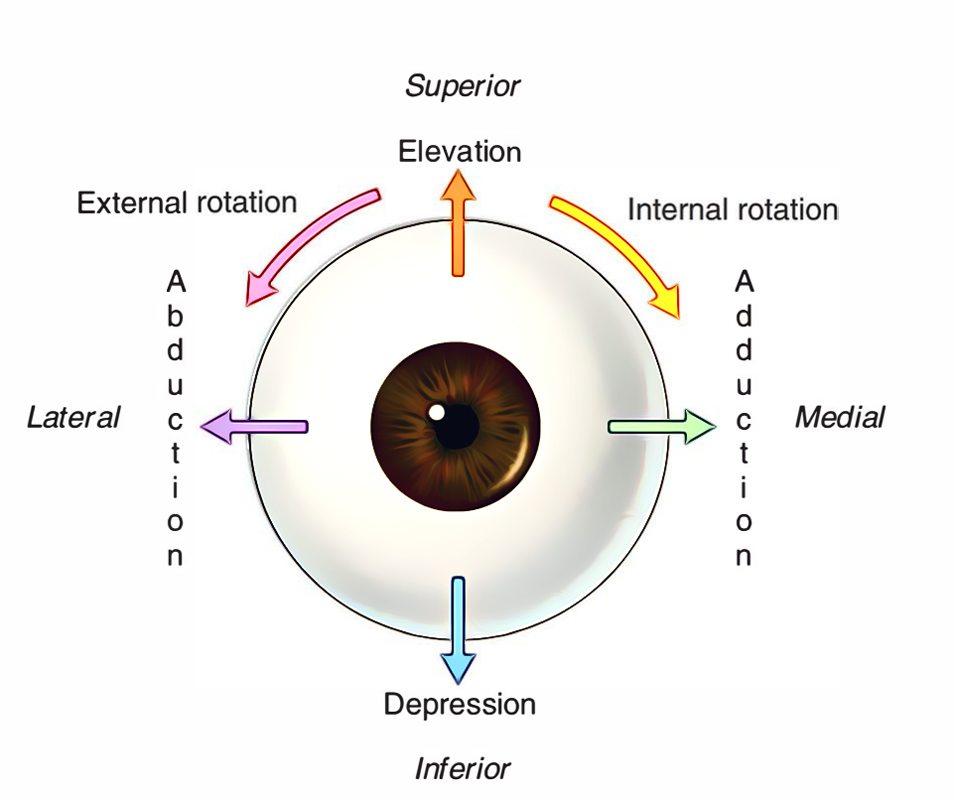
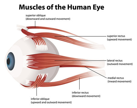
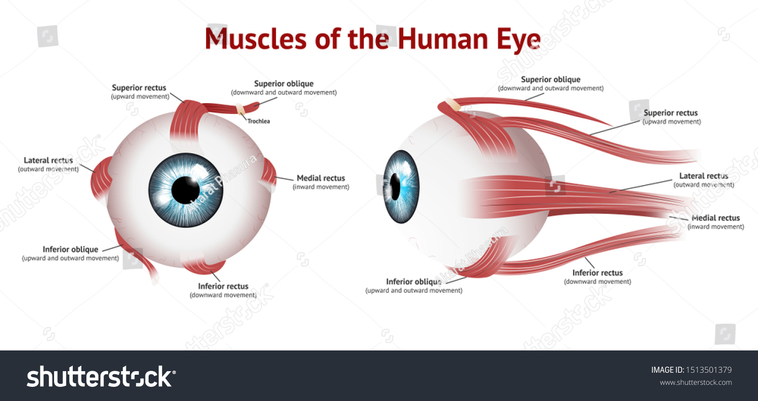
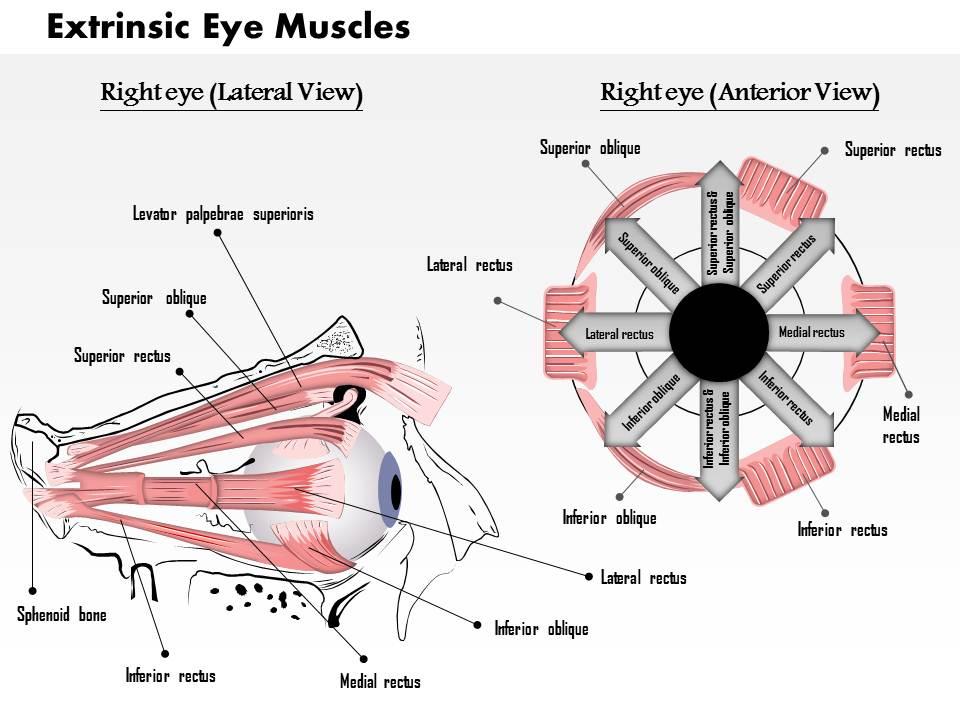
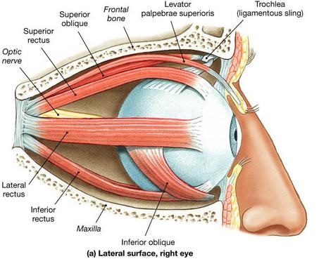
The Human Eye (Eyeball) Diagram, Parts and Pictures ... The six extrocular muscles move the eyelids and eyeballs. Levator palpebrae superioris - lifts the upper eyelid Superior oblique - moves eyeball to the outer side (abduct) and downwards (depresses), medial rotation Inferior oblique - moves eyeball to the outer side (abduct) and upwards (elevates), lateral rotation
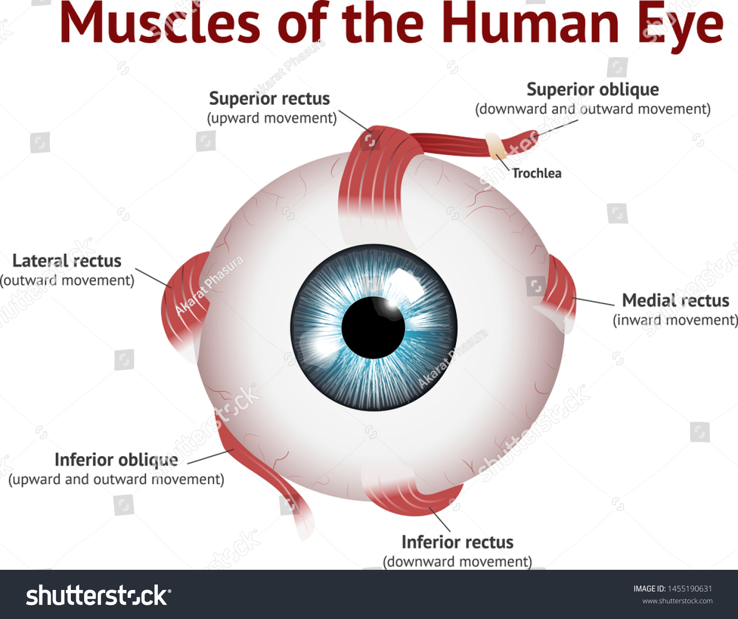
Diagram of eye muscles
Eye Muscles Diagram | Quizlet what two muscles give the eye rotary movement. superior oblique inferior oblique. what kind of visual system does the human brain have. binocular, single-image. what three cranial nerves move the extraocular muscles. VI, IV, III. direction of movement of eye. Sets found in the same folder. ENT. 20 terms. DarcyVanOst PLUS. Muscles of the Eye #2 Diagram | Quizlet Start studying Muscles of the Eye #2. Learn vocabulary, terms, and more with flashcards, games, and other study tools. › anatomy_of_the_eyeHuman Eye Ball Anatomy & Physiology Diagram Feb 04, 2021 · However, the sclera, a tough, leather-like tissue, also extends around the eye. Just like an eggshell surrounds an egg and gives an egg its shape, the sclera surrounds the eye and gives the eye its shape. The extraocular muscles attach to the sclera. These muscles pull on the sclera causing the eye to look left or right, up or down, and diagonally.
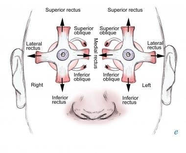
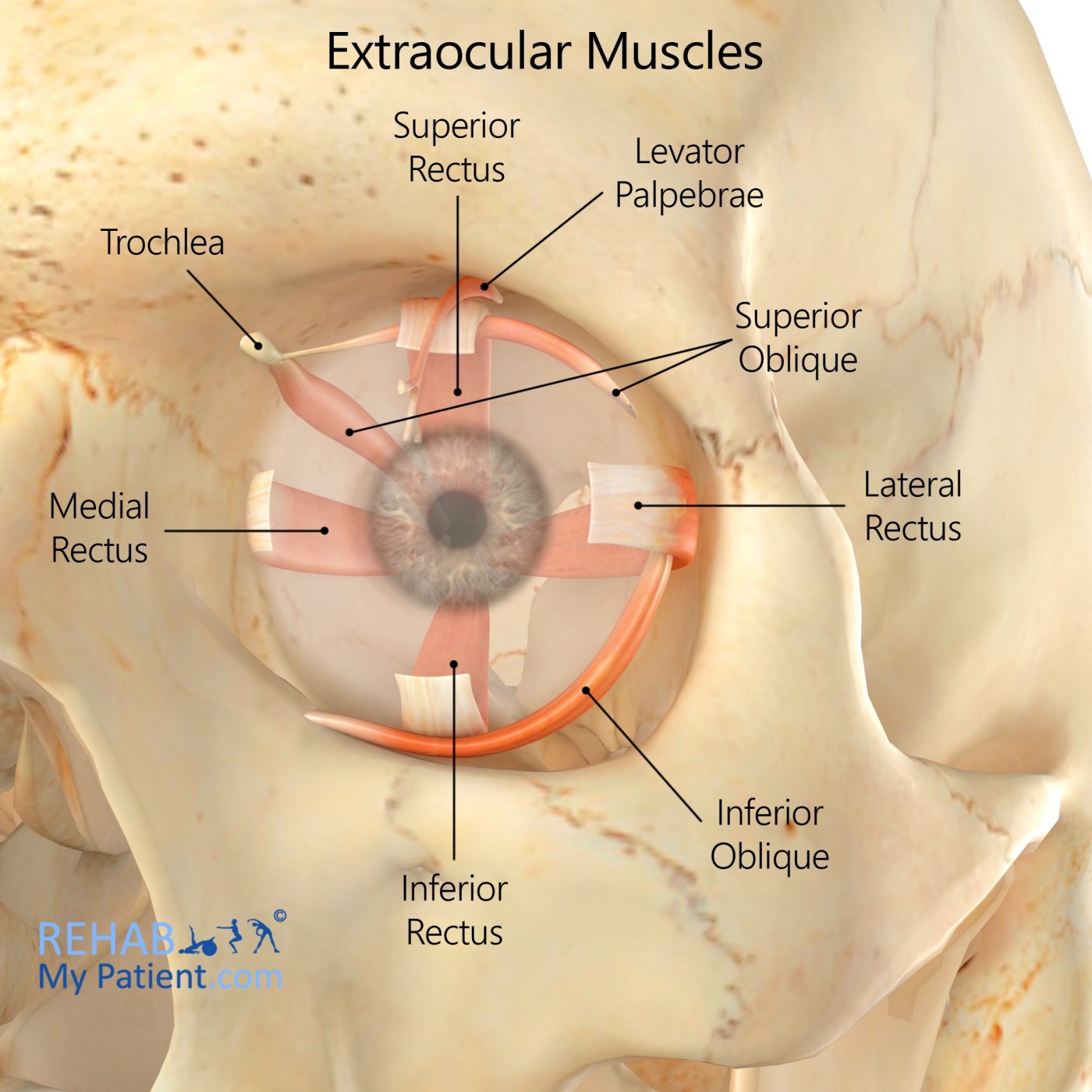
Diagram of eye muscles. Eye Muscles : Attachment, Nerve Supply & Action - Anatomy Info Six skeletal muscles surround and produce various eye movements. Four of the extraocular muscles originated from a tendinous band surrounding the optic nerve. This band, known as the annulus of Zinn. The superior rectus is a thin muscle and forms a straight muscular band between the eye and the annulus of Zinn. Extrinsic Muscles of Eye Diagram | Quizlet Start studying Extrinsic Muscles of Eye. Learn vocabulary, terms, and more with flashcards, games, and other study tools. Trigeminal Nerve: Function, Anatomy, and Diagram 9.11.2021 · It also stimulates movement of the muscles in the jaw and some of the muscles within the inner ear. Diagram The image below shows the location of the 12 cranial nerves, including the trigeminal nerve. Extraocular Muscle Anatomy - Ophthalmology Review The oblique muscles abduct the eye ("abduction is an oblique crime ... in abduction the superior rectus is the primary elevator and the inferior rectus muscle is the depressor. Motility diagrams reflect this relationship between the various actions of the extraocular muscles:
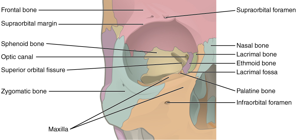
Diagram of the Eye - Lions Eye Institute Instructions. Click the parts of the eye to see a description for each. Hover the diagram to zoom. Iris. The iris is the coloured part of the eye which surrounds the pupil. It controls light levels inside the eye, similar to the aperture on a camera. The iris contains tiny muscles that widen and narrow the pupil size. Anatomy of the Eye - American Association for Pediatric ... There are six muscles that attach to the eye to move it. These muscles originate in the eye socket (orbit) and work to move the eye up, down, side to side, and rotate the eye. The superior rectus is an extraocular muscle that attaches to the top of the eye. It moves the eye upward. Muscles of the eye Diagram | Quizlet Muscles of the eye study guide by landin_sorenson includes 7 questions covering vocabulary, terms and more. Quizlet flashcards, activities and games help you improve your grades. Cat Anatomy | Diagrams & Images of a Cats Body and Skeleton A human has 206 bones, however a cat has around 290 bones and 517 separate muscles, this makes them very agile animals, they use more than 500 muscles to leap, jump and sprint. A cat can jump over 7 times its own height. A cat has 13 ribs in its body. Take a look below at the diagram of a cats skeleton.
Eye muscles - All About Vision Recti muscles The eye has four recti muscles, all of which attach to the front half of the eye (anterior to the equator of the eye). These muscles are: Superior rectus muscle Medial rectus muscle Lateral rectus muscle Inferior rectus muscle Eye Pictures, Anatomy & Diagram | Body Maps A series of muscles helps the eye move. The first set is the superior and inferior rectus muscles, which allow upward and downward motion. The medial and lateral rectus muscles allow the eye to... › physics › human-eyeThe Human Eye - Diagram, Parts, Working, Function and Work of ... Six muscles are in the eye. They are responsible for controlling the movement of the eye. The most common kinds of muscles that are in the eye are the lateral rectus, medial rectus, inferior oblique, or superior rectus. (Image will be Uploaded soon) Parts of the Human Eye. Pupil: The pupil is a small opening in the iris. The iris controls the ... Vision and Eye Diagram: How We See - AARP Small elastic muscles, known as ciliary muscles, which are attached to the lens, help it change its shape in order to focus at various distances. When these muscles contract, the curvature of the lens increases, allowing us to see objects that are close up. When these muscles relax, the lens becomes flattened, helping with long-range vision.
Muscles Human Eye Eye Muscle Anatomy Stock Vector (Royalty ...
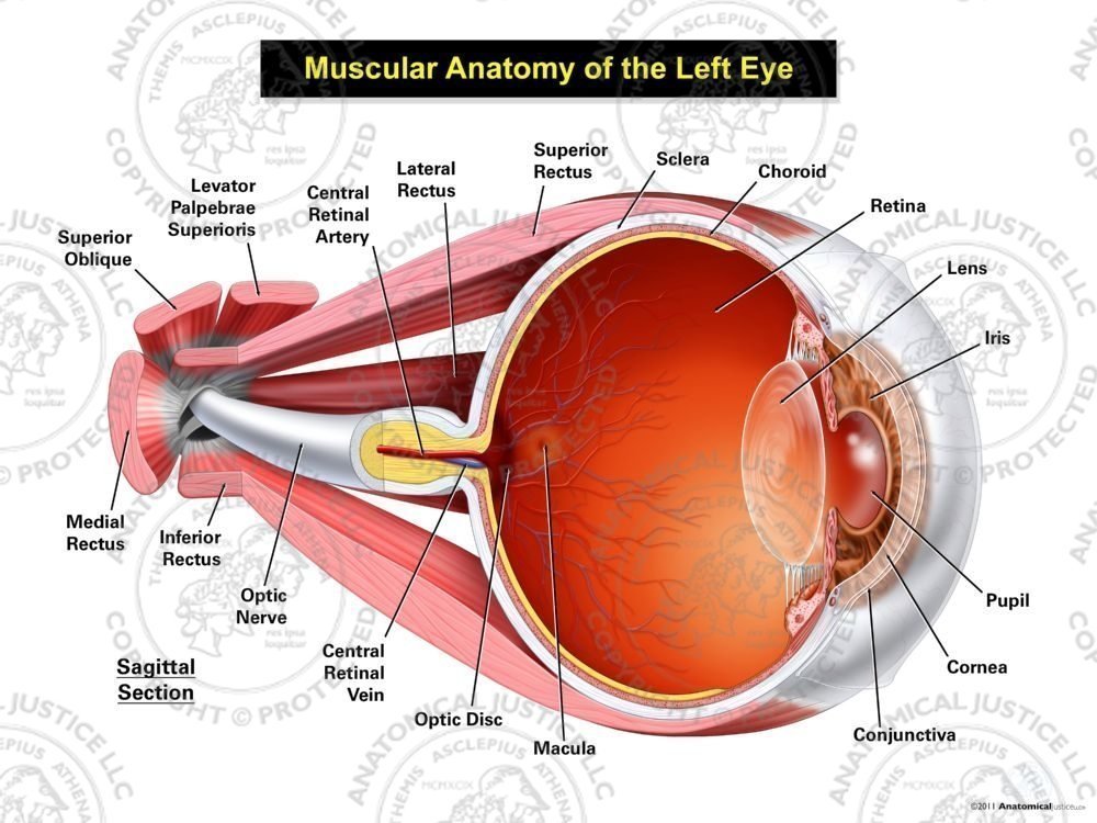
Eye Anatomy: Parts of the Eye and How We See - American ... The eye sits in a protective bony socket called the orbit. Six extraocular muscles in the orbit are attached to the eye. These muscles move the eye up and down, side to side, and rotate the eye. The extraocular muscles are attached to the white part of the eye called the sclera. This is a strong layer of tissue that covers nearly the entire ...

human eye - Movements of the eyes | Britannica
Control of Breathing - an overview | ScienceDirect Topics Andrew M. Dylag MD, Richard J. Martin MD, in Updates on Neonatal Chronic Lung Disease, 2020 Control of Breathing and Physiologic Contributions to Immature Respiratory Control. Respiratory control and its maturation is under tight regulation, with interplay from the central and peripheral nervous systems and feedback from the lung parenchyma and airway musculature.
Extraocular Muscles – Earth's Lab
Have One Pupil Bigger than the Other? - Anisocoria Adie’s tonic pupil is a dilated pupil caused by damage to nerve fibers that control muscles in the eye that constrict the pupil. The affected pupil also reacts poorly to light. Adie’s tonic pupil occurs primarily in women between the ages of 20 to 40 years.

Anatomy and Actions of the Extra-ocular (Eye) Muscles ...
The Six Muscles of the Eye - Brimhall Eye The primary job of this muscle is to turn the eye inward. Every eye muscle does multiple jobs, so the superior oblique does contribute to other motions. Inferior Oblique. The inferior oblique has a similar job to the inferior rectus, but it is the muscle that moves the eye upward when the eye is looking in toward the nose, rather than away.
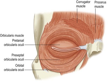
Eyelid Anatomy and Function | Ento Key
PDF Eye Anatomy Handout - National Eye Institute of light entering the eye. Lens: The lens is a clear part of the eye behind the iris that helps to focus light, or an image, on the retina. Macula: The macula is the small, sensitive area of the retina that gives central vision. It is located in the center of the retina. Optic nerve: The optic nerve is the largest sensory nerve of the eye.

2 Surgical Anatomy for Strabismus Surgery | Ento Key
› articles › 326621What are the 12 cranial nerves? Functions and diagram Oct 10, 2019 · The functions of the cranial nerves are sensory, motor, or both: Sensory cranial nerves help a person to see, smell, and hear. Motor cranial nerves help control muscle movements in the head and neck.
Extraocular Muscle Actions: Eye Movements, Rectus Muscles ...
The Penis (Human Anatomy): Diagram, Function, Conditions ... WebMD's Penis Anatomy Page provides a diagram of the penis and describes its function, parts, and conditions that can affect the penis.

Muscles human eye eye muscle anatomy on white Vector Image
anatomysystem.comAnatomy System - Human Body Anatomy diagram and chart images ... diagram anatomy of the eye. Diagram Anatomy Of The Eye Diagram - Diagram Anatomy Of The Eye Chart - Human anatomy diagrams and charts explained. This anatomy system diagram depicts Diagram Anatomy Of The Eye with parts and labels. Best diagram to help learn about health, human body and medicine.

Alila Medical Media | Muscles of the eye, labeled diagram ...
Muscles of the Eye - Innerbody The inferior, medial, and lateral rectus muscles are almost identical to the superior rectus muscle, except they insert on the inferior, medial, and lateral edges, respectively, of the eye. Compared to the four rectus muscles of the eye, the two oblique muscles follow unique paths and insert on the eye at oblique angles.

llustration of the extraocular muscle anatomy. Orientation ...
9.6 Forces and Torques in Muscles and Joints – College ... Muscles, for example, exert far greater forces than we might think. Figure 1 shows a forearm holding a book and a schematic diagram of an analogous lever system. The schematic is a good approximation for the forearm, which looks more complicated than it is, and we can get some insight into the way typical muscle systems function by analyzing it.
Eye Muscles Images – Browse 28,624 Stock Photos, Vectors, and ...
The Extraocular Muscles - The Eyelid - Eye Movement ... There are seven extraocular muscles - the levator palpebrae superioris, superior rectus, inferior rectus, medial rectus, lateral rectus, inferior oblique and superior oblique. Functionally, they can be divided into two groups: Responsible for eye movement - Recti and oblique muscles.

Pinterest Log in Download Terminology – Ophthalmology ...
› anatomy_of_the_eyeHuman Eye Ball Anatomy & Physiology Diagram Feb 04, 2021 · However, the sclera, a tough, leather-like tissue, also extends around the eye. Just like an eggshell surrounds an egg and gives an egg its shape, the sclera surrounds the eye and gives the eye its shape. The extraocular muscles attach to the sclera. These muscles pull on the sclera causing the eye to look left or right, up or down, and diagonally.

Eye Part 1 Extraocular Muscles
Muscles of the Eye #2 Diagram | Quizlet Start studying Muscles of the Eye #2. Learn vocabulary, terms, and more with flashcards, games, and other study tools.
Eye Anatomy | Blood supply - Orbit - Extraocular muscles ...
Eye Muscles Diagram | Quizlet what two muscles give the eye rotary movement. superior oblique inferior oblique. what kind of visual system does the human brain have. binocular, single-image. what three cranial nerves move the extraocular muscles. VI, IV, III. direction of movement of eye. Sets found in the same folder. ENT. 20 terms. DarcyVanOst PLUS.
2020–2021 BCSC Basic and Clinical Science Course™

Solved EXTRINSIC EYE MUSCLES LABEL THE DIAGRAM BELOW WITH ...

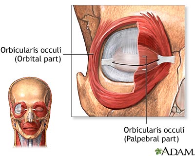
Muscles of the Eyelid - Prohealthsys

The Eye | Biology of Aging

Eye muscles responsible for eye movements and pupil dilation ...
0514 Anatomy Of Eye Muscles Medical Images For PowerPoint ...

The extrinsic muscles of the eye come from the bones of the ...

Eye and orbit - Knowledge @ AMBOSS
Extraocular Muscles | Rehab My Patient
Eye muscles: MedlinePlus Medical Encyclopedia Image
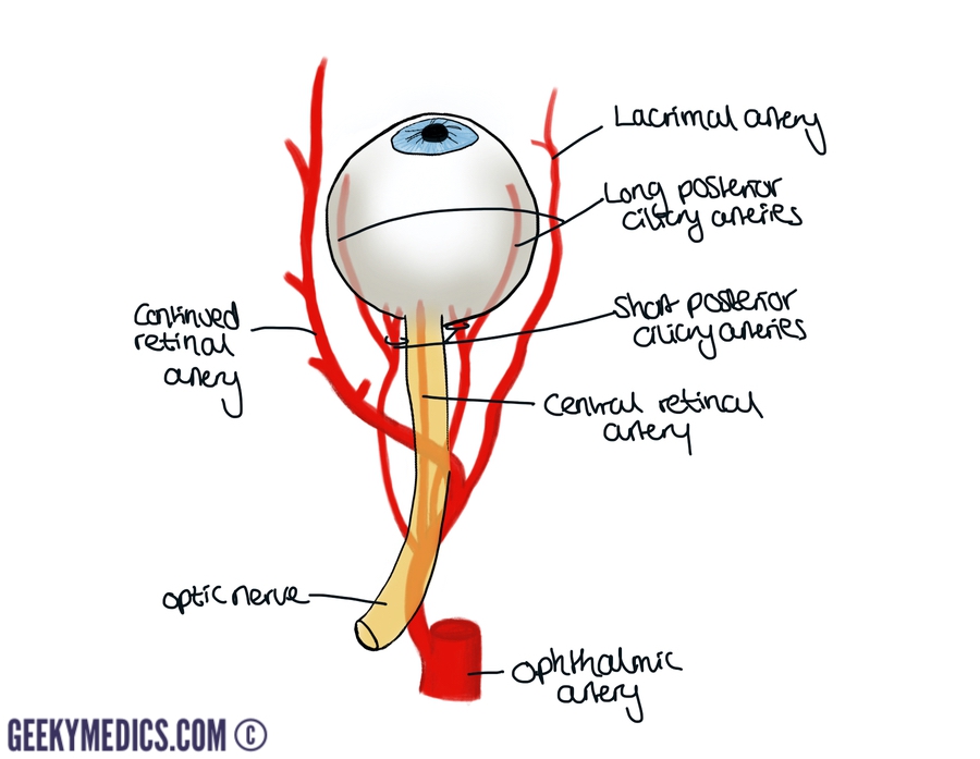
Eye Anatomy | Blood supply - Orbit - Extraocular muscles ...
![Extraocular muscles [8]. | Download Scientific Diagram](https://www.researchgate.net/publication/340572016/figure/fig1/AS:879018110877697@1586585657485/Extraocular-muscles-8.png)
Extraocular muscles [8]. | Download Scientific Diagram
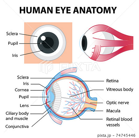
Diagram of human eye anatomy with label - Stock Illustration ...

ophthalmoplegia | eye disorder | Britannica
Extraocular Muscles – Earth's Lab
Muscles Human Eye Eye Muscle Anatomy Stock Vector (Royalty ...

Eyes, Vision Gallery - Free Medical Images for Education by ...
0514 The Extrinsic Eye Muscles Medical Images For PowerPoint ...

Eye Anatomy - Integral Eyesight Improvement

Extraocular Eye Muscles (Lateral View) Diagram | Quizlet

Eye Muscle Clip Art - Royalty Free - GoGraph

Extrinsic eye muscles Diagram | Quizlet
Eye Muscles : Attachment, Nerve Supply & Action - Anatomy Info
Muscular Anatomy of the Left Eye
0 Response to "38 diagram of eye muscles"
Post a Comment