40 spinal nerve root diagram
The first nerve root exits between S1 and S2. One pair of coccygeal (Co1) nerves meets in the area of the tailbone. By way of the peripheral nervous system (PNS), nerve impulses travel to and from the brain through the spinal cord to a specific location in the body. The PNS is a complex system of nerves that branch off from the spinal nerve ... Spinal Nerve Roots. Create healthcare diagrams like this example called Spinal Nerve Roots in minutes with SmartDraw. SmartDraw includes 1000s of professional healthcare and anatomy chart templates that you can modify and make your own.
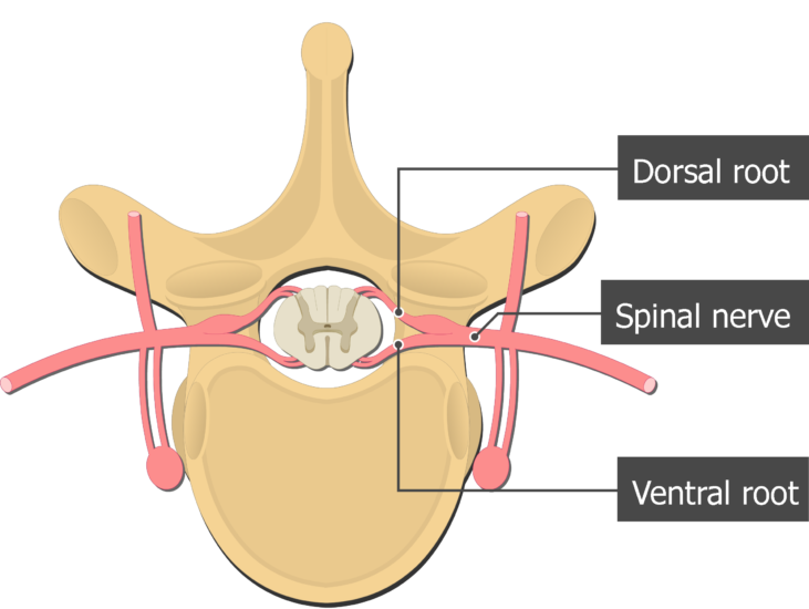
Spinal Nerves . There are 31 pairs of spinal nerves. Again, they are named according to where they each exit in the spine (see figure below). Each spinal nerve is attached to the spinal cord by two roots: a dorsal (or posterior) root which relays sensory information and a ventral (or anterior) root which relays motor information.Therefore, once the two roots come together to form the spinal ...

Spinal nerve root diagram
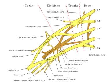
Derived from two Greek root words in “derma” (skin) and “tomos” (a slice), dermatomes refers to a section of skin supplied by a spinal nerve, starting with the cervical spine (in the neck) down to the sacrum (the base of our pelvis). These areas are not perfectly exact, and multiple nerves may innervate a single general area. The relevant anatomy of the spinal nerve-muscular innervation of the back is centered around the lumbar spinal nerves, peripheral nerves of the lumbar plexus, spinal cord, and lumbar vertebral column. Within the lumbar region, the vertebral bodies are larger than in the thoracic and cervical regions due to the lumbar spine being designed for weight-bearing purposes. The cervical nerves arise from the spinal cord in the form of rootlets, or fila radicularia, smaller neuron bundles that coalesce to form roots. For each spinal nerve, an anterior and posterior root join to form the completed nerve. Shortly after branching out of the spinal cord, the cervical nerves form the cervical and brachial plexuses.
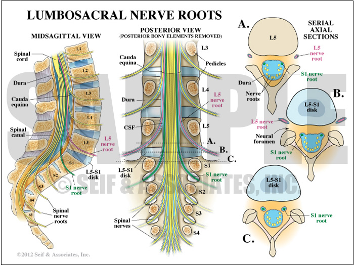
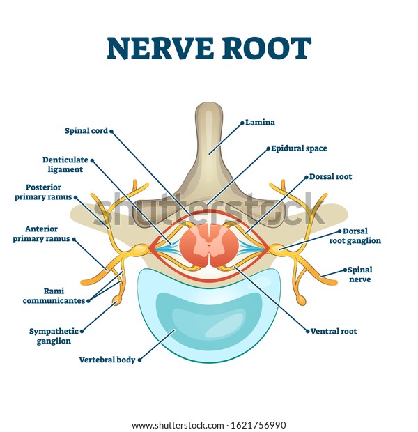
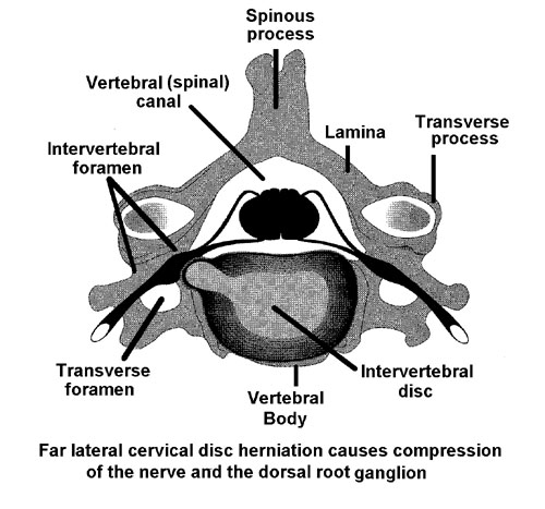
Spinal nerve root diagram. (tissue, organs, etc.), which may cause irritation and/or compression of nerve roots and affect these components.1 The nervous system controls and coordinates all organs and structures of the human body. Many nerves come from the spinal cord, pass through foramina The parenthesis next to the spinal nerve root means this level contributes to the innervation but is not the primary nerve root. For example: the serratus anterior muscle is innervated by the long thoracic nerve with contributions from spinal nerve root C5, 6, 7 (8). •31 pairs of spinal nerves arise from cervical, thoracic, lumbar, sacral and coccygeal regions of the cord. •Cauda Equina (resemble a horse's tail) is the highly branched part of the spinal cord from L2 to S5 which is composed of nerve roots. Structure . The spinal nerves are relatively large nerves that are formed by the merging of a sensory nerve root and a motor nerve root. These nerve roots emerge directly from the spinal cord—sensory nerve roots from the back of the spinal cord and the motor nerve roots from the front of the spinal cord. As they join, they form the spinal nerves on the sides of the spinal cord.
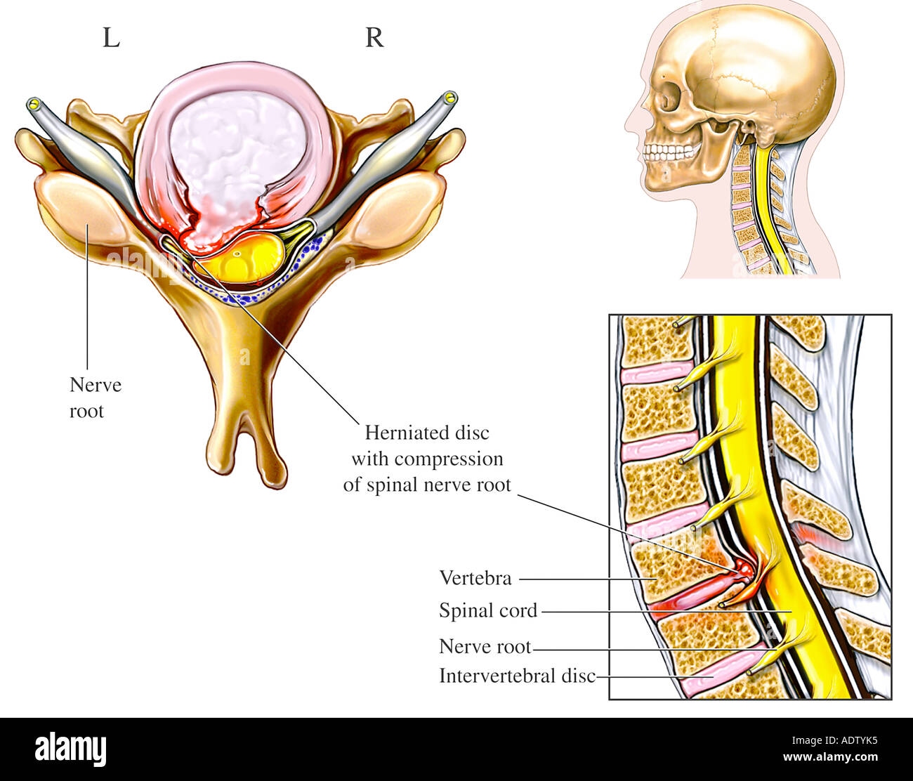
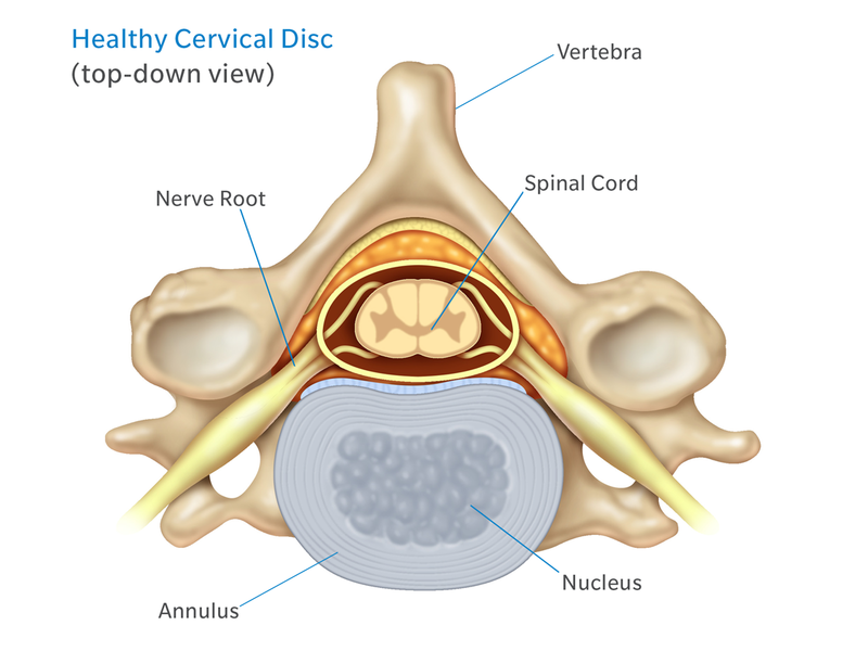
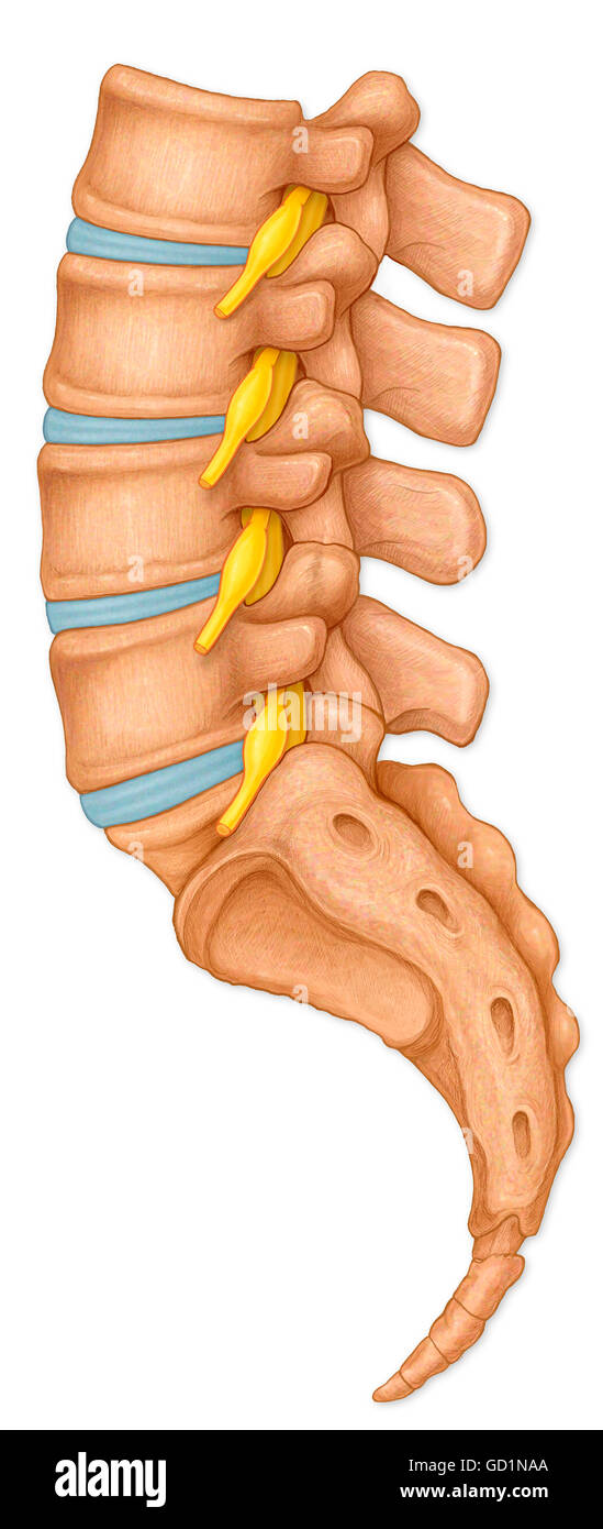
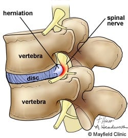
The pudendal nerve originates from the sacral spinal nerves and innervates the descending colon, rectum, urinary bladder, and genitals. Coccygeal Spinal Nerves. There is 1 coccygeal spinal nerve pair. 1 coccygeal nerve; For most spinal segments, the nerve roots run through the bony canal, and at each level a pair of nerve roots exits from the spine. Cervical spine nerve roots. In the neck, the nerve root is named for the lower segment that it runs between (e.g. C6 nerve root at C5-C6 segment). Lumbar spine nerve roots. Spinal meninges (diagram) The spinal cord and spinal nerve roots are wrapped within three layers called meninges. The outermost is the dura mater, underneath it is the arachnoid mater, and the deepest is the pia mater. Dura mater has two layers (periosteal and meningeal), between which is the epidural space. The spinal nerve travels a short distance inside the intervertebral foramen, after which it branches off into several nerves that innervate different parts of the body. Doctors may sometimes refer to the part of the spinal nerve exiting the intervertebral foramen as the nerve root or use the terms nerve root and spinal nerve interchangeably. In ...
Anatomy Of The Spine And Back Spine Muscles Diagram. In this image, you will find 1st cervical vertebrae, atlus, cervical plexus, 7th cervical vertebrae, 1st thoracic vertebrae, brachial plexus, spinal dura mater, filaments of spinal nerve roots, 12th thoracic vertebra, 1st lumber vertebra, iliohypogastric nerve, ilioinguinal nerve, lumbar ... On the chart below you will see 4 Columns (Vertebral Level, Nerve Root, Innervation, and Possible Symptoms). Under ‘Vertebral Level’: C1-C7 is the NECK, ; T1-T12 is the UPPER BACK/rib cage area, and ; L1-L5 is the LOWER BACK.; Simply line up the “Vertebral Level” with the “Possible Symptoms” and you will see some surprising connections of symptoms that relate to your spine. Silver stained spinal cord Silver stained cross section of spinal cord. The white matter surrounds the central grey matter, which show large anterior horns where motor neurons (bottom) are located. At the top, the entry of dorsal roots into the spinal cord is observed. nerve root stock pictures, royalty-free photos & images The 10 spinal laminae of the spinal cord are shown on a second diagram about the grey matter of the spinal cord. ... two axial sections of the spinal cord and adjacent structures allow the organisation of a spinal nerve to be displayed with its various branches (sensitive posterior root, anterior motor root, meningeal branch, muscular branches ...
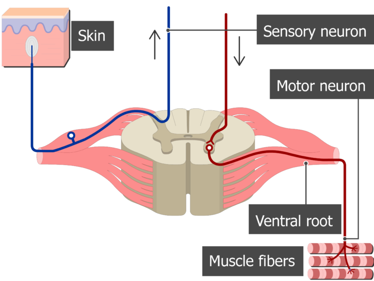
Sympathetic nerve Sympathetic ganglion Spinal nerve Ventral ramus Dorsal ramus Dorsal root ganglion Dorsal root Visceral motor Somatic Ventral root The distribution of motor neurons in the spinal cord and motor fibers within the spinal nerve and its branches. Although the gray ramus is typically proximal to the white ramus,
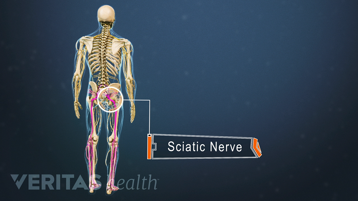
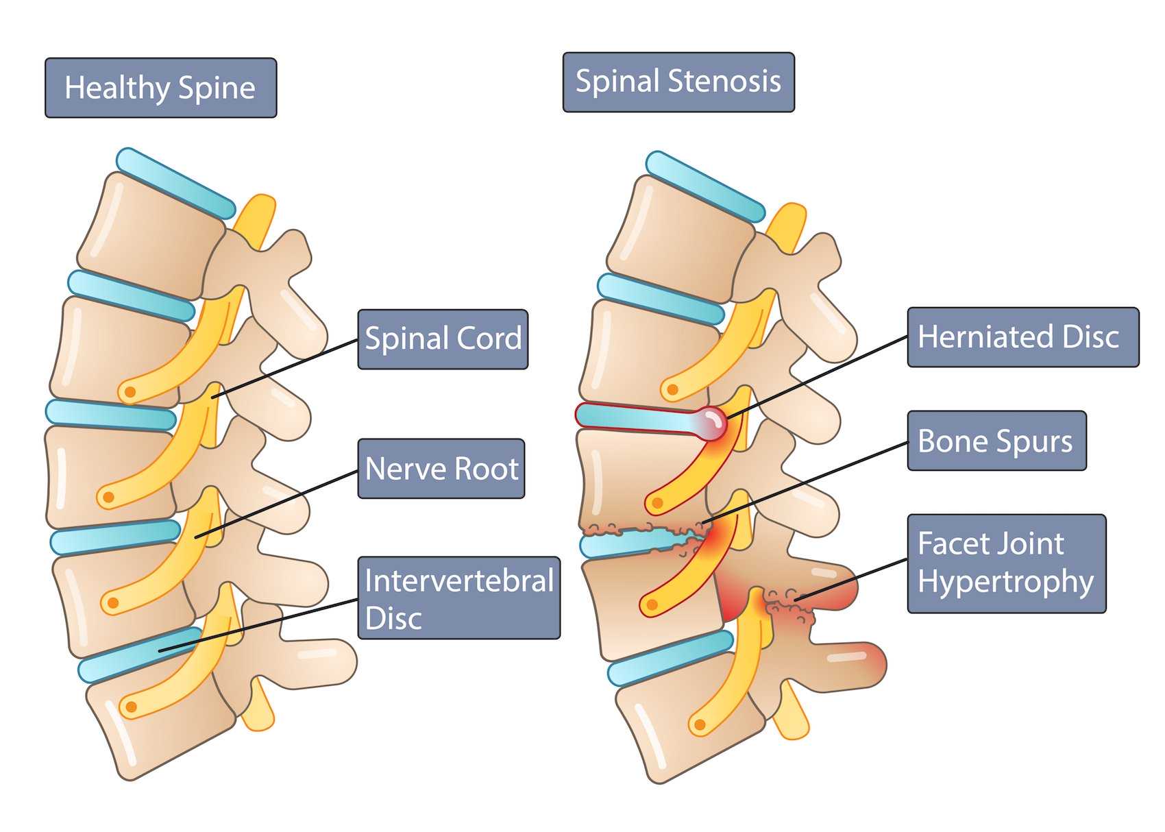
Nerve root pain originates from nerves that have been damaged or are compressed in the spine. Nerves carry information that control body movements and sensations to the brain. When a nerve in the spine is damaged it can cause pain, increased sensitivity, numbness and muscle weakness.

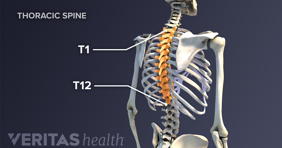
The spine is divided into 4 segments. Cervical: 7 vertebral segments, 8 nerve roots. Thoracic:12 vertebral segments,12 nerve roots. Lumbar: 5 vertebral segments,5 nerve roots. Sacral: 5 fused segments,5 nerve roots . The spinal cord is connected to the brain stem and carries a number of motor and sensory tracts.

The nerve roots exit the spinal canal through the intervertebral foramen, small hollows between each vertebra. The brain and the spinal cord make up the Central Nervous System (CNS). The nerve roots that exit the spinal cord/spinal canal branch out into the body to form the Peripheral Nervous System (PNS). Type of Neural Structure. Role/Function.

The rootlets unite to form an anterior (ventral) or posterior (dorsal) root of a spinal nerve. The anterior/ventral root contains efferent nerve fibres, which carry stimuli away from the CNS towards their target structures.The cell bodies of the anterior root neurons are located in the central grey matter of the spinal cord.

The spinal nerves consist of a group of 31 nerves. These nerves are attached to the spinal cord by two roots- dorsal sensory root and ventral motor root. The sensory root fibres carry sensory impulses to the spinal cord. The motor roots, on the contrary, carry impulses from the spinal cord.

Spine and Nerves. The vertebral column’s most important physiologic function is protecting the spinal cord, which is the main avenue for communication between the brain and the rest of the body ...
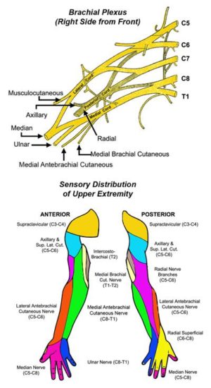
C5 is the nerve "root" that exits the spinal cord above the fifth vertebra in the neck. It travels into the brachial plexus and eventually becomes the nerves that feed muscles around the shoulder and chest. It also provides sensation to parts of the upper arm. C6 is the nerve "root" that exits the spinal cord above the sixth vertebra in the neck.
spine on sitting. Chart of Spinal Nerve Supply and The Effect of Spinal Misalignment Every area of the body is controlled by nerves. The normal function of these can be disturbed by misalignments of the vertebrae effecting the disease conditions shown below. 4 3 2 1 12 11 9 10 8 7 6 5 4 2 1 7 6 3 5 4 1 2

A dermatome map is a diagram that identifies dermatomes, or areas of skin that are innervated by a single nerve, and their corresponding nerve roots along the length of the spinal . Dermatomes are areas of skin that receive sensations from sensory nerves exiting the spinal cord. Sensory nerves provide the feeling of hot, cold, pain, etc.

spinal nerve root (see the following image). Dermatomes of the head, face, and neck. There are 31 segments of the spinal cord, each with a pair (right and left) of ventral (anterior) and dorsal (posterior) nerve roots that innervate motor and sensory function, respectively. The anterior and posterior nerve roots combine on each side to form the

This is because the C1 spinal nerve typically doesn’t have a sensory root. As a result, dermatomes begin with spinal nerve C2. Dermatomes have a segmented distribution throughout your body.

Drag the labels onto the diagram to identify the spinal nerve roots and meninges. 1. ventral rootlets of spinal nerve 2. ventral root 3. dorsal root 4. dorsal rootlets of spinal nerve 5. dorsal root ganglion 6. spinal nerve 7. pia mater 8. arachnoid mater 9. dura mater.
The cervical nerves arise from the spinal cord in the form of rootlets, or fila radicularia, smaller neuron bundles that coalesce to form roots. For each spinal nerve, an anterior and posterior root join to form the completed nerve. Shortly after branching out of the spinal cord, the cervical nerves form the cervical and brachial plexuses.
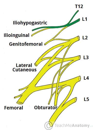
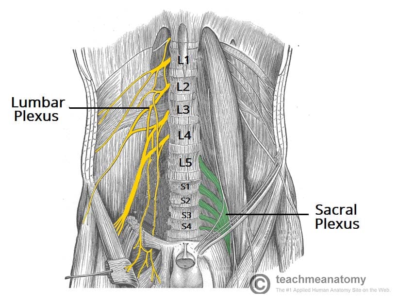
The relevant anatomy of the spinal nerve-muscular innervation of the back is centered around the lumbar spinal nerves, peripheral nerves of the lumbar plexus, spinal cord, and lumbar vertebral column. Within the lumbar region, the vertebral bodies are larger than in the thoracic and cervical regions due to the lumbar spine being designed for weight-bearing purposes.
Derived from two Greek root words in “derma” (skin) and “tomos” (a slice), dermatomes refers to a section of skin supplied by a spinal nerve, starting with the cervical spine (in the neck) down to the sacrum (the base of our pelvis). These areas are not perfectly exact, and multiple nerves may innervate a single general area.
/spinal-column--illustration-487736937-5a6e4ceceb97de0037ea5bb5.jpg)


0 Response to "40 spinal nerve root diagram"
Post a Comment