39 wiggers diagram cardiac cycle
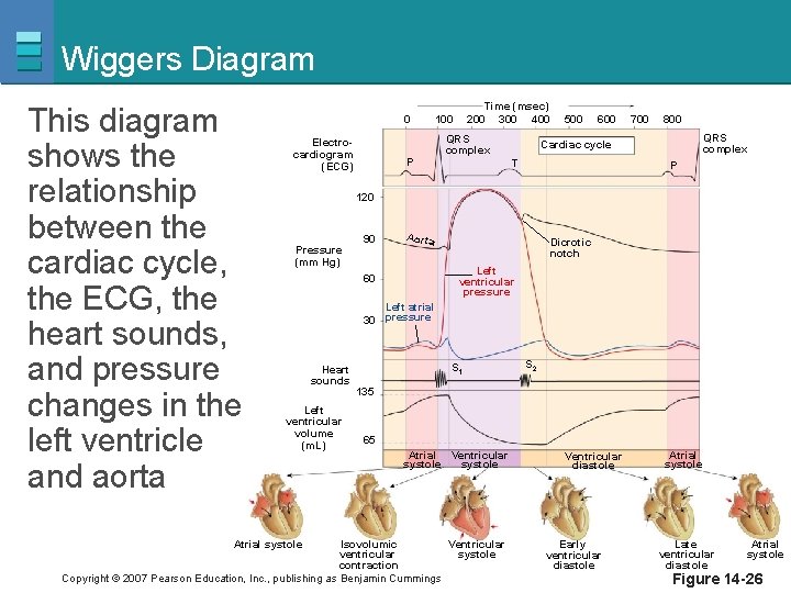
A Wiggers diagram illustrate events and details of the cardiac cycle with electrographic trace lines, which depict (vertical) changes in a parameter's value as time elapses left-to-right. The ventricular "Diastole", or relaxation, begins with "Isovolumic relaxation", then proceeds through three sub-stages of inflow, namely: "Rapid inflow ... Wiggers diagram records are most commonly used in cardiac care units. The primary benefit of the Wiggers diagram is that it shows several aspects of the cardiac cycle together over time so that doctors can ensure the heart is beating properly. In a healthy heart, electrical impulses from the nervous system control the four chambers.
12/11/2021 · The cardiac cycle refers to a series of physiological events making up a single heartbeat. The cycle involves contraction (systole) and relaxation (diastole) of the atria and ventricles to effectively pump blood. ... A Wiggers diagram showing the events of the cardiac cycle. 2.
Wiggers diagram cardiac cycle
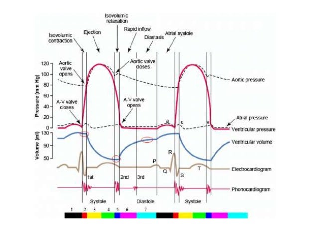
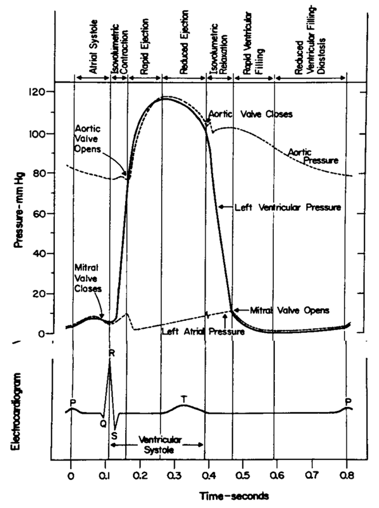
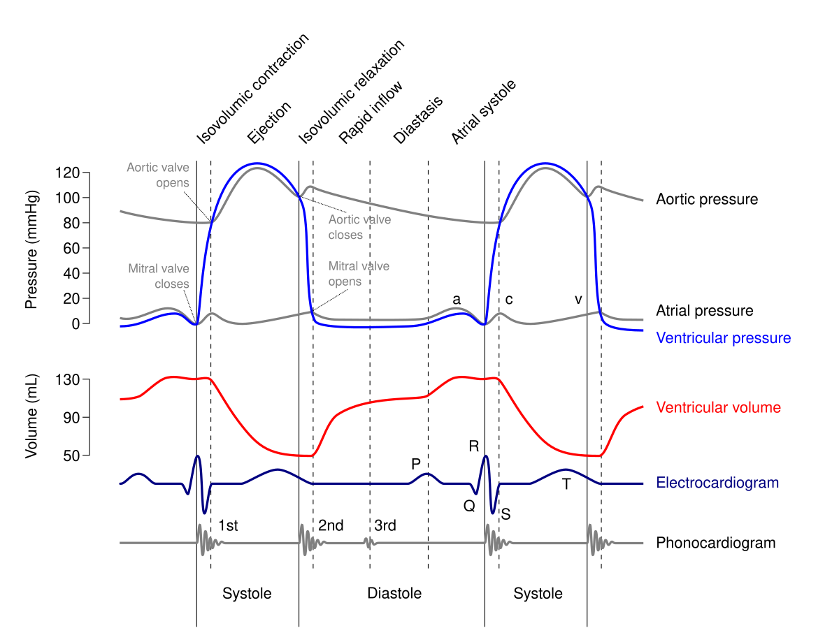
by BE Wright · 2020 — Heart sounds, which in the Wiggers diagram have their own line separate from the valve closures line, are shown within the Wright table both for ...The Wright table of the car...The Wright Table of the Car...Using the Wright Table as a...1 of 3Bruce E. Wright; ,; Gretchen LeFever Watson; , and; Nancy J. Selfridge. Bruce E. Wright. Department of Medical Foundations, Ross University School of Medicine, St. Michael, Barbados. Correspondence: B...Continue on journals.physiology.org »2 of 3The Wright table of the cardiac cycle is a teaching tool specifically designed to illustrate how the heart acts as a pump and how the related events of the cardiac cycle all work together to achieve t...Continue on journals.physiology.org »3 of 3The Wright table shows students when, how, and why blood flows as it does during each phase of the cardiac cycle. The role of the ECG in driving the heart into different phases is shown as transitions...Continue on journals.physiology.org » Diagram - Wiggers diagram. This shows the cardiac cycle, including the various pressures and volumes around the heart, an ECG trace (showing the electrical activity of the heart) and a phonocardiogram (showing the heart sounds - each sound corresponds to a valve shutting). The cardiac cycle diagram shown to the right depicts changes in aortic pressure (AP), left ventricular pressure (LVP), left atrial pressure (LAP), left ventricular volume (LV Vol), and heart sounds during a single cycle of cardiac contraction and relaxation. These changes are related in time to the electrocardiogram.
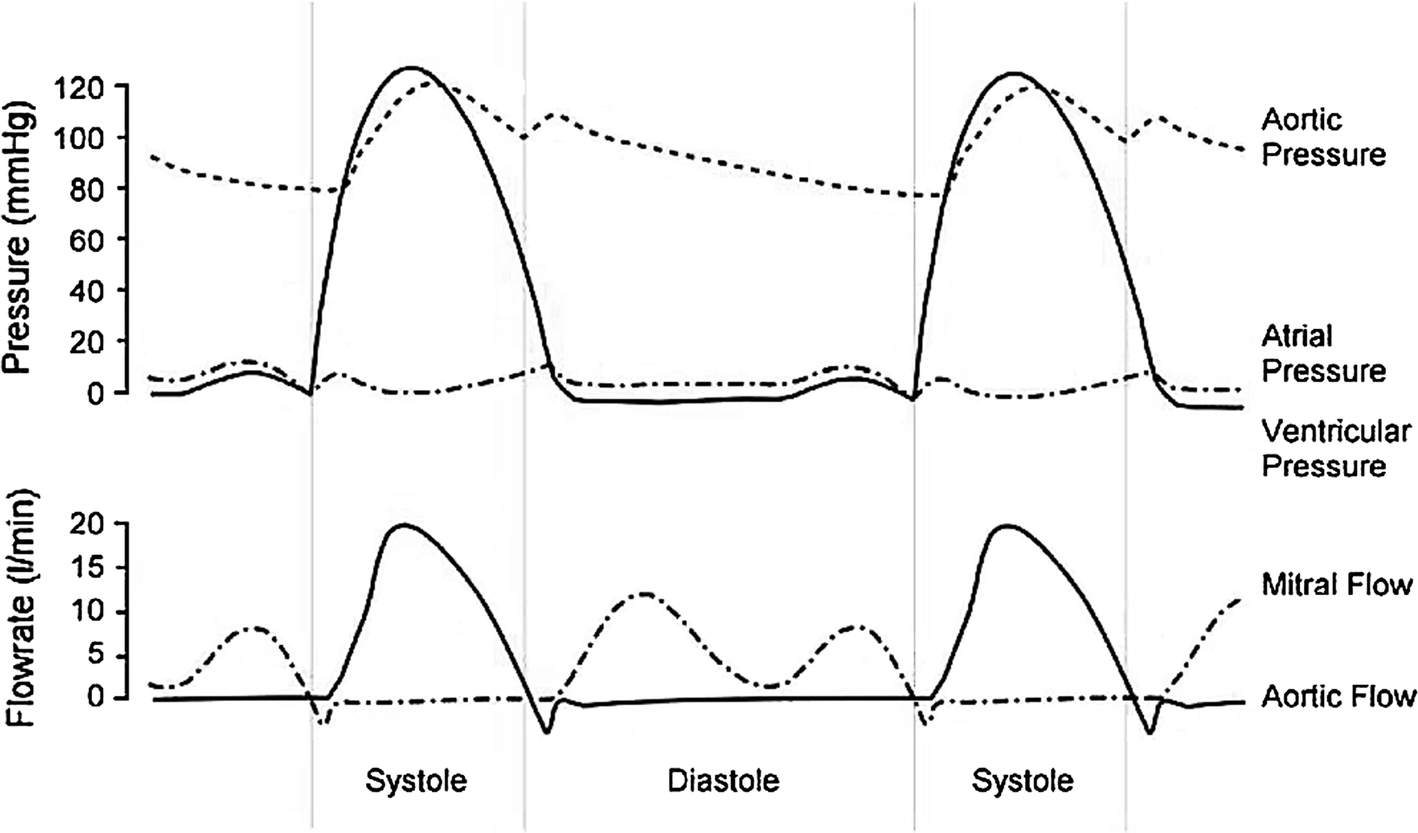
Wiggers diagram cardiac cycle. These didactic lectures usually use PowerPoint slides, with pictures and animations. For instance, the Wiggers diagram is often used to teach the concept of the cardiac cycle in a cardiovascular physiology class. Teaching the cardiac cycle to students can be challenging for many teachers. Consider the Wiggers diagram, for example. Simple Explanation Of Wiggers Diagram Understanding the limits. All quotes from pre-screened, qualified and licensed vendors that. In what portion of the cardiac cycle is there a brief negative aortic flow in the Wiggers diagram and what is the explanation for that?Personal Statement Examples Wiggers Diagram Explanation Comments. Wigger's diagram: This seemingly complex diagram (Don't Panic!) is actually a very useful tool to learn the components of the cardiac cycle and how they relate to cardiac exam findings, which I have indicated in blue. To start, identify the aortic, LV and LA pressure lines and talk yourself through the sequence of A Wiggers diagram, named after its developer, Dr. Carl J. Wiggers, is a standard diagram that is used in teaching cardiac physiology. In the Wiggers diagram, the . Wiggers Diagram Explained Health Education, Ultrasound, Physiology, . Ekg en 3D Más Heart Anatomy, Ekg Leads, Nursing Students, Ekg Interpretation. The cardiac cycle diagram shown ...
Jan 11, 2017 — Wiggers Diagram. 36,897 views36K views. Jan 11, 2017 ... Cardiac Cycle - Wiggers Diagram. Wallace's Anatomy and Physiology. Cardiac Cycle - Summary and Wigger's Diagram. Aortic valve stenosis. Summary of Cardiac Cycle How to draw Wigger's Diagram of Cardiac Cycle? Epomedicine is a source of concise notes on medical topics with relevant details and mnemonics which makes them easy to conceptualize. This is the 1st first video in the Cardiology section dedicated to an important topic.Wiggers Cardiac Diagram.We go through different curves of the Wigger's ... Description. A PV diagram plots the change in pressure P with respect to volume V for some process or processes. Typically in thermodynamics, the set of processes forms a cycle, so that upon completion of the cycle there has been no net change in state of the system; i.e. the device returns to the starting pressure and volume. [citation needed]The figure shows the features of an idealized PV ...
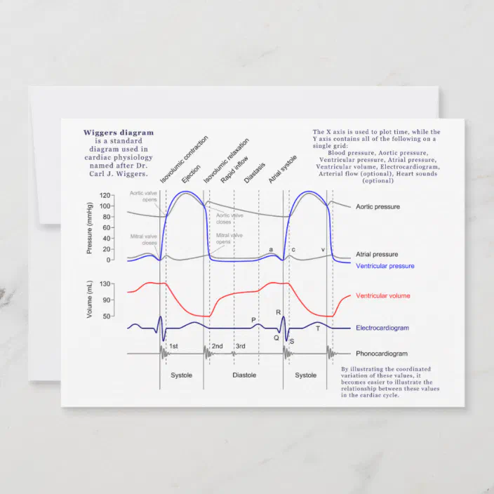
A Wiggers diagram is a standard diagram used in cardiac physiology named after Dr. Carl J. Wiggers who did important work in circulatory physiology in the early part of the 20th century. This diagram is a graphical representation of the cardiac cycle. Wiggers' diagrams can vary in detail and number of variables presented. The Wiggers diagram is a synchronous tracing of aortic pressure, left atrial pressure, left ventricular pressure, left ventricular volume, and EKG throughout the cardiac cycle.Which of the following is the best description the events that occur during diastole on a Wiggers diagram, during the course of S2 on the phonocardiogram - marking the beginning and end of isovolumetric relaxation? THE CARDIAC CYCLE Objectives: Identifying Factors which affect heart rate Describe Cardiac Functional Anatomy (including a review of blood flow and valves) Understand the Wiggers Diagram of Cardiac Cycle Differentiate between Wiggers Diagram and the Pressure Volume Curve Review the electrical basis of excitable cardiac tissue The cardiac cycle - The "Wiggers diagram" Prof. J Swanevelder 4 - 2 The cardiac cycle can be examined in detail by considering the ECG trace, intracardiac pressure and volume curves, and heart valve function. Fig. 1 The "Wiggers Diagram" - Cardiac cycle, showing ventricular volume, ventricular pressure, aortic pressure and atrial pressure
May 5, 2014 — An in-depth look at Wiggers Diagram. Examining the cardiac cycle, Electrocardiogram, Aortic Pressure, Ventricular Pressure, Atrial Pressure, ...

This is well-illustrated on a Wiggers diagram where the QRS complex on the ECG directly precedes ventricular systole (represented on the diagram by increased ventricular pressure). Function. Concerning the events of the cardiac cycle, it is important to compartmentalize their sequence.
by JR Mitchell · 2014 · Cited by 53 — All Wiggers' diagrams that portray PAO throughout a cardiac cycle provide an opportunity to introduce the concept of energy waves and their possible ...AbstractDISCLOSURESAUTHOR CONTRIBUTIONSREFERENCES1 of 4for >90 years, the Wiggers diagram has been a fundamental tool for teaching cardiovascular (CV) physiology, with some of his earliest descriptions of the heart and circulation published in 1915 (18). ...Continue on ncbi.nlm.nih.gov »2 of 4No conflicts of interest, financial or otherwise, are declared by the author(s). Go to:Continue on ncbi.nlm.nih.gov »3 of 4Author contributions: J.R.M. and J.-J.W. conception and design of research; J.R.M. and J.-J.W. analyzed data; J.R.M. and J.-J.W. interpreted results of experiments; J.R.M. and J.-J.W. prepared figures...Continue on ncbi.nlm.nih.gov »4 of 4Abstract; DISCLOSURES; AUTHOR CONTRIBUTIONS; ACKNOWLEDGMENTS; REFERENCES. Articles from Advances in Physiology Education are provided here courtesy of American Physiological Soci

Oct 9, 2021 — This rhythmic sequence causes changes in pressure and volume that are often seen graphically in the form of a Wiggers diagram or venous ...
A description of the cardiac cycle using the Wiggers Diagram.
The Cardiac Cycle III: Volumes in the Left Ventricle: 6 mins: 0 completed: Learn. The Cardiac Cycle IV: Heart Sounds: 7 mins: 0 completed: Learn. The Cardiac Cycle V: The Wiggers Diagram: 10 mins: 0 completed: Learn. The Cardiac Cycle VI: The Ventricular Pressure-Volume Loop: 9 mins: 0 completed: Learn. Cardiac Output I: Definitions: 5 mins: 0 ...

The Wright table is introduced as a novel tool for teaching and learning the cardiac cycle. It supplements the nearly 100-yr-old Wiggers diagram, which is information rich but difficult for many students to learn. The Wright table offers a compact presentation of information, viewable both in terms …

28/10/2021 · Wiggers Diagram. The American-born physiologist Dr. Carl J Wiggers has provided many health care students over the past 100 years with a unique tool to understand the cardiac cycle. The Wiggers diagram highlights the relationship between pressure and volume over time, along with the electrical activity of the heart. The diagram uses the left ...

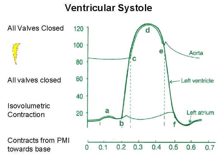
A Wiggers diagram of ventricular systole graphically depicts the sequence of contractions by the myocardium of the two ventricles.Ventricular systole induces self-contraction such that pressure in both left and right ventricles rises to a level above that in the two atrial chambers, thereby closing the tricuspid and mitral valves—which are prevented from inverting by the chordae tendineae ...
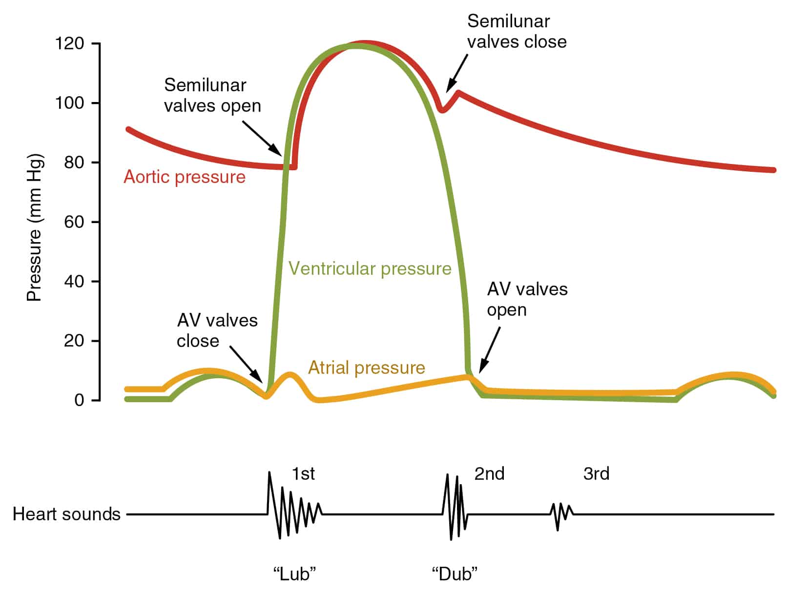
Aug 30, 2016 — How to draw Wigger's Diagram of Cardiac Cycle? · 1/3rd of ejection period (pressure rises from 80 to 120 mmHg): rapid ejection occurs (70% volume ...Summary of Cardiac CycleHeart soundsNormal Intracardiac and Vas...1 of 3When the valve opens, different compartments act as a single chamber (atrio-ventricle or aorto-ventricle). For a blood to flow, pressure in “giver” must be higher then that in “receiver”. Pressure dif...Continue on epomedicine.com »2 of 3First (S1): Closure of atrioventricular valves. Split S1 results from asynchronous closure of mitral (M1) and tricuspid (T1) valves. M1 is usually the earlier component, with T1 normally occuring ~20-...Continue on epomedicine.com »3 of 3Notice, the pressure of atria is the diastolic pressure of ventricles. The systolic pressure of ventricles and outflow tracts are the same. The diastolic pressure of ventricles is less compared to tha...Continue on epomedicine.com »

Basically, a "Wiggers Diagram" is a description of the events which take place over the cardiac cycle and which a plotted on a time scale. Yes, it's the Wiggers diagram, not Wigger's diagram or Wiggers' diagram, because a guy called Wiggers was responsible for the development of its most important components.
09/10/2021 · The cardiac cycle is a series of pressure changes that take place within the heart. These pressure changes result in the movement of blood through different chambers of the heart and the body as a whole. These pressure changes originate as conductive electrochemical changes within the myocardium that result in the concentric contraction of cardiac muscle.
2. Understand the relationship between volume, pressure, and flow during a single cardiac cycle (Wiggers diagram) 3. Be able to draw a Wiggers diagram from scratch 4. Identify and understand the basic function of cardiac valves and associated heart sounds. 5. Introduce Pressure-Volume loops and Frank-Starling curves as tools to express cardiac ...

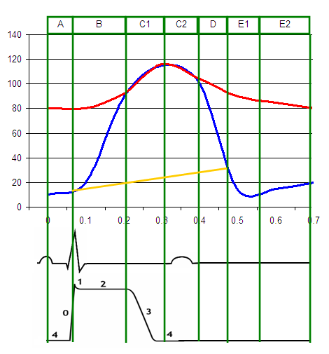
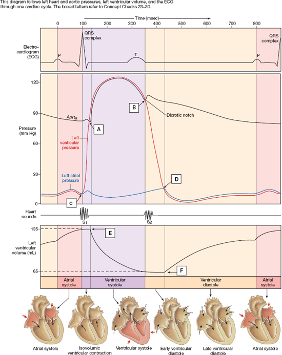
This diagram remains the most commonly used model to teach the cardiac cycle today. A typical Wiggers diagram is shown in Fig. 1. Fig. 1.The Wiggers diagram. From top to bottom, the lines show: 1) aortic pressure, 2) ventricular pressure, 3) atrial pressure, 4) electrocardiogram, 5) mitral and aortic valve opening and closing, and 6) heart sounds.
The Wiggers diagram clearly illustrates the coordinated variation of these values as the heart beats, assisting one in understanding the entire cardiac cycle.EventsAdditional imagesSee alsoReferences1 of 4Note that during isovolumetric/isovolumic contraction and relaxation, all the heart valves are closed. At no time are all the heart valves open. S3 and S4 heart sounds are associated with pathologies ...Continue on en.wikipedia.org »2 of 4Ventricular systole. Cardiac diastole. ECG. The EKG complex. P=P wave, PR=PR interval, QRS=QRS complex, QT=QT interval, ST=ST segment, T=T wave. Wiggers with jugular venous waveformContinue on en.wikipedia.org »3 of 4Pressure volume diagramContinue on en.wikipedia.org »4 of 4Stub icon. This cardiovascular system article is a stub. You can help Wikipedia by expanding it. v · t · e. Retrieved from "https://en.wikipedia.org/w/index.php?title=Wiggers_diagram&oldid=1029541282"...Continue on en.wikipedia.org »

English: A Wiggers diagram, showing the cardiac cycle events occuring in the left ventricle. In the atrial pressure plot: wave "a" corresponds to atrial ...Description: English: A Wiggers diagram, sho...Source: Wikimedia Commons, File:Wiggers Di...
cardiac cycle and wiggers diagram. STUDY. Flashcards. Learn. Write. Spell. Test. PLAY. Match. Gravity. Created by. Agnesch PLUS. Terms in this set (29) what's the wigger's diagram. it is a diagram that shows the sequence of electrical and mechanical events from the beginning of one heartbeat to the next.
Wiggers Diagram, Daniel Chang, CC-SA 2.5. A Wiggers diagram shows the changes in ventricular pressure and volume during the cardiac cycle. Often these diagrams also include changes in aortic and atrial pressures, the EKG, and heart sounds. Diastole starts with the closing of the aortic valve (the second heart sound).

A Wiggers diagram, named after its developer, Dr. Carl J. Wiggers, is a standard diagram that is used in teaching cardiac physiology. In the Wiggers diagram, the X-axis is used to plot time, while the Y-axis contains all of the following on a single grid: Blood pressure.Wiggers Diagrams | Printable DiagramCardiac Cycle - Summary and Wigger's ...
The cardiac cycle diagram shown to the right depicts changes in aortic pressure (AP), left ventricular pressure (LVP), left atrial pressure (LAP), left ventricular volume (LV Vol), and heart sounds during a single cycle of cardiac contraction and relaxation. These changes are related in time to the electrocardiogram.

Diagram - Wiggers diagram. This shows the cardiac cycle, including the various pressures and volumes around the heart, an ECG trace (showing the electrical activity of the heart) and a phonocardiogram (showing the heart sounds - each sound corresponds to a valve shutting).
by BE Wright · 2020 — Heart sounds, which in the Wiggers diagram have their own line separate from the valve closures line, are shown within the Wright table both for ...The Wright table of the car...The Wright Table of the Car...Using the Wright Table as a...1 of 3Bruce E. Wright; ,; Gretchen LeFever Watson; , and; Nancy J. Selfridge. Bruce E. Wright. Department of Medical Foundations, Ross University School of Medicine, St. Michael, Barbados. Correspondence: B...Continue on journals.physiology.org »2 of 3The Wright table of the cardiac cycle is a teaching tool specifically designed to illustrate how the heart acts as a pump and how the related events of the cardiac cycle all work together to achieve t...Continue on journals.physiology.org »3 of 3The Wright table shows students when, how, and why blood flows as it does during each phase of the cardiac cycle. The role of the ECG in driving the heart into different phases is shown as transitions...Continue on journals.physiology.org »


0 Response to "39 wiggers diagram cardiac cycle"
Post a Comment