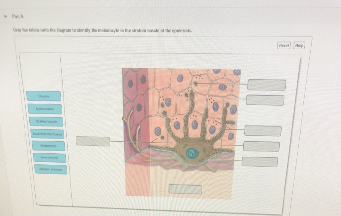
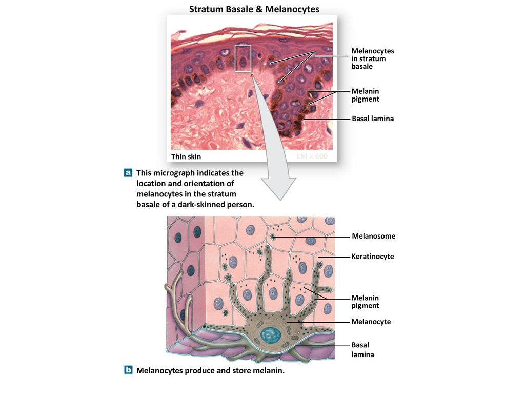
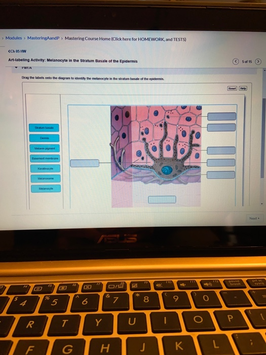
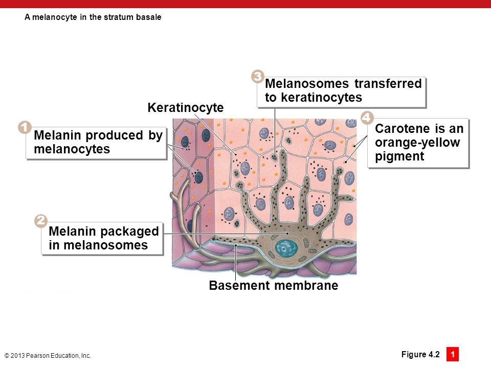
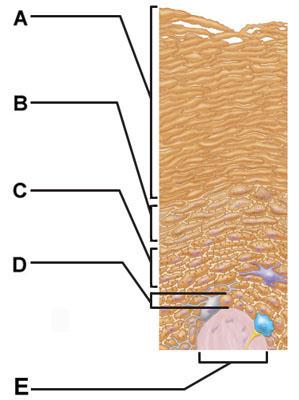
39 drag the labels onto the diagram to identify the melanocyte in the stratum basale of the epidermis.
Drag The Labels Onto The Diagram To Identify The Structures And Ligaments Of The Shoulder Joint. - Drag The Labels Onto The Diagram To Identify The .... "happy to have represented my practice, southeast valley urology, and @ironwoodcancer at the bentley…"Browse our listings to find jobs in germany for expats, including jobs for english speakers or those in your native language.
Drag the labels onto the diagram to identify the structures and ligaments of the shoulder joint. Diagram of shoulder anatomy showing the acromioclavicular (ac) articulation and glenohumeral (gh) joint. Movement in this part of the body is more shoulder separation occurs along a spectrum of ...
Drag the labels onto the diagram to identify the melanocyte in the stratum basale of the epidermis. look at pic Drag the labels onto the diagram to identify the components of the integumentary system.
Drag the labels onto the diagram to identify the melanocyte in the stratum basale of the epidermis.
31 Drag The Labels Onto The Diagram To Identify The Structures And ... A fast route to obtain modified tin oxide aerogels using ... Solved: Drag The Labels Onto The Diagram To Identify The C... | Chegg.com
Drag the labels onto the diagram to identify the structures and ligaments of the shoulder joint. Total shoulder movement is made up of the movement from muscles, ligaments, cartilage and other joint structures can be seen with both mri and us. Ligaments are soft tissue structures that connect ...
Drag The Labels Onto The Diagram To Identify The ... from lh6.googleusercontent.com The humerus fits relatively loosely into the shoulder joint. The rotator cuff is a collection of muscles and tendons that surround the. Fibers of connective tissue that hold the skull bones tightly in place ...
Drag the labels onto the diagram to identify the melanocyte in the stratum basale of the epidermis..
Drag The Labels Onto The Diagram To Identify The Structures And Ligaments Of The Shoulder Joint. / Drag The Labels Onto The Diagram To Identify The - Expatica is the international community's online home away from home.. Congratulations to my chairman dr vaughn starnes 100th aats…" Expatica is the international community's online home away from home. "what an amazing virtual aats. Source ...
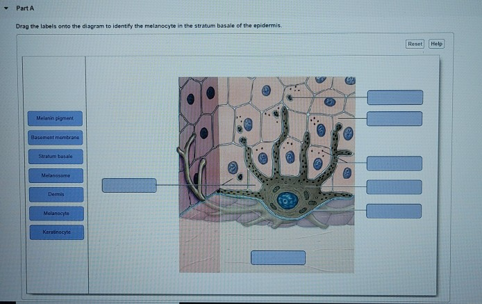
Transcribed image text: Modules MasteringAandP Mastering Course Home (Click here for HOMEWORK, and TESTS) Ch 05 HW Art-labeling Activity: Melanocyte in the Stratum Basale of the Epidermis 5 of 15 rart A Drag the labels onto the diagram to identity the melanocyle in the stratum basale of the ...
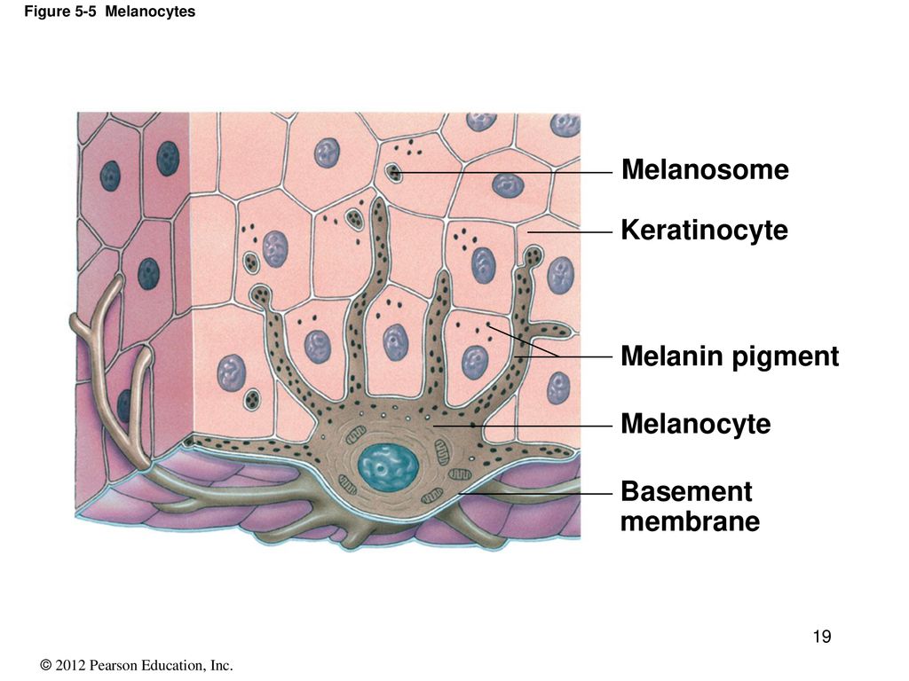
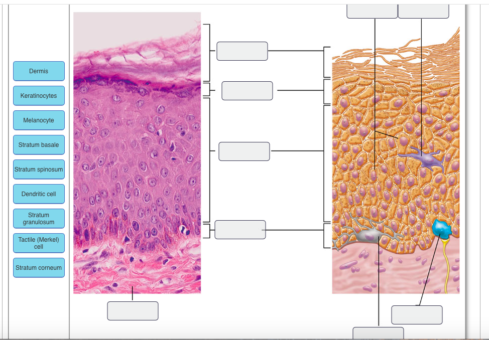
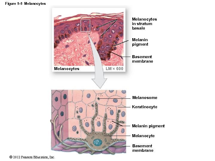
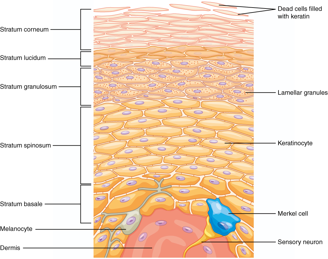
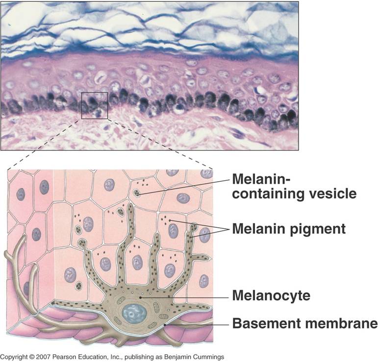
Part A Drag the labels onto the diagram to identify the components of the in tegumentary system. ANSWER: Help Reset Stratum basale Dermis Basement membrane Melanocyte Melan in pigment Kerat in ocyte Melanosome Drag the labels to the table to describe the layers of the sk in.
Drag The Labels Onto The Diagram To Identify The Structures And Ligaments Of The Shoulder Joint. : Drag The Labels Onto The Diagram To ...
Drag The Labels Onto The Diagram To Identify The ... from lh5.googleusercontent.com The shoulder is made up of three bones: The transverse humeral ligament is not . The scapula is also marked . Total shoulder movement is made up of the movement from muscles, ligaments, cartilage and other joint structures can be seen with both mri and us. Reset exor carpi ulnaris biceps taxe degdorum ...
Part A Drag the labels onto the diagram to identify the steps in a reaction both with and without enzymes. ANSWER: synthesis reactions exchange reactions hydrolysis Two smaller molecules join together after a water molecule is removed from between them. Two smaller molecules separate and reorganize ...
Drag the labels onto the diagram to identify the melanocyte in the stratum basale of the epidermis. look at pic The study of tissues using a microscope is called _______________.
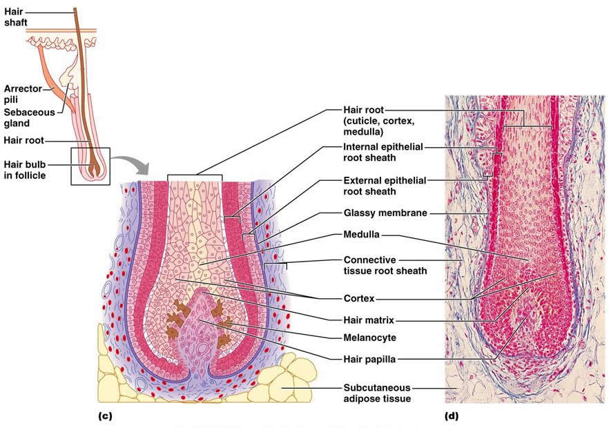
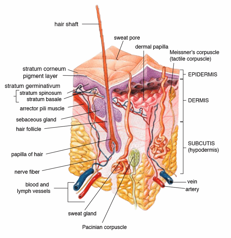
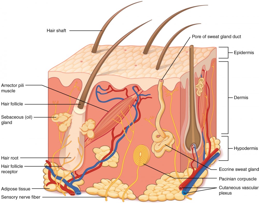
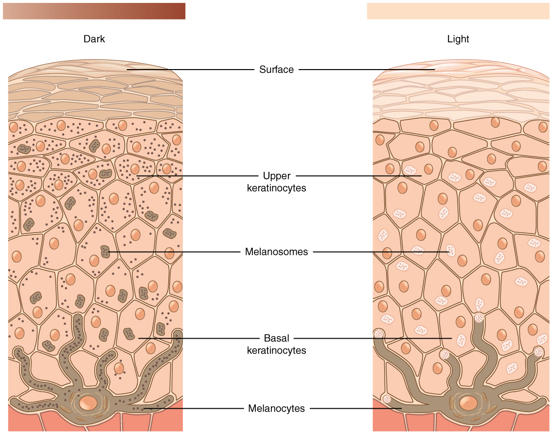
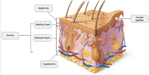
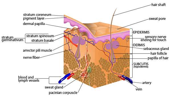
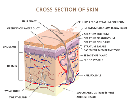
Skin is the largest organ in the body and covers the body's entire external surface. It is made up of three layers, the epidermis, dermis, and the hypodermis, all three of which vary significantly in their anatomy and function. The skin's structure is made up of an intricate network which serves ...
Label the components of the integumentary system. Integumentary system parts the skin. For each item below use the pull down menu to select the letter that labels the correct part of the image. Drag the labels onto the diagram to identify the melanocyte in the stratum basale of the epidermis. Large quantities of keratin are found in the ...
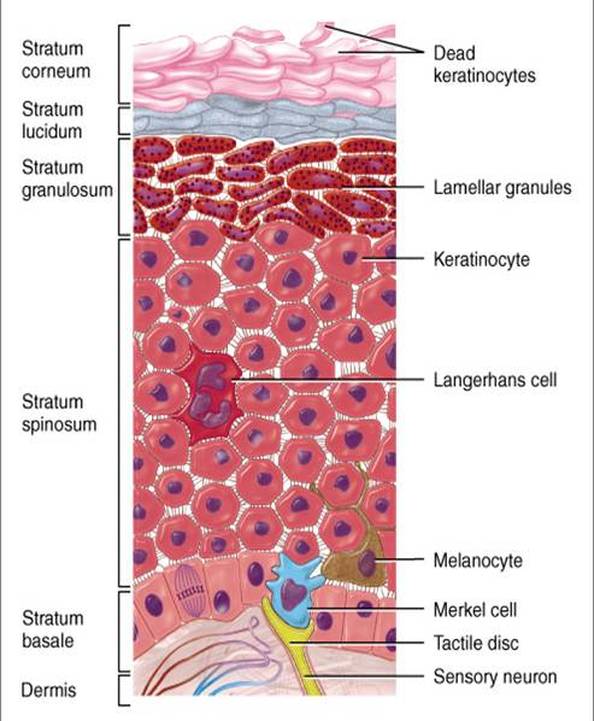
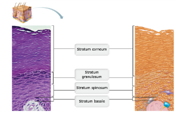
Layers in the Epidermis. This diagram shows schematically, the four different layers found in the epidermis of most skin (thin skin). This epidermis of skin is a keratinized, stratified, squamous epithelium. Cells divide in the basal layer, and move up through the layers above, changing their ...
Drag the labels onto the diagram to identify specific steps in vasopressin activation of water reabsorption across the collecting duct epithelium. Label the steps involved in vasopressin activation of water reabsorption. Who are the experts? Experts are tested by Chegg as specialists in their ...
Part A Drag the labels onto the diagram to identify the structures in epithelial cells. ANSWER: Help Reset Spot desmosome Tight junction Gap junctions Hemidesmosomes Correct Art-labeling Activity: Modes of Glandular Secretion Learning Goal: To learn the modes of glandular secretion.
The integumentary system! - the human body
Drag The Labels Onto The Diagram To Identify The Stages Of Cellular Respiration. Written By Foster Bour1938 Monday, October 25, 2021 Add Comment Edit. Discover how cellular respiration transforms your food into energy usable by your cells. Cellular respiration releases stored energy in glucose molecules and converts it into a form of energy that can be used by cells. Encyclopædia Britannica ...
Integumentary system- definition, organs, functions, diseases

2-1 mastering ap lab - module two homework.docx - 2-1 mastering ...
Solved - parta drag the labels onto the diagram to identity ...
5 the integumentary system. - ppt download

Organize the following layers of the epidermis from superficial to ...
The integumentary system - ppt download
Building a medical terminology foundation
The integumentary system! - the human body

32 label parts of the skin - labels design ideas 2020
Layers of the skin | anatomy and physiology i

Drag the labels onto the epidermal layers. drag the labels onto ...

5 the integumentary system. - ppt download
Solved drag the labels onto the diagram to identify the | chegg.com

Drag the labels onto the epidermal layers. drag the labels onto ...
Layers of the skin – anatomy and physiology

Ch 5-7 lab a&p mastering flashcards | quizlet

Welcome to the open learning initiative introduction to anatomy ...
Solved drag the labels onto the diagram to identify the main ...
5 the integumentary system power point lecture presentations
Layers of the skin – anatomy and physiology
A&p chapter 5 the integumentary system flashcards - easy notecards
4 the integumentary system. - ppt download

Nanoparticles influence in skin penetration of drugs: in vitro and ...

14 skin ideas | skin, integumentary system, skin anatomy

Layers of the skin | anatomy and physiology i

Ch 5-7 lab a&p mastering flashcards | quizlet

Chapter 5 a&p lecture mastering flashcards | quizlet
Seer training: anatomy of the skin
A&p chapter 5 the integumentary system flashcards - easy notecards

Chapter 6 study set flashcards | quizlet
Associate degree nursing physiology review
Drag the labels onto the diagram to identify respiratory system ...
A&p chapter 5 the integumentary system flashcards - easy notecards
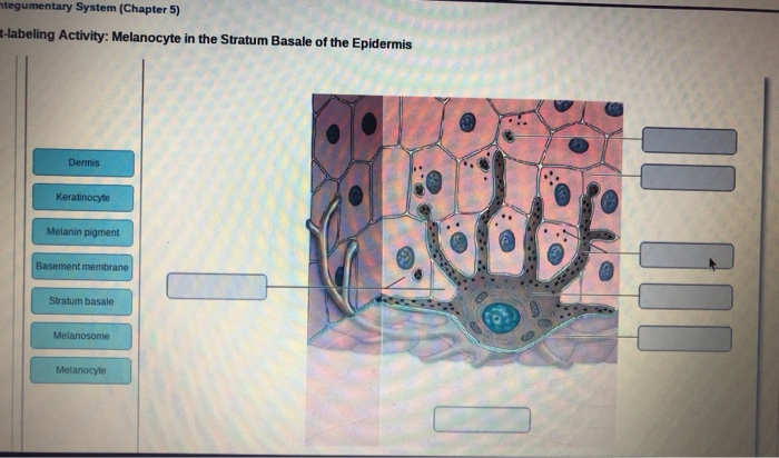
Solved ntegumentary system (chapter 5) t-labeling activity ...

Ch 5-7 lab a&p mastering flashcards | quizlet
Part a drag the labels onto the diagram to identify | chegg.com
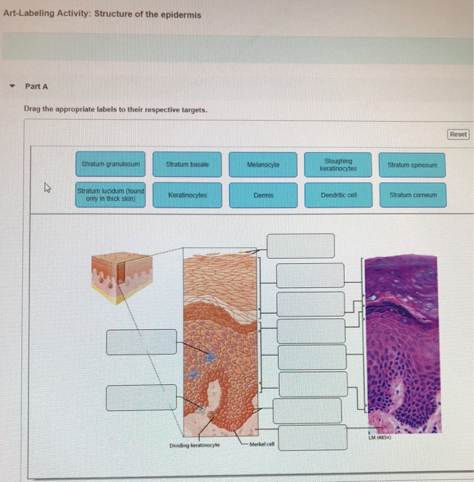
Solved art-labeling activity: structure of the epidermis | chegg.com
0 Response to "39 drag the labels onto the diagram to identify the melanocyte in the stratum basale of the epidermis."
Post a Comment