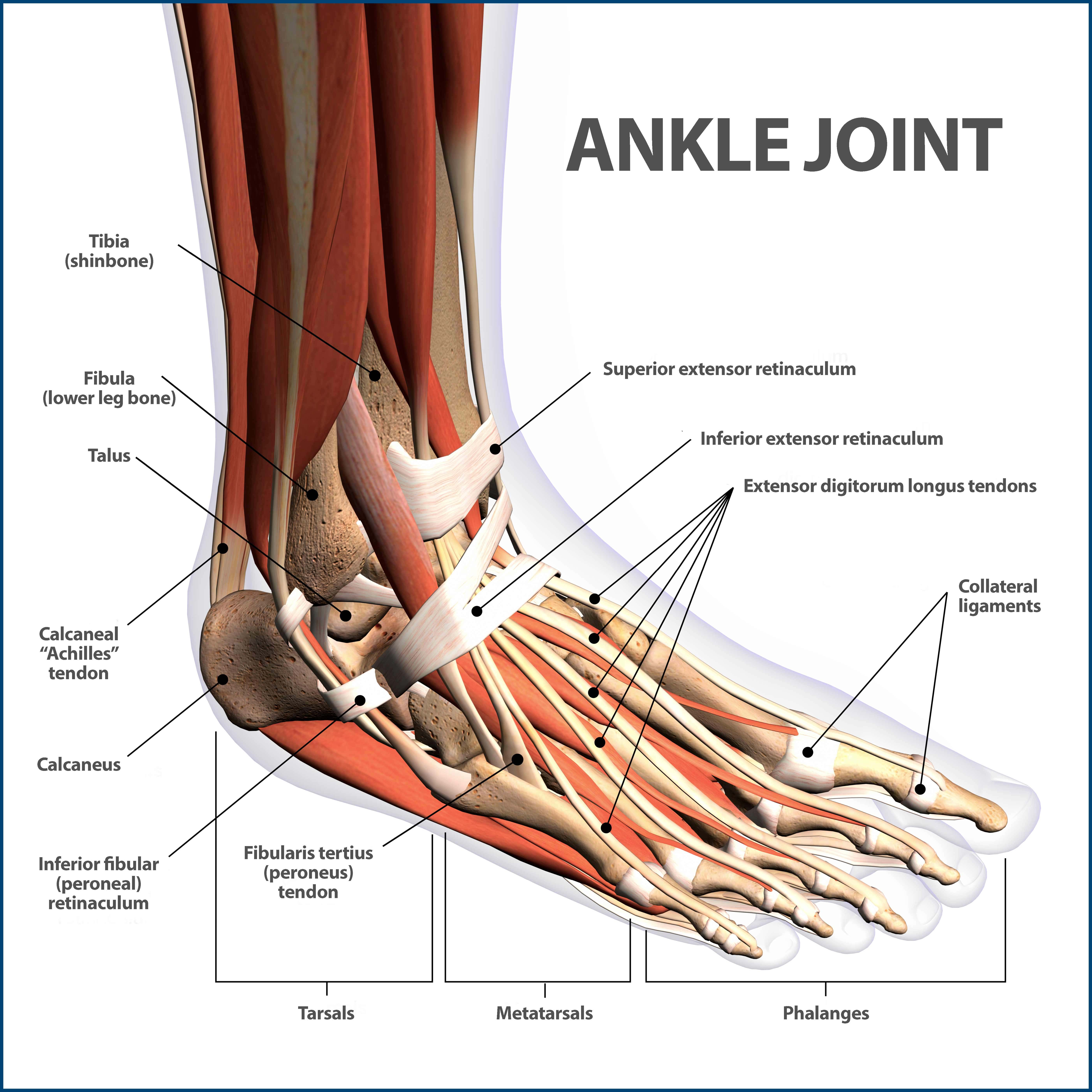
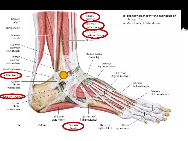
39 ankle muscles and tendons diagram
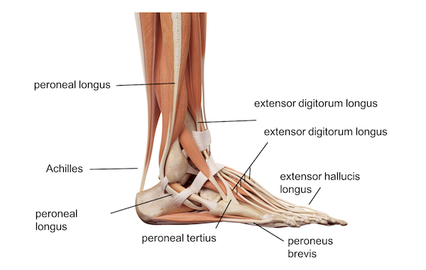
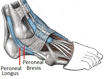
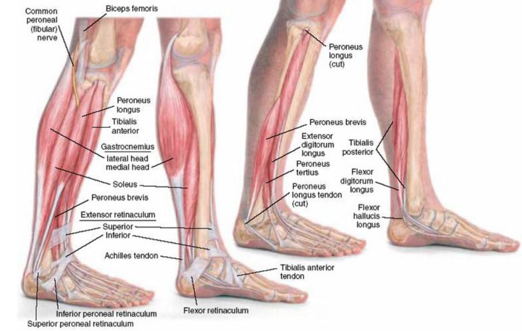
The peroneal muscles (peroneus longus and peroneus brevis), on the outside edge of the ankle and foot. · The calf muscles (gastrocnemius and soleus), which are ...
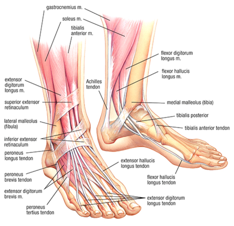
Jul 6, 2020 — The anterior tibial tendon allows us to raise the foot. Two tendons run behind the outer bump of the ankle (lateral malleolus) and attach to ...
5. Knee Tendon Anatomy. Tendons are often overlooked as part of knee joint anatomy. They are they soft tissues found at the end of muscles which link the muscle to bone. The main tendon found at the knee is the patellar tendon which links the quads muscles to the shin bone. The knee cap actually sits inside the patellar tendon.
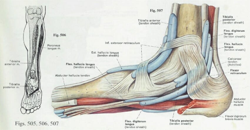
Ankle muscles and tendons diagram
Bones & Joints of the Shoulder. The bones of the shoulder consist of the humerus (the upper arm bone), the scapula (the shoulder blade), and the clavicle (the collar bone). The clavicle is the only bony attachment between the trunk and the upper limb. It forms the front portion of the shoulder girdle and is palpable along its entire length with ...
Eccentric exercises are when the muscle contracts but also lengthens at the same time. This occurs to the quadriceps muscles during the downwards phase of a squat. Eccentric strengthening exercises are thought to be beneficial in treating chronic tendon injuries including quadriceps tendinopathy.
Iliotibial band syndrome - aftercare. The iliotibial band (ITB) is a tendon that runs along the outside of your leg. It connects from the top of your pelvic bone to just below your knee. A tendon is thick elastic tissue that connects muscle to bone. Iliotibial band syndrome occurs when the ITB becomes swollen and irritated from rubbing against ...
Ankle muscles and tendons diagram.
Other Knee Muscles. 1. Popliteus. This is another of the muscles in the back of the knee called popliteus which helps the knee to twist, aids stability of the knee and helps protect the lateral meniscus. 2. Calf Muscles. Just below the knee on the back of the shin are the calf muscles, soleus and gastrocnemius.
For lower leg pain that goes beyond general shin soreness, a more aggressive approach must be taken. Shin Splints Anatomy. To better understand shin splints an understanding of the muscles, tendons and bones involved is required. As you can see from the diagram, there are many muscles and tendons that make up the lower leg, or calf region ...Chronic lower leg pain is part of a larger ...
Acute Achilles tendon (AT) tears are common. Even when managed optimally these injuries result in impaired muscle strength and endurance and may lead to major functional impairment and retirement from sport [1, 2].There are no clear guidelines for the management of AT ruptures, and treatment is often based on surgeons and patient preferences [2,3,4,5].
Devices which passively stiffen muscle-tendon pathways have been shown to be effective at reducing muscle activity at the ankle, knee, hip, and lumbosacral joints across a variety of tasks 31,32 ...
Muscles and Tendons — Anatomy of the Foot and Ankle. Bones and Joints Ligaments Muscles and Tendons Nerves ...
Muscles Purpose In this activity you will look at the microanatomy of muscle cells and you will identify muscle locations on your body Background Tendons can be felt through the skin. Chapter 5 lab investigation muscles quizlet. Simple Skin Model - Hair Follicle. 0-1 chapter 12 Nervous Tissue 0-1 Chapter 13 the Spinal Cord 0-1 Chapter 14 ...
A acetabulum In dinosaurs, the acetabulum (plural: acetabula) or hip socket is an opening in the pelvis formed by the ilium, pubis, and ischium that is visible in lateral and medial views. It accommodates the head of the femur, forming the hip joint.Most tetrapods show a closed acetabulum, in which the socket is completely filled with bone, forming a depression.
In PD MRI muscle tendons are shown as black, while muscles are displayed as gray. Locate the biceps femoris, sartorius, semimembranosus, plantaris, popliteus, and gastrocnemius muscles. Look at the lateral and medial heads of the gastrocnemius muscle, nestled between the heads you will see familiar circular structures representing the blood ...
Lower Body Bone Diagram / BIOSC 222 Study Guide (2013-14 Cummings) - Instructor - In human anatomy, the lower leg is the part of the lower limb that lies between the knee and the ankle. Written By Weber Laway1986 Monday, November 29, 2021 Add Comment Edit
Muscles Purpose In this activity. 131-132 of your lab manual to complete the following. Muscles For Lab Muscles Of The Upper Leg Posterior View Diagram Diagram Quizlet . A P Lab Muscles Knee Ankle Muscles Pictures Flashcards Quizlet . Lab Practical 3 Muscles Locations Flashcards Quizlet Flashcards Muscle Book Images
The supraspinatus muscle's tendon is most commonly affected. Impingement syndrome typically presents with pain , weakness and restricted shoulder movement . Patients with impingement syndrome often complain of pain when their arms are raised (this is particularly common in mechanics and manual labourers who work with their arms overhead).
Physical Therapy in Louisiana for Ankle · Talus Works Like a Hinge · Mortise and Tenon · Cartilage · Collagen · Three Main Ligaments · Joint Capsule · Achilles Tendon ...
Foot (anatomy): bones, ligaments, muscles, tendons, arches and skin
Leg Muscles Diagram : Muscles Of The Human Body Art Rocket - If you're hit with a muscle cramp, it will get your attention right away. From here, different types of crabs are divided into different infraorders, but they all share the. The lower leg muscles are essential bodily structures. They allow you to move and provide support for your ...

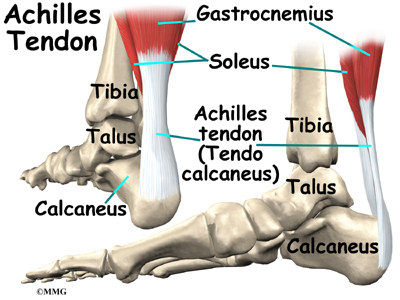
Anatomy of the achilles, posterior heel view and ankle view
Chapter 5 Lab Investigation Muscles Quizlet. Fingerprints and Facial Recognition is a new chap-ter focusing on the application of fingerprint iris and facial biometrics used to create biometric databases. Osteoblasts lay down bone around the cartilage spicules in the bones interior. Lab 5 Muscles Of The Torso Upper Torso Anterior View Diagram ...

Anatomy of the foot and ankle | orthopaedia
Use the pictures in the Lab 5 Exercise Image Library on pp. Anatomy And Physiology Chapter 13-The Spinal Cord - 55 cards. Choose from 500 different sets of muscle test chapter 5 anatomy physiology flashcards on Quizlet. Anatomy of the Nervous System. Identify the following muscles or muscle groups on a diagram.

Muscles and tendons of the ankle-foot complex | download ...
Difference. Ligament. Tendon. Connections. Connect bones to bones. Connect bones to muscles. Classifications. Classified into three categories: peritoneal ligaments (which form the abdominal cavity lining), articular ligaments (which connect bone-bone) and fetal remnant ligaments (which are present from the time of being a fetus, eventually developing into other ligament tissue).
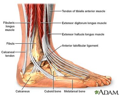
Diagram - the ankle
The abdomen and pelvic regions are continuous with each other, making up the distal part of the trunk. Bar the brain, heart and lungs, this region contains virtually all your body organs, including those involved in the digestive, endocrine, lymphatic, urinary and reproductive systems. So, it is crucial that you cover this section thoroughly.

Regenerative stem cell treatment for ankle tendon tears
The main function of the gastrocnemius muscle is to plantarflex your ankle. This means that as your gastroc contracts, your ankle and toes point down. When walking, running, or climbing stairs, the muscle works to flex your ankle and propel you forward. The muscle is considered one of the "anti-gravity" muscles.

Anatomy of the foot and ankle | orthopaedia
All of these muscles stem from the heel of the foot through the calcaneal tendon. There are two tendons involved that help to cut down on the friction caused by the movements. Gastrocnemius; This muscle has two parts, the lateral and the medial and they both meet in the middle to form a single muscle.

Ankle anatomy
Muscle and Ligament Attachments. The only muscle that gets attached to the navicular is the tendon of the tibialis posterior. It gets inserted into the bone medially on the navicular tuberosity. There are several ligaments that attach to this bone. The talonavicular ligament attaches the dorsal surface of the bone to the neck of the talus.

Benefit pt's anatomy series: the ankle
The ankle is a synovial joint formed by the bones of the lower leg tibia and fibula ... This bone inserts the Achilles tendon arising from the calf muscles ...

All about ankle sprains and strains
Leg Muscles Diagrams Human Anatomy | 101 Diagrams Apr 08, 2019 · In the leg muscles diagram above, there are many muscles that make up your legs and support it to move. One of the most important tendons in terms of mobility of the leg is the Achilles tendon. This important tendon in the back of the calf and ankle stores the

Page not found | ankle tendonitis, foot anatomy, ankle anatomy
All foot muscles and tendons Leg Muscles Anatomy, Ankle Anatomy, Foot Anatomy, Human. Developing Strength & Stability in the Foot, Ankle, and Lower Leg ...
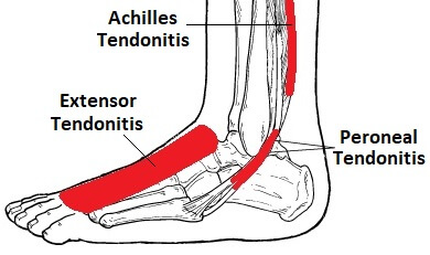
Foot & ankle tendonitis: causes, symptoms & treatment
The muscles of the lower leg are many and they allow the ankle to plantar flex (pointing the toes away from the nose), dorsiflex (pulling the toes toward the nose), invert (twisting medial aspect ...

A patient's guide to foot anatomy | 2020 orthonorcal, los gatos ...
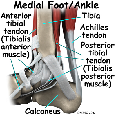
TENDONS AND LIGAMENTS · Achilles tendon: attaches the calf muscle to the heel bone. · Posterior tibial tendon: attaches one of the smaller muscles of the calf to ...
Ankle anatomy - be in motion physiotherapy
The anterior tibial tendon allows us to raise the foot. Two tendons run behind the outer bump of the ankle (the lateral malleolus). These two tendons, called ...
Ankle anatomy - eorthopod.com
Biceps femoris. The biceps femoris is the most lateral muscle in the posterior compartment of the thigh and, as the term bicep suggests, has two heads: the long head and the short head.. These muscle heads have different origins but join to form a palpable tendon on the lateral distal thigh inserting on to the head of the fibula. The long head of biceps femoris protects the sciatic nerve as it ...

Tibialis posterior: pain on the inside of the ankle - pure sports ...
It is a dorsiflexor of the ankle. Origin: Upper 1/2 of lateral and anterior surfaces of the tibia. Insertion: Inner surface of the medial cuneiform and 1st metatarsal. Actions: Inversion & Dorsiflexion. Innervation: Deep peroneal nerve. Daily uses: Walking - to lift the foot up and clear the ground.

Foot anatomy - podiatrist san angelo, tx
The flexor digitorum longus (FDL) tendon originates in the deep posterior compartment of the leg, crosses the ankle medially, and flexes all three joints of the lesser toes, though it acts ...
Peroneal tendon syndromes: practice essentials, epidemiology ...
(A) Achilles tendon contracture and inversion of the right ankle; (B) schematic diagram of the surgical incision design of the modified percutaneous Achilles tendon lengthening for the right ankle joint: the distal incision was marked at 0.5 cm of the calcaneal insertion of Achilles tendon; the middle incision was marked at 5-6 cm away from ...

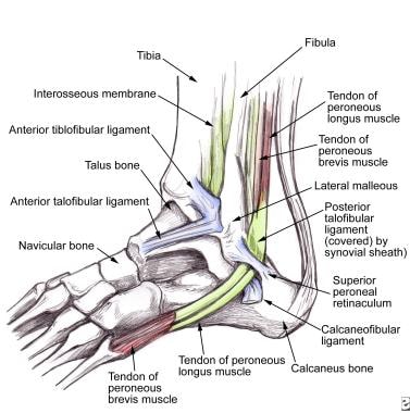
Anatomy of the lateral ankle (reproduced with permission from ...
The system based on the legs of a peregrine falcon has a 3D printed structure in place of bones, as well as motors and fishing lines that play the role of muscles and tendons. Inspired by the way these tendons pass around the ankle in birds, a similar mechanism absorbs the energy of the landing impact and converts it into force when grabbing an ...

Heel muscles and tendons online sale, up to 68% off
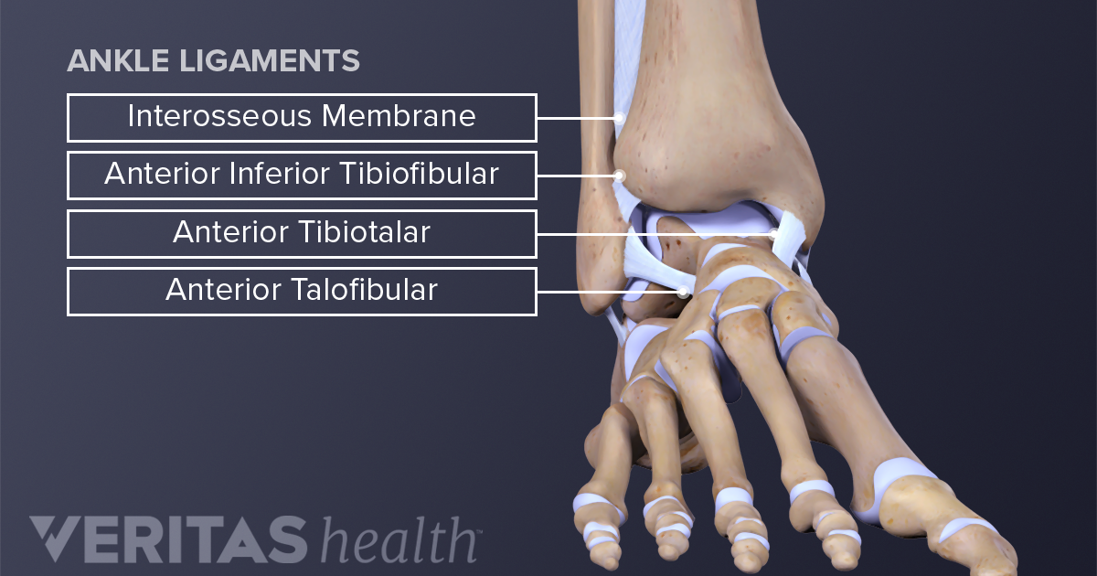
Ankle anatomy: muscles and ligaments
Ankle foot anatomy
Foot & ankle tendons: anatomy, function & injuries

Physical therapy in olmos park for foot - anatomy
Developing strength & stability in the foot, ankle, and lower leg ...
Developing strength & stability in the foot, ankle, and lower leg ...

Runner's web and triathlete's web, a running, track and field and ...

Ankle muscle images, stock photos & vectors | shutterstock

Extensor muscle | anatomy | britannica

Ankle strain germantown,collierville, tn - pittman physical therapy
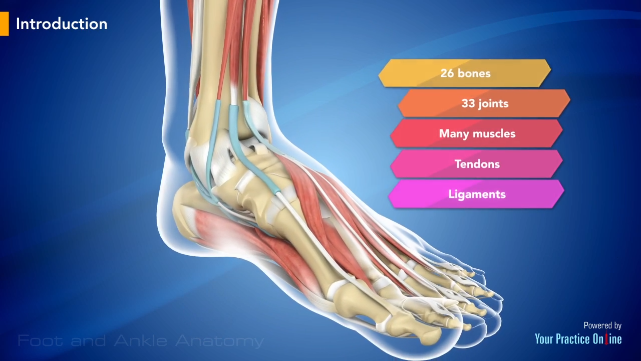
Foot and ankle anatomy video | foot & ankle

Tendons in the foot, tendonitis - remember to stretch, feet too ...
Ankle fractures broken ankle | florida orthopaedic institute

Muscles in the ankle - joi jacksonville orthopaedic institute

Tendons in the foot | ankle anatomy, foot anatomy, sprained ankle

Anatomy of the ankle – elliot's site
Anatomy of the foot and ankle bones muscles

Benefit pt's anatomy series: the ankle
0 Response to "39 ankle muscles and tendons diagram"
Post a Comment