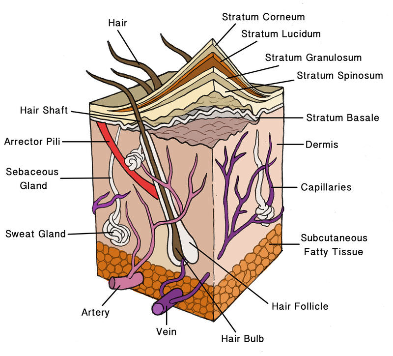
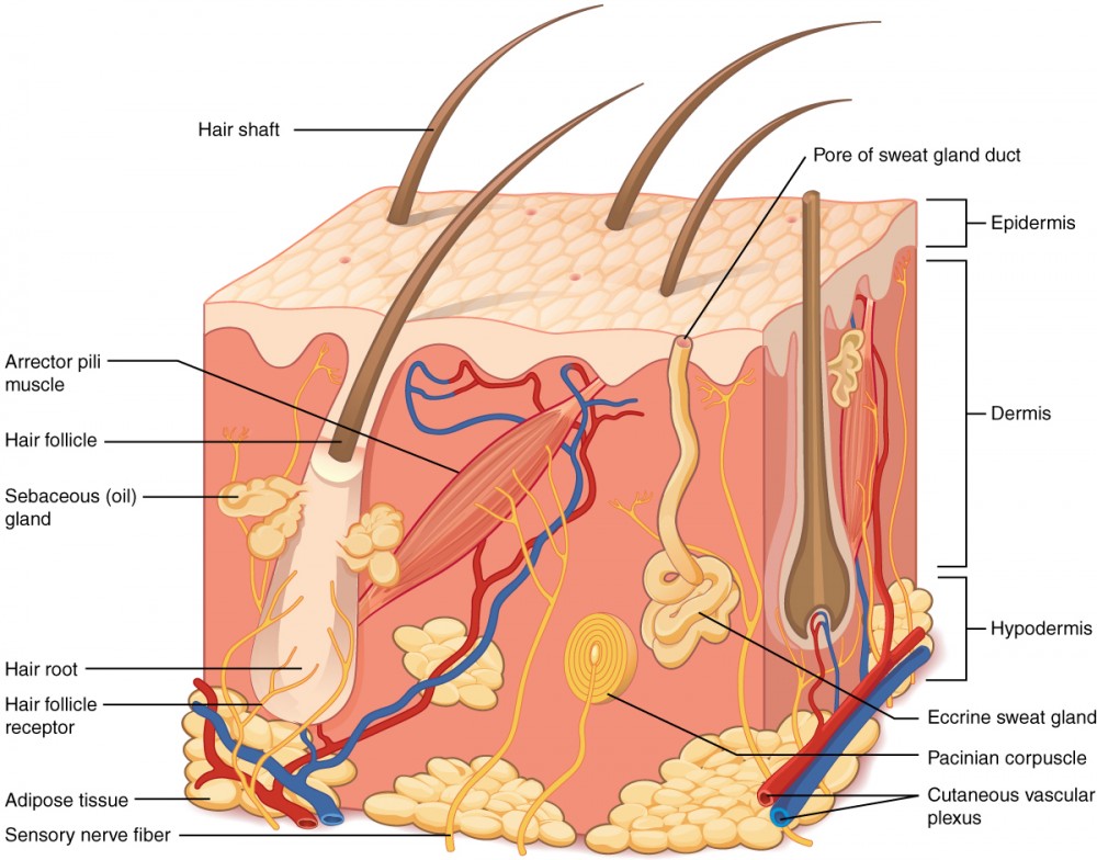
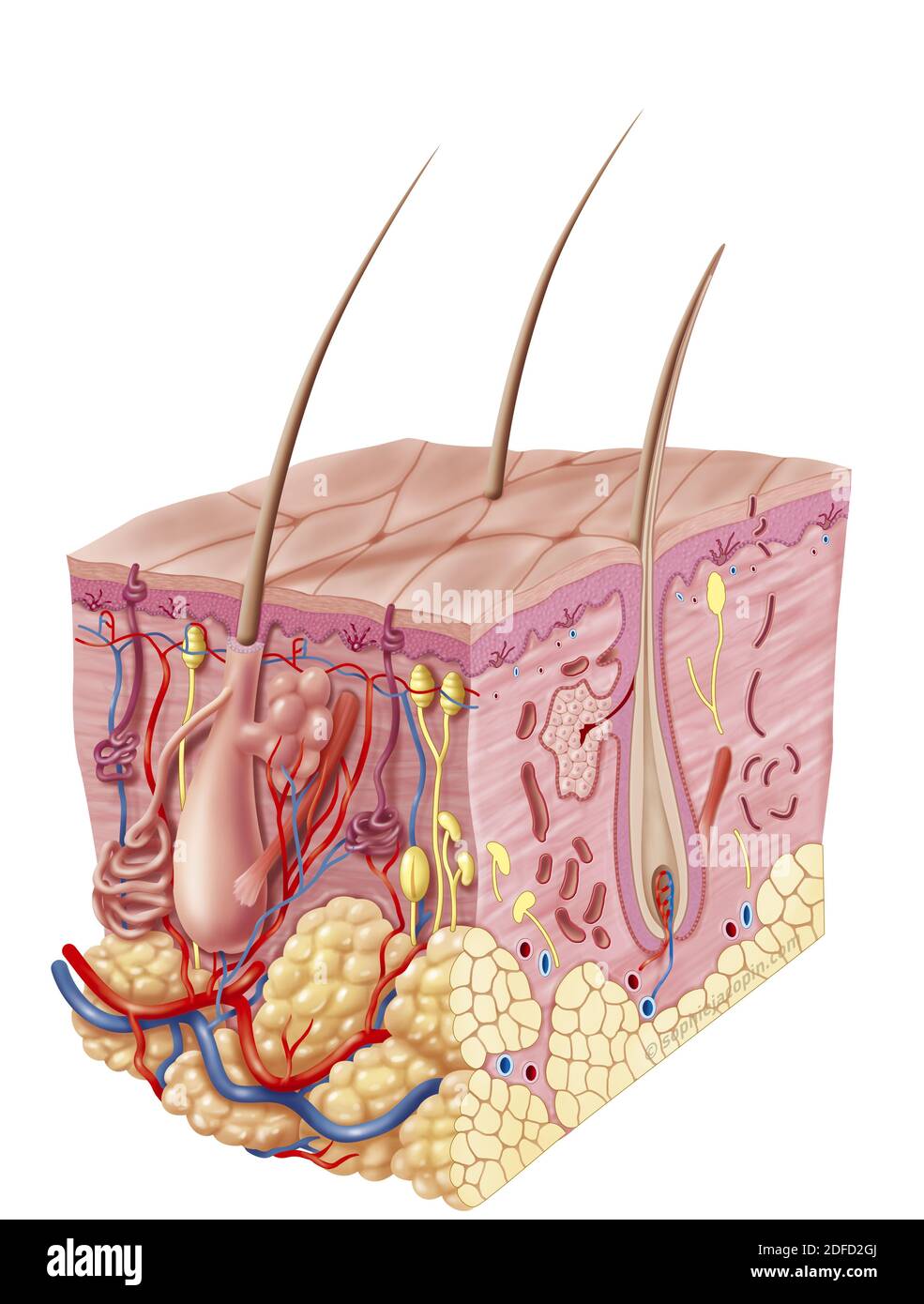
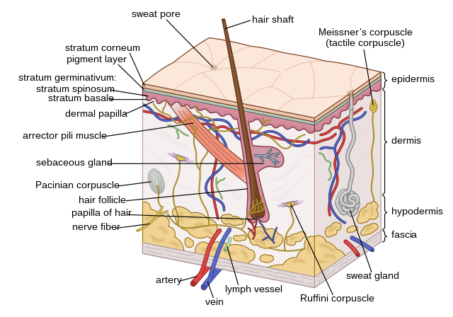
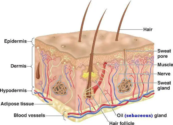
38 integumentary system diagram labeled
INTEGUMENTARY SYSTEM PART III: ACCESSORY STRUCTURES Integumentary Accessory Structures • Hair, hair follicles, sebaceous glands, sweat glands, and nails: – are made of epithelial tissue (part of epidermis) – are located in dermis – project through the skin surface The Hair Follicle • Is located deep in dermis – (made of epithleial tissue) The integumentary system is a system complete to the brim with amazing structures. Yet which of castle are an initial to spring to your mind? You'll likely think the the skin. Well, being the largest organ in the person body, skin anatomy is certainly an essential part of the integumentary system. However, it's not the only part. From nails ...
Oct 28, 2021 · Integumentary system quiz and answers. One of the best ways to start learning about a new system, organ or region is with a labeled diagram showing you all of the main structures found within it. Not only will this introduce you to several new structures together, it will also give you an overview of the relations between them.

Integumentary system diagram labeled
Start studying Integumentary System Labeling. Learn vocabulary, terms, and more with flashcards, games, and other study tools. The Integumentary System The integumentary system consists of the skin, hair, nails, the subcutaneous tissue below the skin, and assorted glands. Functions of the Integumentary System • Protection against injury and infection • Regulates body temperature • Sensory perception • Regulates water loss • Chemical synthesis Anatomy. Function. Interactions. The integumentary system is made up of several organs and structures including the skin, hair, nails, glands, and nerves. The primary function of the integumentary system is to protect the inside of the body from elements in the environment—like bacteria, pollution, and UV rays from the sun.
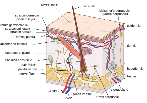
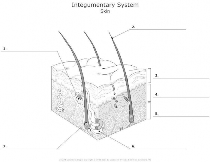
Integumentary system diagram labeled. The Integumentary System . 1. Label the diagram in the spaces provided. Artery Sweat Gland. Hair Shaft Epidermis. Sebaceous (Oil) Gland Vein. Melanin Subcutaneous. Sweat Pore Erector Muscle. Dermis Nerve. Name the three parts of the integumentary system. Describe the types of glands in the skin. 3. HASPI Medical Anatomy and Physiology 07a Lab Activity MOD EJO 2016 Station 3: Skin, Hair, and Nails Examine the features of the integumentary system. You will collect skin, hair, and nail samples to observe under the microscope. Skin 1. Use a Q-tip to gently swab the inside cheek of your mouth. Rub the Q-tip on Nov 28, 2021 · Integumentary system definition, organs, structure, physiology and functions, integumentary system diseases. Figure 1 shows a diagram of the skin structure. The skin is composed of three layers: Hair, nails, and cutaneous glands. Oct 04, 2021 · The integumentary system is the body system which surrounds you, both literally and metaphorically speaking. If you look in the mirror you see it, if you look anywhere on your body you see and if you look around you in the outside world, you see it. It is the system that can instantly tell us whether someone is young or old, someone’s ...
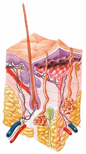
Diagram of the skin and hair (includes sweat gland) Skin: Tissue creating an external covering of the body. Epidermis: The upper layer of skin composed of the Stratum Corneum, stratum Lucidum, Stratum Granulosum, Stratum Spinosum, and Stratum Germinativum. The Stratum Corneum: The outermost layer of skin consisting of dead and Keratinization cells. integumentary system. Consists of the skin, mucous membranes, hair, and nail, largest organ of the human body. epidermis. The outer layer of the skin. dermis. A layer tissue underneath the epidermis of the skin which contains blood vessels, lymphatic vessels, nerves, sensory receptors, and oil and sweat glands. hypodermis. Feb 10, 2016 - integumentary system diagram - Google Search. Mons pubis, pad of fatty tissue lying in front of the pubic symphysis. The mons pubis is a rounded eminence made by fatty tissue beneath the skin. The integumentary system, or skin, is the largest organ in the body. Besides the skin, it comprises the hair and nails as well, which are appendages of the skin. Labeled Skin Diagram/ Parts of the Integumentary System. Parts of the Integumentary System. STUDY. PLAY. Skin. The Integumentary System, Part 1 - Skin Deep: Crash Course A&P #6
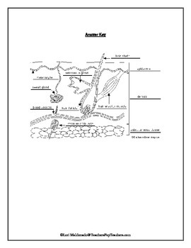
We are pleased to provide you with the picture named Integumentary system diagram.We hope this picture Integumentary system diagram can help you study and research. for more anatomy content please follow us and visit our website: www.anatomynote.com. Anatomynote.com found Integumentary system diagram from plenty of anatomical pictures on the internet. Human Physiology/Integumentary System 2 Layers The skin has two major layers which are made of different tissues and have very different functions. Diagram of the layers of human skin Skin is composed of the epidermis and the dermis. Below these layers lies the hypodermis or subcutaneous adipose layer, which is not usually classified as a layer ... Functions of the Integumentary system 1. protection a) chemical factors in the skin: Sebum (or oil) from the sebaceous glands is slightly acidic, retarding bacterial colonization on the skin surface. Sweat from the sudoriferous glands is slightly hypertonic and can flush off most bacteria on the skin surface. Integumentary System: Skin Diagram to Label. Students will read the definitions and label the skin anatomy diagram. Answer key included. This diagram had been modified from Enchanted Learning. I have used this worksheet as an in-class assignment as well as a homework assignment. You can also view a preview of this diagram :) *****...
Anatomy and physiology INTEGUMENTARY SYSTEM. STUDY. PLAY. Epidermis. consists of a stratified squamous epithelium organized into many distinct layers, or STRATA, of cells. Dermis. a thick layer of irregularly arranged connective tissue that supports and nourishes the epidermis and secures the integument to the underlying structures.
Deeper layer of the dermis that supplies the skin with oxygen and nutrients, 80% of the thickness of dermis, collagen run parallel to skin (surgeons use to reduce scarring from incision), dermis folds and creases to accommodate the joint movements ex. knuckles. Functions of integumentary system. Protection, body temperature regulation ...
Diagram of the Human Integumentary System (Infographic) By Ross Toro 05 August 2013. The skin is the largest organ of the body, and helps protect it from the environment.
Start studying Integumentary System: Anatomy. Learn vocabulary, terms, and more with flashcards, games, and other study tools.
Anatomy. Function. Interactions. The integumentary system is made up of several organs and structures including the skin, hair, nails, glands, and nerves. The primary function of the integumentary system is to protect the inside of the body from elements in the environment—like bacteria, pollution, and UV rays from the sun.
The Integumentary System The integumentary system consists of the skin, hair, nails, the subcutaneous tissue below the skin, and assorted glands. Functions of the Integumentary System • Protection against injury and infection • Regulates body temperature • Sensory perception • Regulates water loss • Chemical synthesis
Start studying Integumentary System Labeling. Learn vocabulary, terms, and more with flashcards, games, and other study tools.
(118).jpg)






:background_color(FFFFFF):format(jpeg)/images/article/en/think-you-know-the-integumentary-system-quiz-yourself/7MaCXPGKwYQW4RTCZCf3kg_integumentary_system_unlabeled.png)


:max_bytes(150000):strip_icc()/skin_layers_2-592309585f9b58f4c01554be.jpg)


0 Response to "38 integumentary system diagram labeled"
Post a Comment