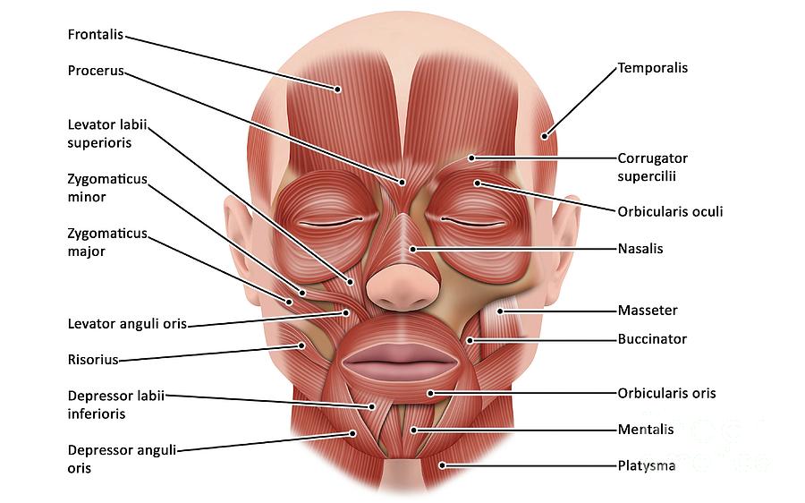
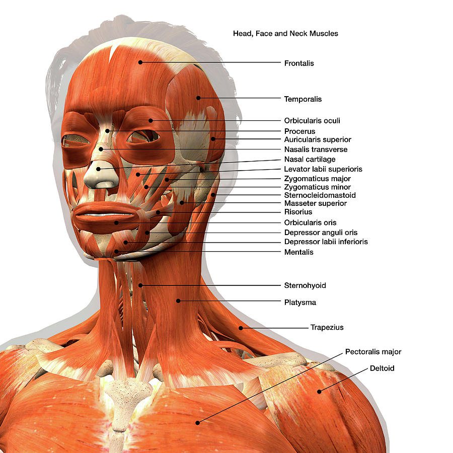
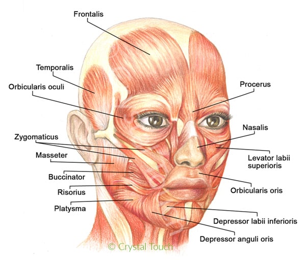
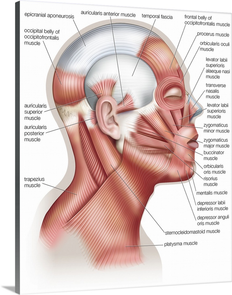
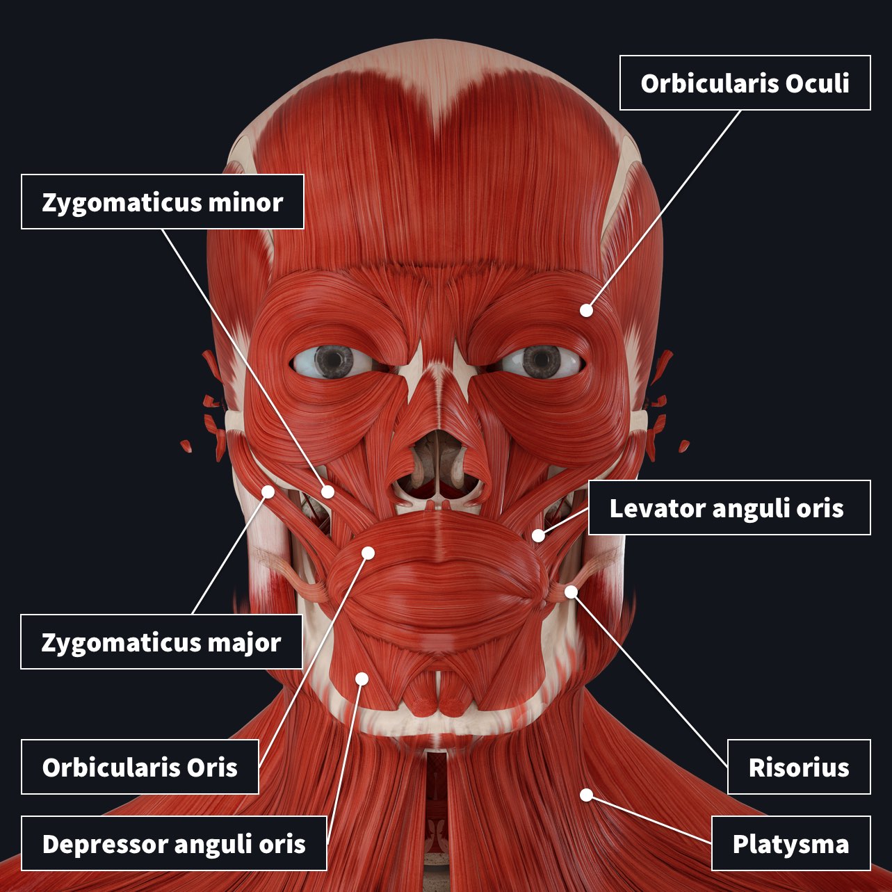
36 muscles of the face diagram
The muscles of facial expression (also known as the mimetic muscles) can generally be divided into three main functional categories: orbital, nasal and oral. These muscles are all innervated by the facial nerve (CN VII).¹. These striated muscles broadly originate from the surface of the skull and insert onto facial skin.
Muscle Charts of the Human Body For your reference value these charts show the major superficial and deep muscles of the human body. Superficial and deep anterior muscles of upper body
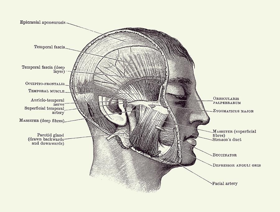
Muscles of the Face and Head Produce movement for facial expressions Vital for nonverbal communication Vary in shape and strength Tend to be fused together Many do not attach to bone. Many muscles in the face and head attach to the skin or other muscles. 9 Lateral View of the Head.

Muscles of the face diagram
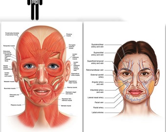
Facial anatomy at-a-glance. Dec 06, 2017. Knowing the facial anatomy is fundamental to performing more than aesthetic surgery. A provider's lack of understanding of the intricate web of facial muscles, nerves, arteries and more can turn a relatively simple injection technique, with botulinum toxin or a filler, into a serious complication.
The muscles of the face overlap and crisscross over each other, creating a mask of muscle over the skull and jawbone. They attach to various parts of the skull and other muscles, allowing for a ...
Function: This facial muscle helps to hold food inside the mouth in proper position and aids in chewing. Flattening the cheeks and pulling the angle of the mouth backwards is supported by this muscle. #5. Mentalis Muscle of the Face: The furrow between the lower lip and chin is formed by this muscle of the face. In other words, it can be said that this facial muscle is located at the tip of ...
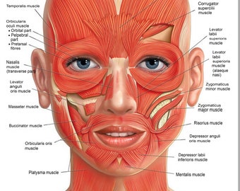
Muscles of the face diagram.
Doing facial exercises, or facial yoga, is a natural way to make your face look younger by firming muscles and reducing wrinkles. These are also good exercises to do if you have a muscle problem on your face, creating stronger muscles for a toned and more confident look.
May 6, 2015 - Explore Edward Boyle's board "Face Anatomy" on Pinterest. See more ideas about face anatomy, anatomy, muscle anatomy.
Page 3. Labelled diagram of Bones of the face. Page 4. Labelled diagram of the bones neck, chest & shoulder. Task 2 Page 5. Labelled diagram showing the position of the Muscles of the face. Page 6. Labelled diagram showing the Muscles that move the head Page 7 .Chart showing the action and location of the muscle of the face. Task 3 Page 8 (A).
The muscles of the head and neck perform many important tasks, including movement of the head and neck, chewing and swallowing, speech, facial expressions, and movement of the eyes. These diverse tasks require both strong, forceful movements and some of the fastest, finest, and most delicate adjustments in the entire human body.
Muscles of the Head and Neck. Humans have well-developed muscles in the face that permit a large variety of facial expressions. Because the muscles are used to show surprise, disgust, anger, fear, and other emotions, they are an important means of nonverbal communication. Muscles of facial expression include frontalis, orbicularis oris, laris ...
Joints of the Body (Advanced) Coloring Page. Joints of the Body (Basic) Coloring Page. Knee Joint Anatomy Coloring Page. Large Intestine Coloring Page. Liver Function Organs (Labeled) Coloring Page. Lower Limb (Thigh, Leg and Foot) Coloring Page. Lungs Coloring Page. Mouth, Pharynx and Esophagus Coloring Page.
8,582 anatomy face muscle stock photos, vectors, and illustrations are available royalty-free. See anatomy face muscle stock video clips. of 86. muscles of the face face muscle vector face muscles muscle of the face face anatomy face muscle muscle face woman face muscle female anatomy face women face muscles. Try these curated collections.
This medical illustration depicts the following muscles of the face (facial muscles) ... Anatomy Of Facial Muscles - Human Anatomy Diagram. More information.
Start studying Muscles of the face and head - epicranius. Learn vocabulary, terms, and more with flashcards, games, and other study tools.
Facial Muscle Chart. $9.00. translation missing: en.products.notify_form.description: Notify me when this product is available: Simply put, this Facial Muscle Chart shows the muscle structure of the face and help boost confidence in your MyoLift session. For beginners, people often wonder where to apply the microcurrent applicators.
4 Aug 2021 — A human face has 20 main facial muscles, or craniofacial muscles, which are essential to chewing and making facial expressions.
The facial muscles can broadly be split into three groups: orbital, nasal and oral. The orbital group of facial muscles contains two muscles associated with the eye socket. These muscles control the movements of the eyelids, important in protecting the cornea from damage. They are both innervated by the facial nerve.
Start studying Muscles of the Face. Learn vocabulary, terms, and more with flashcards, games, and other study tools.
Zygomaticus minor is a thin paired facial muscle extending horizontally over the cheeks. It belongs to a large group of muscles of facial expression. The zygomaticus major is a muscle of the human body. It is a muscle of facial expression which draws the angle of the mouth superiorly and posteriorly to allow one to smile. Frontalis
2) Occipitalis - not shown on diagram (in back): a. Actions: pulls scalp back b. Innervation: Facial Nerve (Vll) c. Origin: from posterior back of skull d. Insertion: to anteriorly with the galea aponeurotica 3) Orbicularis oculi (sphincter muscle of the eyelid): a. Actions: squinting, closes eye, crowsfeet, eye bags b. Innervation: Facial ...
The muscles of facial expression are embedded in the superficial fascia of the face. The majority of them originate from bones of the skull and are added into the skin. They bring about various types of facial expressions, thus the name muscles of facial expression, the activities of many are indicated by their names.
Anatomical Facial Muscles of a Woman on White Background 3D Rendering of the facial muscle anatomy of a woman, frontal view on a white background. Includes eyes. face diagram stock pictures, royalty-free photos & images
The facial muscles are the only group of muscles that insert into the skin. They are located in the subcutaneous tissue, originating from bone or fascia. When contracting, the muscles pull on the skin and exert their effects ?. This group of muscles comes from the same embryonic origin, the 2 nd pharyngeal arch.
Face muscle anatomy. Found situated around openings like the mouth, eyes and nose or stretched across the skull and neck, the facial muscles are a group of around 20 skeletal muscles which lie underneath the facial skin.The majority originate from the skull or fibrous structures, and connect to the skin through an elastic tendon.
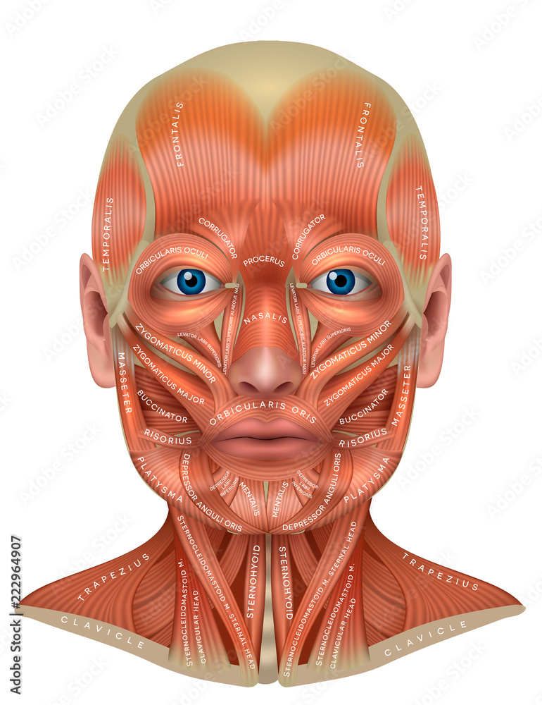
Human facial muscle diagram. In this image, you will find galea aponeurotica, frontalis, corrugator, levator labii superiors alaeque nasi, levator superons, obicularis oris, risonus, mentalis, platysma in it. You may also find depressor labii inferioris, depressor anguli oris, masseter, zygomarticus major, zygomaticus minor, nasalis, procerus ...
Facial muscles (Musculi faciales) The facial muscles, also called craniofacial muscles, are a group of about 20 flat skeletal muscles lying underneath the skin of the face and Most of them originate from the bones or fibrous structures of the skull and radiate to insert on the. Contrary to the other skeletal muscles they are not surrounded by a fascia, with the exception of the ...
Feb 7, 2020 - Explore Susan Torrey's board "face diagrams" on Pinterest. See more ideas about facial anatomy, facial aesthetics, face anatomy.
Muscles of the face - superficial facial muscles - anterior view - human anatomy diagram Join am-medicine Group T he muscles of the face - head and neck perform many important tasks, including movement of the head and neck, chewing and swallowing, speech, facial expressions, and movement of the eyes.
Posted December 30, 2010 in BOTOX® Cosmetic and Dysport®, Face. In order to understand what to expect from Botox or Dyport treatment, a basic knowledge of muscle anatomy really helps. Look at the diagram below: Anatomy diagram: A. Frontalis muscle: contraction raises the eyebrows and causes horizontal brow wrinkles. Injection of Botox or ...
The muscles of the face. In this article, we'll recap the main muscle groups covered in the previous step. Rating: 4,8 · 245 reviews
Temporalis, Sternocleidomastoid, Orbicularis Oris, Orbicularis Oculi, Masseter, Zygomaticus, Mentalis, Frontalis, Corrugator, Buccinator, Trapezius, Risorius.
:watermark(/images/watermark_5000_10percent.png,0,0,0):watermark(/images/logo_url.png,-10,-10,0):format(jpeg)/images/overview_image/241/SPJ2HZkcfjFlJC7JVNp4g_facial-muscles_english.jpg)


:background_color(FFFFFF):format(jpeg)/images/library/14066/Facial_muscles.png)








0 Response to "36 muscles of the face diagram"
Post a Comment