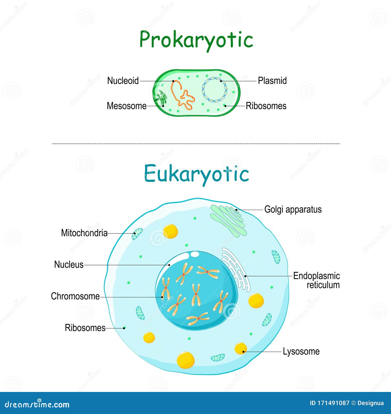
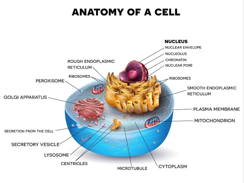
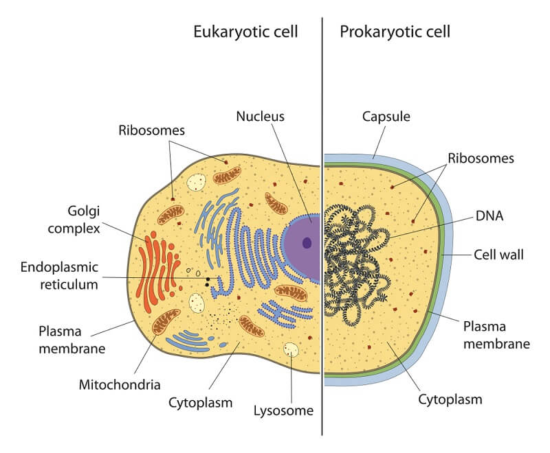
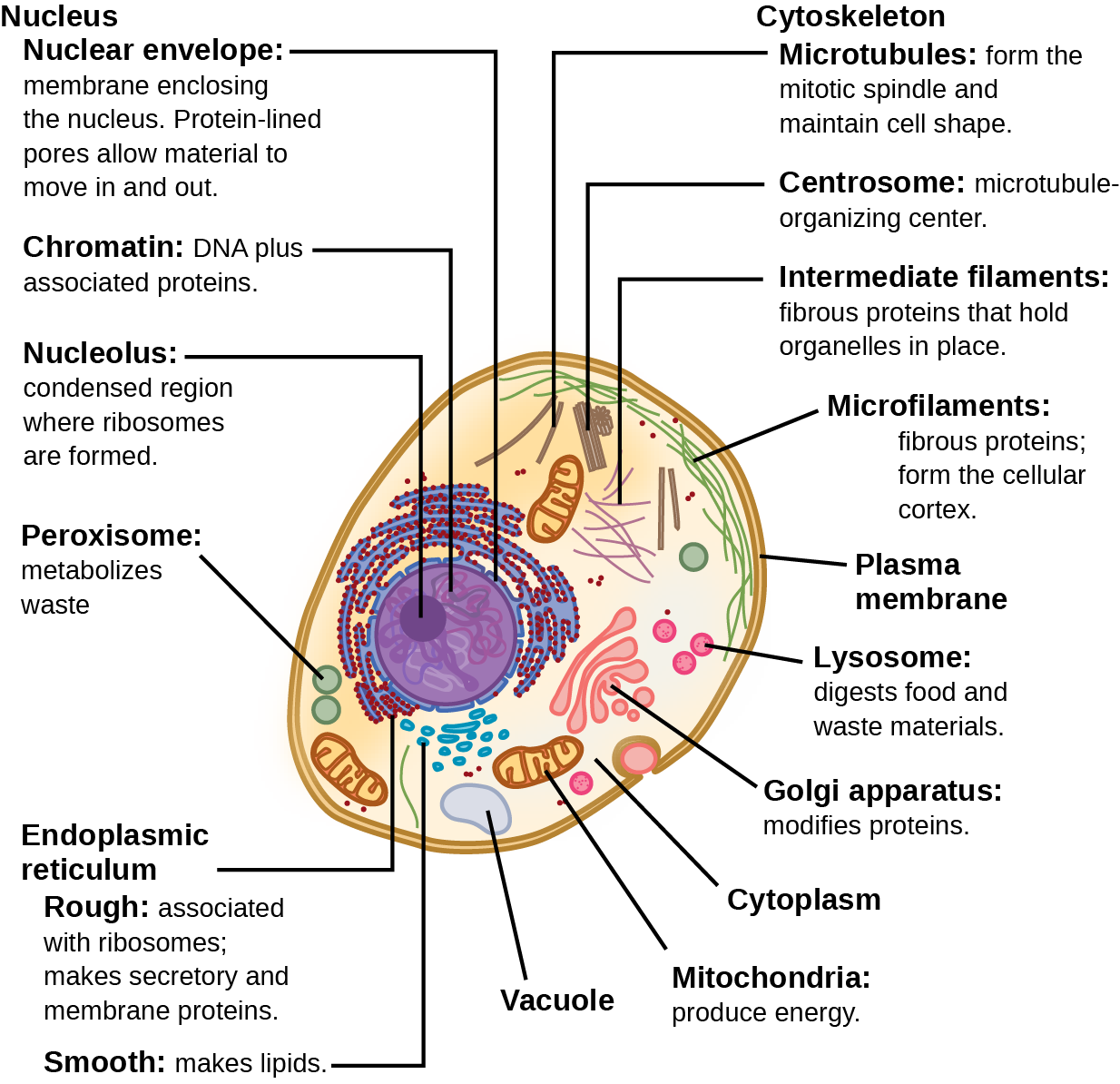
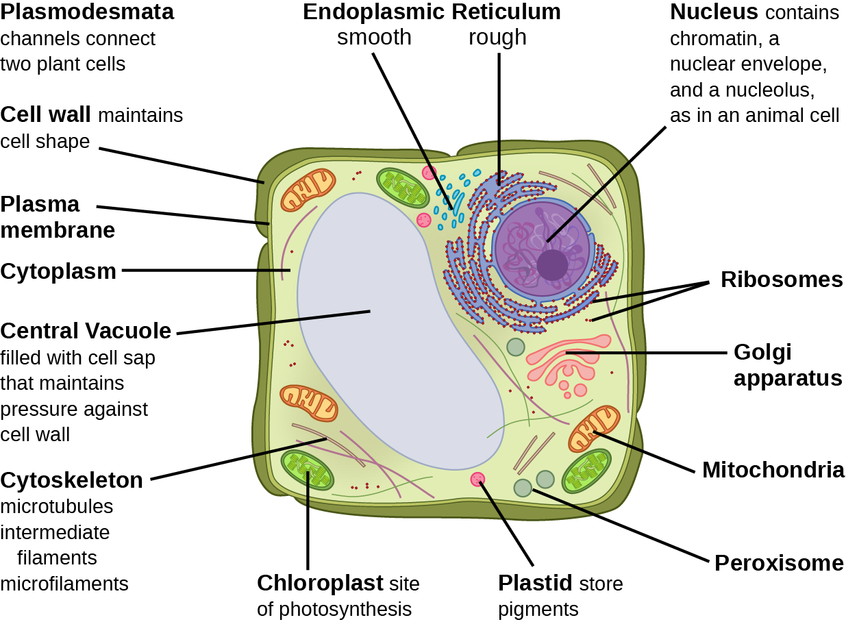
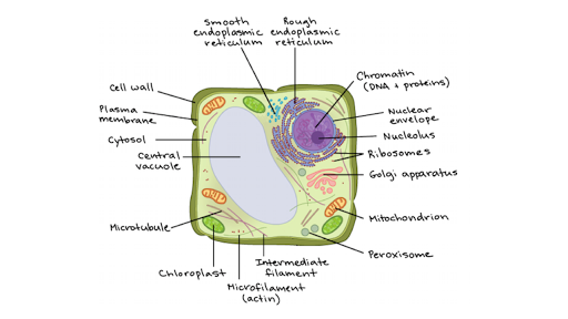
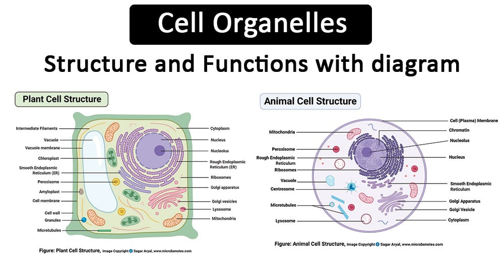
36 eukaryotic cell diagram with labels
Science Olympiad Student Center - Scioly.org eukaryotic organism. (l mark) The diagram shows a mitochondrion. (a) (a) (a) Name the parts labelled X and Y. (2 marks) Scientists isolated mitochondria from liver cells. They broke the cells open in an ice-cold, isotonic solution. They then used a centrifuge to separate the cell organelles. The diagram shows some of the steps in the process of centrifugation. High speed 5 minutes …
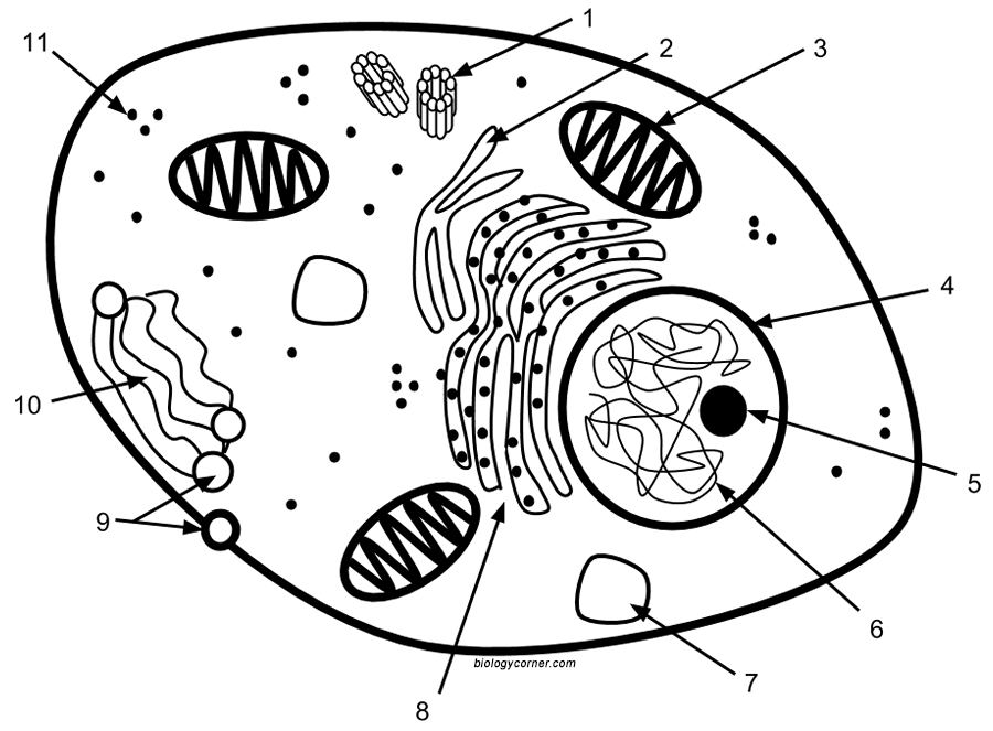
Dec 01, 2021 · Directions: Label the diagram below with the following choices: Nucleotide Deoxyribose Phosphate group Base pair Hydrogen bond Nitrogenous base Directions: Complete each sentence. Why do cells The Plant Cell Worksheet / Eukaryotic Cell Structure : Plants in plant cells, chloroplasts perform photosynthesis, a process that converts light en. 1 .

Eukaryotic cell diagram with labels
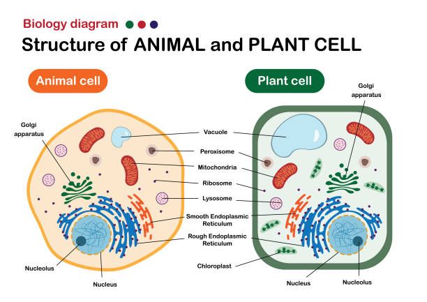
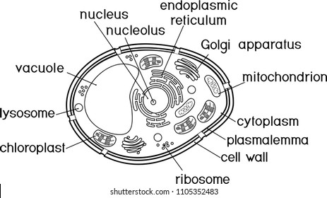
Many plant cell organelles are also found in animal cells. In what follows, I’ll focus on the parts unique to plants, and list the name and function of those organelles shared by both kingdoms. For an overview of animal cells, see the previous tutorial. The cell wall. Part 1 is the cell wall. Cell walls are composed of cellulose, or plant fiber. Eukaryotic promoters are more complex than their prokaryotic counterparts, in part because eukaryotes have the aforementioned three classes of RNA polymerase that transcribe different sets of genes. The diagram below shows a bacterial replication fork and its principal proteins. Drag the labels to their appropriate locations in the diagram to describe the name or function of each structure. Use pink labels for the pink targets and blue labels for the blue targets.
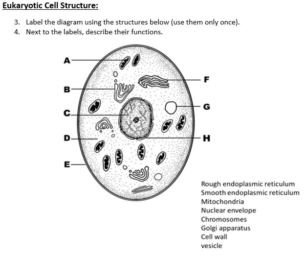
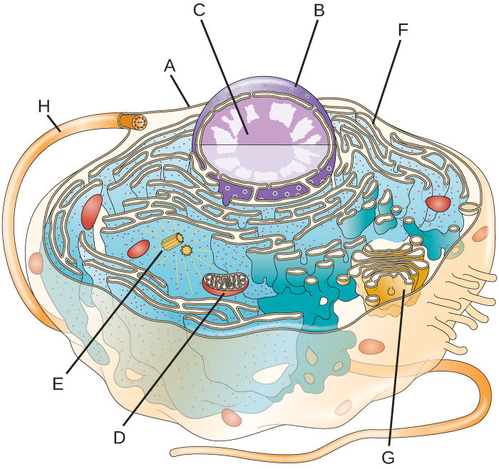
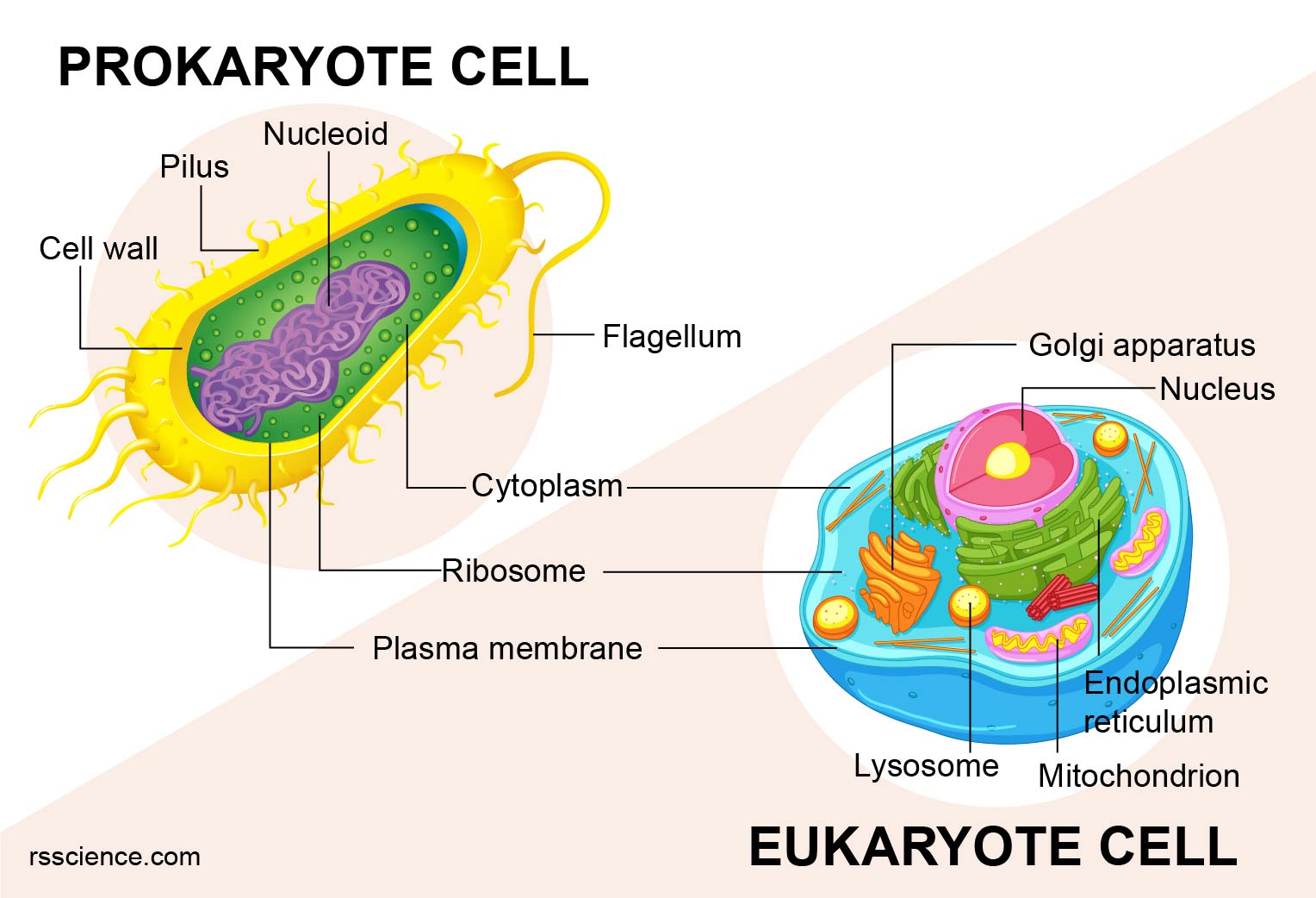
Eukaryotic cell diagram with labels. Proteins that are secreted from a eukaryotic cell must first travel through the endomembrane system. Drag the labels onto the diagram to identify the path a secretory protein follows from synthesis to secretion. Not all labels will be used. Pulse-chase experiments and protein location Scientists can track the movement of proteins through the endomembrane system using an … 01.12.2021 · Label the structures of the prokaryotic cell in the figure below Cell structure Light and electron microscopes allow us to see inside cells. Plant, animal and bacterial cells have smaller components each with a specific function. Outer membrane. The outer nuclear membrane also shares a common border with the endoplasmic reticulum. While it is physically linked, the outer nuclear membrane contains proteins found in far higher concentrations than the endoplasmic reticulum. All four nesprin proteins (nuclear envelope spectrin repeat proteins) present in mammals are expressed in the outer …
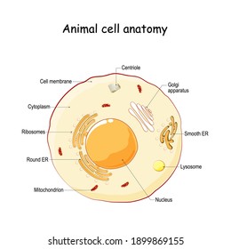
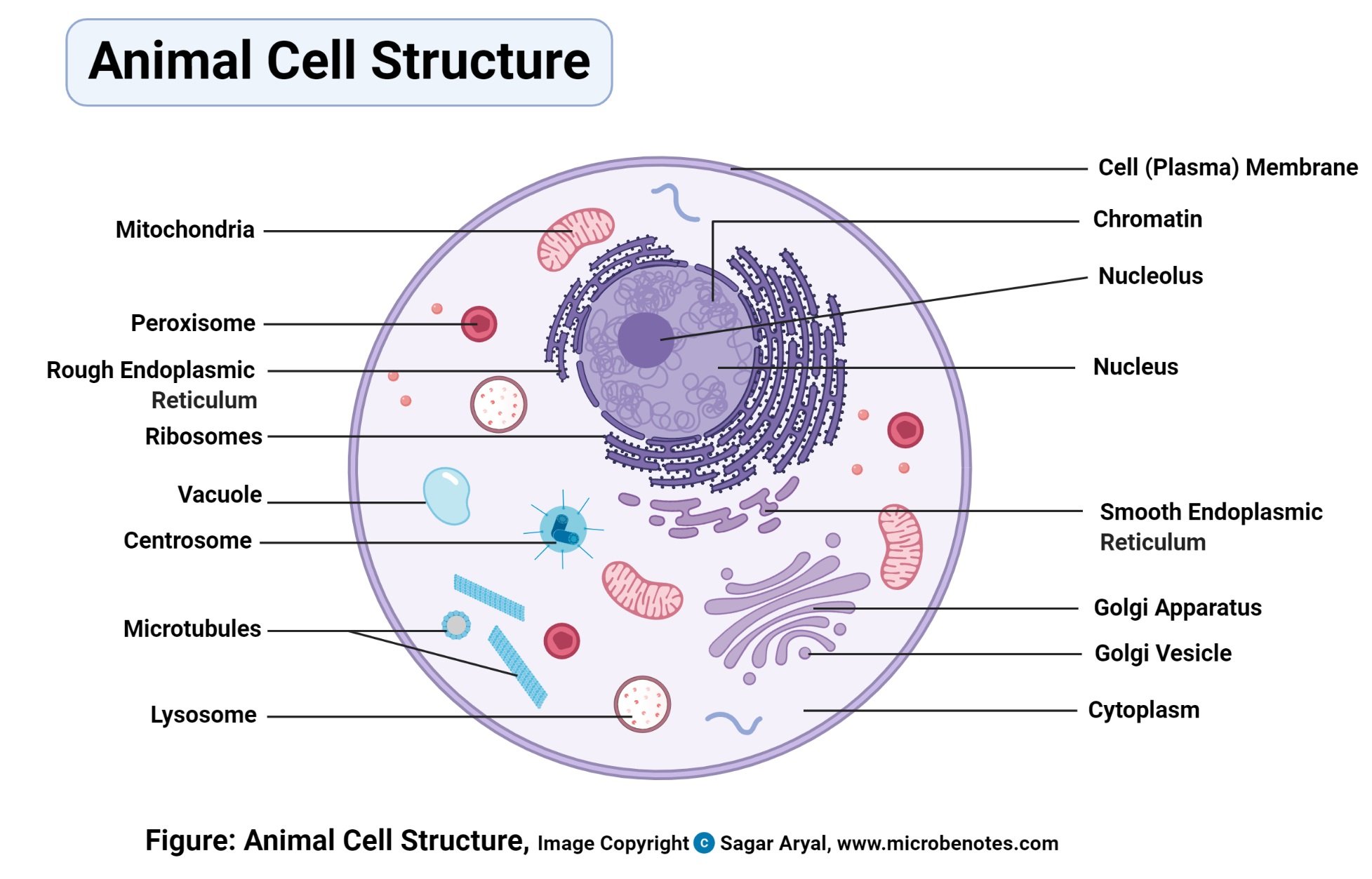
Eukaryotic pre-mRNA processing. Practice: Transcription. Next lesson. Translation. Sort by: Top Voted. Overview of transcription. Eukaryotic pre-mRNA processing. Up Next. Eukaryotic pre-mRNA processing. Biology is brought to you with support from the Amgen Foundation. Biology is brought to you with support from the. Our mission is to provide a free, world-class … Animal Cell Illustration With Hyperlinked Labels. Click On Each Label For More Information. Illustration of a generalized animal cell. Eukaryotic Cell Definitions: = Typically Found Only In Plant Cells = Typically Found In Animal Cells. Golgi Apparatus: A series (stack) of flattened, membrane-bound sacs (saccules) involved in the storage, modification and secretion of … INTERPHASE. Gap 0. Gap 1. S Phase. Gap 2. MITOSIS . ^ Cell Cycle Overview Cell Cycle Mitosis > Meiosis > Get the Cell Division PowerPoints The diagram below shows a bacterial replication fork and its principal proteins. Drag the labels to their appropriate locations in the diagram to describe the name or function of each structure. Use pink labels for the pink targets and blue labels for the blue targets.
Eukaryotic promoters are more complex than their prokaryotic counterparts, in part because eukaryotes have the aforementioned three classes of RNA polymerase that transcribe different sets of genes. Many plant cell organelles are also found in animal cells. In what follows, I’ll focus on the parts unique to plants, and list the name and function of those organelles shared by both kingdoms. For an overview of animal cells, see the previous tutorial. The cell wall. Part 1 is the cell wall. Cell walls are composed of cellulose, or plant fiber.










:background_color(FFFFFF):format(jpeg)/images/library/12788/histology-eukaryotic-cell_english.jpg)


0 Response to "36 eukaryotic cell diagram with labels"
Post a Comment