36 drag the labels onto the diagram to identify the blood vessels of the kidneys.
The study of kidney function is an example of ______. organ physiology ... Drag the labels onto the diagram to identify the organ systems. Rating: 5 · 1 review Pink labels may be used more than once drag the blue labels to the blue targets to indicate whether the genotype in each box confers hemophilia normal or carrier status. Part a identifying the structures of the kidney. Drag the labels onto the diagram to identify the structures and functions ...
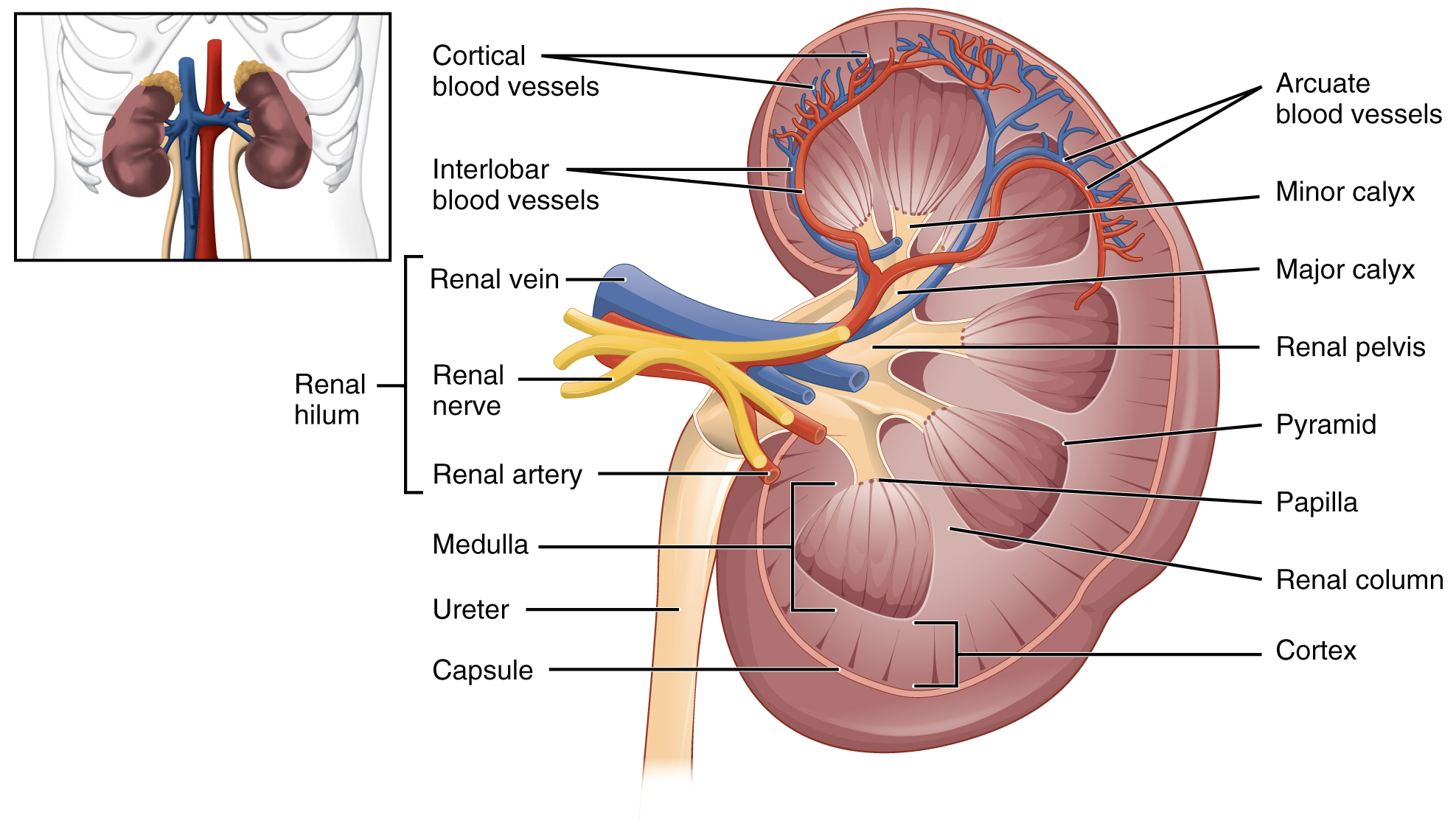
Despite their relatively small size, the kidneys receive approximately 20% of the cardiac output. Each renal artery branches into segmental arteries, dividing further into interlobar arteries, which penetrate the renal capsule and extend through the renal columns between the renal pyramids. The interlobar arteries then supply blood ...
Drag the labels onto the diagram to identify the blood vessels of the kidneys.
Drag the labels onto the diagram to identify the effects of isotonic hypotonic and hypertonic solutions on red blood cells. Drag the labels onto the diagram to identify respiratory system structures. Lungs are to the respiratory system as the liver is to the system. Label the structures of the lower respiratory tract. Drag the labels onto the diagram to identify the lymphoid tissues and organs of the lymphatic system. Drag the labels onto the diagram to identify the structural features of the spleen. Which of the labels indicates a structure through which lymph flows? the small, dark structure labeled A. Drag The Labels Onto The Diagram To Identify Structures And Students can also color the image to identify the major structures of the nephron. Label the major structures of the nephron and associated structures. This is the currently selected item. General overview of the raas system. There are about 1000000 nephrons in each human kidney.
Drag the labels onto the diagram to identify the blood vessels of the kidneys.. Question: Drag the labels onto the diagram to identify the parts of a spermatozoon. This problem has been solved! See the answer See the answer See the answer done loading. Drag the labels onto the diagram to identify the parts of a spermatozoon. Show transcribed image text Expert Answer. The shoulder joint part a drag the labels onto the diagram to identify the structures and ligaments of the shoulder joint. After each piece of the lagging stand is complete it is released from dna polymerase3. Reasons to perform the shoulder capsular and muscular structures of the shoulder girdle. Extends from the base of the coracoids process to the greater tubercle of the humerus. Reset help ... renal system - renal system - Renal vessels and nerves: The renal arteries arise, one on each side, from the abdominal aorta at a point opposite the upper border of the second lumbar vertebra (i.e., a little above the small of the back). Close to the renal hilus each artery gives off small ... May 21, 2019 - The kidneys are important to the body’s production of urine. They also play a role in regulating important components of the blood. Oxygenated blood comes to the kidneys from the right and left renal arteries off the abdominal aorta. Deoxygenated blood leaves the kidneys via the right and ...
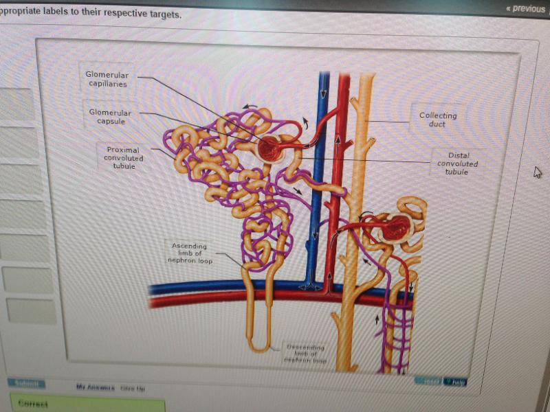
Transcribed image text: Part A Drag the labels onto the diagram to identify the arteries,that supply blood to the kidney Reset H Afferent arterioles ... The vital structural components of a kidney are enclosed in a smooth but tough fibrous capsule called renal capsule.; Inside this capsule, two distinct regions can be observed: a pale outer region called renal cortex, and a dark inner portion called renal medulla.; The renal medulla comprises a set of 8-18 conical structures called renal pyramids that are surrounded by the cortex. Show transcribed image text drag the labels onto the diagram to identify structural features associated with skeletal muscle. Label this diagram. Answer Correct Chapter Test Chapter 10 Question 4 Endomysium Numbers to the left identify the spinal nerves and indicate where the nerve roots leave the vertebral canal. Transcribed image text: Drag the labels onto the diagram to identify the blood vessels of the kidneys. Reset Help Renal vein Renal artery Glomerulus Interlobar artery Afferent arteriole Efferent arteriole Segmental artery Peritubular capillaries Interlobar vein Cortical radiate artery Cortical radiate vein Arcuate vein Arcuate artery Venule NEPHRON
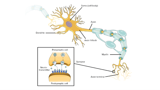
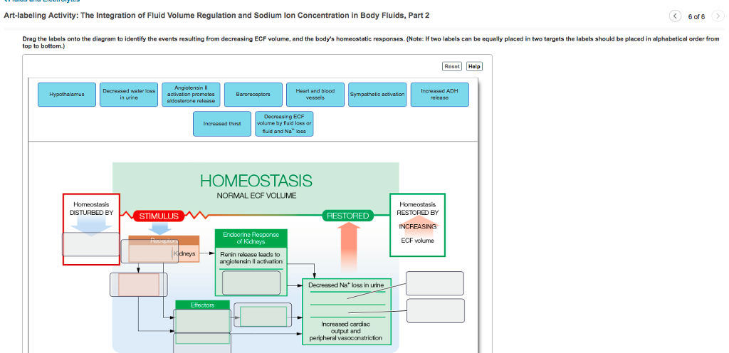
Question: Drag the labels onto the diagram to identify the events ... or fluid and Na loss Heart and blood vessels HOMEOSTASIS NORMAL ECF VOLUME Homeostasis ... Blood vessels line in between the connective tissue coverings in between the fascicles. Chapter 13 Synapses Drag the labels onto the diagram to identify the steps in a reaction both with and without enzymes. Drag the labels onto the diagram to identify parts of the neuromuscular junction. Show transcribed image text label the parts of the ... Anatomy and Physiology. Anatomy and Physiology questions and answers. Drag the labels onto the diagram to identify the parts of the kidney. Reset Help Minor calyx Hilum Ureter Adipose tissue in renal sinus Renal pelvis Renal sinus Major calyx Connection to minor calyx. 41 drag the labels onto the diagram to identify factors that affect mean arterial pressure. Written By Chelsea P. Mariano. Friday, November 26, 2021 Add Comment Edit. Terms in this set (33) Drag the labels onto the diagram to identify aspects of gas transport and exchange. the diaphragm and rib muscles contract. Which statement is correct? In the blood, oxygen is bound to hemoglobin, a protein ...
This is due to its rich blood supply—it houses 90-95% of the kidney's blood vessels. At specific points, extensions of the renal cortex called renal columns pass through the renal medulla to-ward the renal pelvis. The renal columns house blood vessels Figure 24.3 Internal anatomy of the kidney, including the nephron. Renal hilum Renal pelvis
Start studying Blood Vessels. Learn vocabulary, terms, and more with flashcards, games, and other study tools.

This diagram shows the different ions and chemicals that are secreted and reabsorbed along the nephron. solved part a identifying the structures the kidney plete the diagram below using the following steps place the pink labels which indicate interstitial fluid osmolarity in mosm l onto the correct pink tar s note that the numbers inside the ...
... Arterial blood supply to the kidney 21 of 59 > Label the arteries that supply blood to the kidney Part A Drag the labels onto the diagram to identify ...
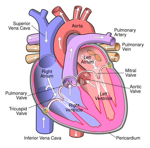
Drag and drop the text labels onto the boxes next to the heart diagram It consists of two periods: one during which the heart muscle relaxes and refills with blood, called diastole, following a period of robust contraction and pumping of blood, dubbed systole Chu, H April 5, 2021 9:30 AM - 5:00 PM ET Register Now!
peritoneal cavity; and the lining of all blood vessels. t Glandular epithelium forms most of the body glands. Epithelia occur at the boundary between two different envi-ronments. The epidermis of the skin, for example, lies between the inside and the outside of the body. Most substances that enter into the body or are released from the body ...

Identify the renal blood vessels and structures of the cortical and juxtamedullary nephrons by clicking and dragging the labels to the correct location.
January 24, 2018 - The kidneys are important to the body’s production of urine, but they also play a role in regulating important components of the blood. Blood comes to the kidneys from the abdominal aorta and inferior vena cava, the large arteries and veins that are part of the ascending aorta.
Drag the labels onto the diagram to identify the parts of the kidney. ... Chronic and acute renal failure impairs all of the functions carried out by the kidneys and, as a consequence, the functions of most other body systems. ... The collection of blood vessels supplying the protective connective tissue layers surrounding the brain may ...
Signal recognition particle SRP binds to the signal peptide as it emerges from the ribosome. part a drag the labels onto the diagram to identify the part a drag the labels onto the diagram to identify the stages of the life cycle not all labels will be used answer chapter 8 reading quiz question 2. Proteins all begin their synthesis in the ...

Drag the labels onto the diagram to identify the origins of the cranial nerves (VII - XII). look at pic The accumulation of blood during an epidural or subdural hemorrhage creates debilitating pressure on the brain and, without help, death is imminent.
Blood Supply and innervation of the Pericardium The pericardium is supplied by the following vessels: a. Pericardiacophrenic arteries. b. Musculophrenic arteries c. Pericardial branches of the bronchial, oesophageal and superior phrenic arteries d. The coronory arteries Pericardial veins drain into the azygos, internal thoracic and cardiac veins.
Drawing and labelling a diagram of the human kidney AND The composition of blood in the renal artery is different from that in the renal vein

Drag the labels onto the diagram to identify the stem cells and stages of white blood cell and platelet production. Drag the labels onto the diagram to identify steps in response to low blood pressure. Maybe venous blood pressure is low. Is his bodys response to the low oxygen. Drag and drop the labels onto the figure in the correct order of events to complete the positive feedback loop. Chapter 1 homework 1. 2 formation of a platelet plug. Harolds doctor noted that he was.
Classifying epithelia identify the types of epithelia. The layers do not contain any blood vessels and one surface of the cells lines the cavity of the organ. Show transcribed image text drag the correct labels onto the diagram to identify the structures and molecules involved in translation. Solved drag the labels onto the diagram to identity ...

Drag the labels onto the diagram to identify the events resulting from decreasing ECF volume, and the body's homeostatic responses. (Note: If two labels can be equally placed in two targets the labels should be placed in alphabetical order from top to bottom.)
This photo about: Drag the Labels Onto the Diagram to Identify Structural Features associated with Skeletal Muscle., entitled as 20 1 Structure And Function Of Blood Vessels - Anatomy And Physiology Drag The Labels Onto The Diagram To Identify Structural Features Associated With Skeletal Muscle. - also describes 20 1 Structure and Function of Blood Vessels - Anatomy and Physiology and ...

Drag The Labels Onto The Diagram To Identify The ... from lh6.googleusercontent.com The humerus fits relatively loosely into the shoulder joint. The rotator cuff is a collection of muscles and tendons that surround the. Fibers of connective tissue that hold the skull bones tightly in place (figure 19.23). This code of practice on how to identify hazardous manual tasks and. 8 name the arteries ...
Show transcribed image text drag the labels onto the diagram to identify structural features associated with skeletal muscle. 20 1 structure and function of blood vessels anatomy and physiology drag the labels onto the diagram to identify structural features associated with skeletal muscle. 5163 x 2217 pixel.

December 3, 2020 - renal artery, one of the pair of large blood vessels that branch off from the abdominal aorta (the abdominal portion of the major artery leading from the heart) and enter into each kidney. (The kidneys are two bean-shaped organs that remove waste substances from the blood and aid in fluid
The tunica intima , or inner-most layer of blood vessels, is made of endothelial tissue. Art-labeling Activity: Figure 32.1 (1 of 2) Label the major types of blood vessels and their layers. Part A Drag the labels onto the diagram to identify the major types of blood vessels and their layers.

Drag the labels onto the diagram to identify the types of epithelia. look at the picture . Exfoliative cytology involves the removal of epithelial cells for examination. Which of the following is NOT a clinical application of exfoliative cytology? examination of cardiac muscle cells after a ...
Answer to Solved Part A Drag the labels onto the diagram to identify. ... and arterial vessels Stroke volume Arterioles Left ventricle Elastic arteries Mean ...

The ureter, blood vessels, and nerves penetrate the kidney on its medial surface. true The fibrous capsule is a layer of adipose tissue that surrounds the kidney.

The force to move the blood The Cardiovascular System WHAT HOW WHY The cardiovascular system delivers oxygen and nutrients to the body tissues and carries away wastes such as carbon dioxide via blood. The heart pumps blood throughout the body in blood vessels. Blood flow requires both the pumping action of the heart and changes in blood pressure.

Check your understanding 2 pts Complete the flow chart showing the flow of blood through the kidneys. Drag and drop the choice labels into the corresponding boxes Afferent arteriole Aorta Interlobar artery Interlobular artery Renal artery Segmental artery Lobar artery 4 Arcuate artery Glomerulus.

Drag the labels onto the diagram to identify the major renal processes and associated nephron structures. nitrogenous . In its excretory role, the urinary system is primarily concerned with the removal of _____ wastes from the body. kidneys. The _____ perform(s) the excretory and homeostatic ...
Label the parts of the neuromuscular junction. Drag the labels onto the diagram to identify the steps in a reaction both with and with out e... Ren in is an enzyme released by the juxtaglomerular cells of the kidneys in response to low blood pressure caus in g the transformation of angiotens in ogen Er cis golgi cisternae medial golgi cisternae.
Drag The Labels Onto The Diagram To Identify Structures And Students can also color the image to identify the major structures of the nephron. Label the major structures of the nephron and associated structures. This is the currently selected item. General overview of the raas system. There are about 1000000 nephrons in each human kidney.
Drag the labels onto the diagram to identify the lymphoid tissues and organs of the lymphatic system. Drag the labels onto the diagram to identify the structural features of the spleen. Which of the labels indicates a structure through which lymph flows? the small, dark structure labeled A.
Drag the labels onto the diagram to identify the effects of isotonic hypotonic and hypertonic solutions on red blood cells. Drag the labels onto the diagram to identify respiratory system structures. Lungs are to the respiratory system as the liver is to the system. Label the structures of the lower respiratory tract.


0 Response to "36 drag the labels onto the diagram to identify the blood vessels of the kidneys."
Post a Comment