38 meniscus diagram of the knee
The knee meniscus: management of traumatic tears and ... In stable knees (intact ACL), about 6% of acutely injured knees sustain a meniscus tear. 10 In chronic ACL-ruptured knees, the rate of meniscal tears is very high, 11 and increases with time with the medial meniscus while it remains the same with the lateral meniscus (around 20%). In traumatic tears, mensicus preservation is the first-line choice. Dog Knee Anatomy with Labeled Diagram » AnatomyLearner ... The dog knee injury is very common in the field. If you want to manage a knee injury, you might have a good piece of knowledge on the dog knee anatomy.Here, I will show you everything on the dog knee, including the bone involvement, ligaments, tendons and their arrangement with a labeled diagram.
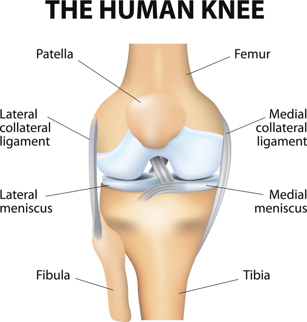
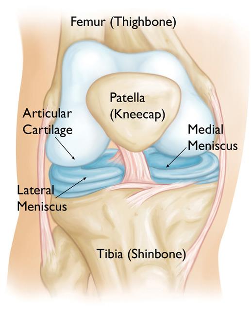
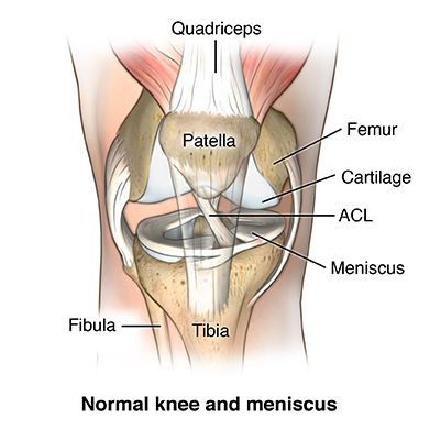
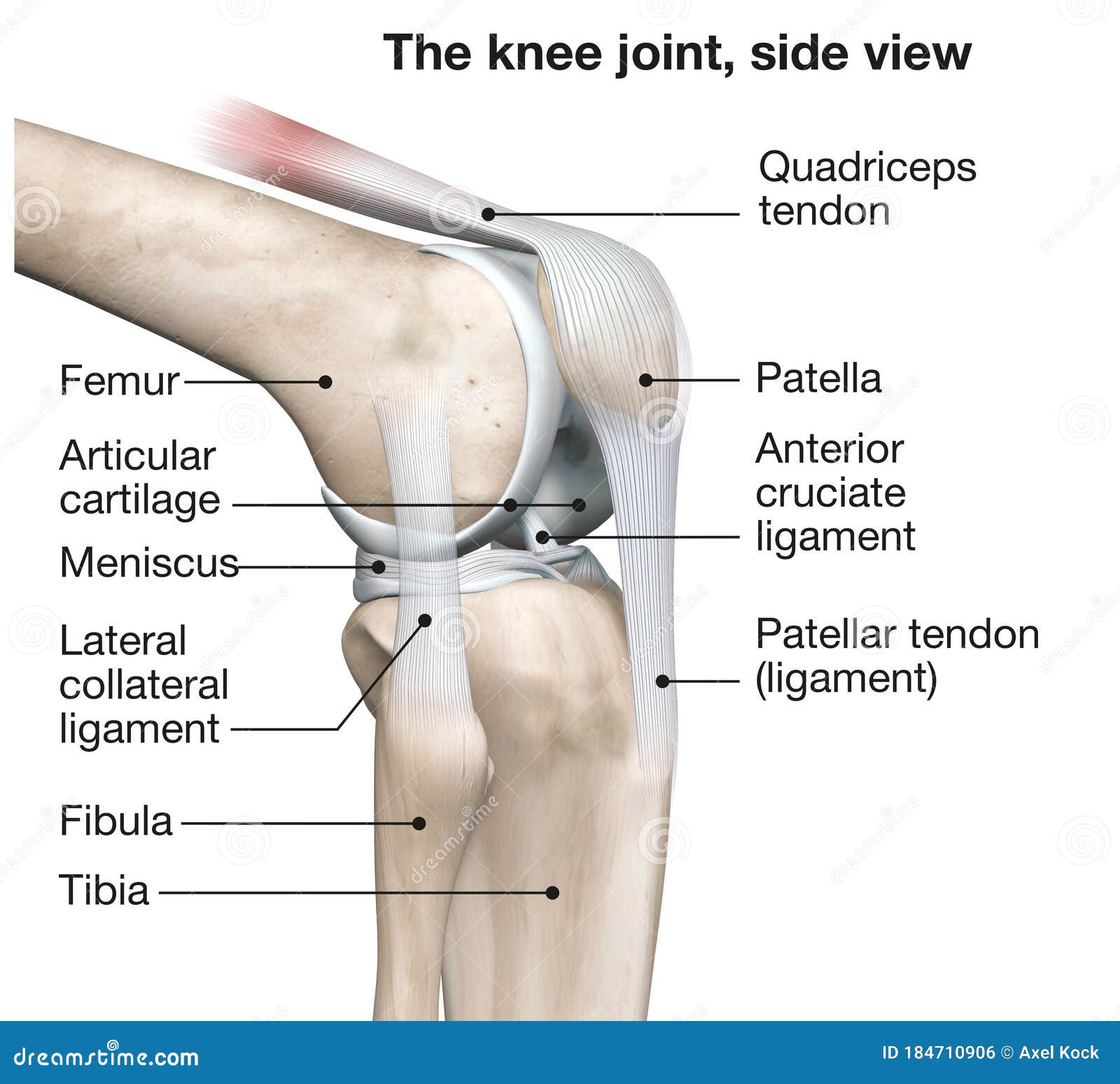
A Labeled Diagram of the Knee With an Insight into Its ... The given diagram of the knee joint can help you to understand its various parts and the description given below will give you an insight of the functioning of the knee. ⚫ Bone There are three bones in the knee namely the femur which is the thigh bone, tibia which is the shin bone and patella which is the knee cap.

Meniscus diagram of the knee
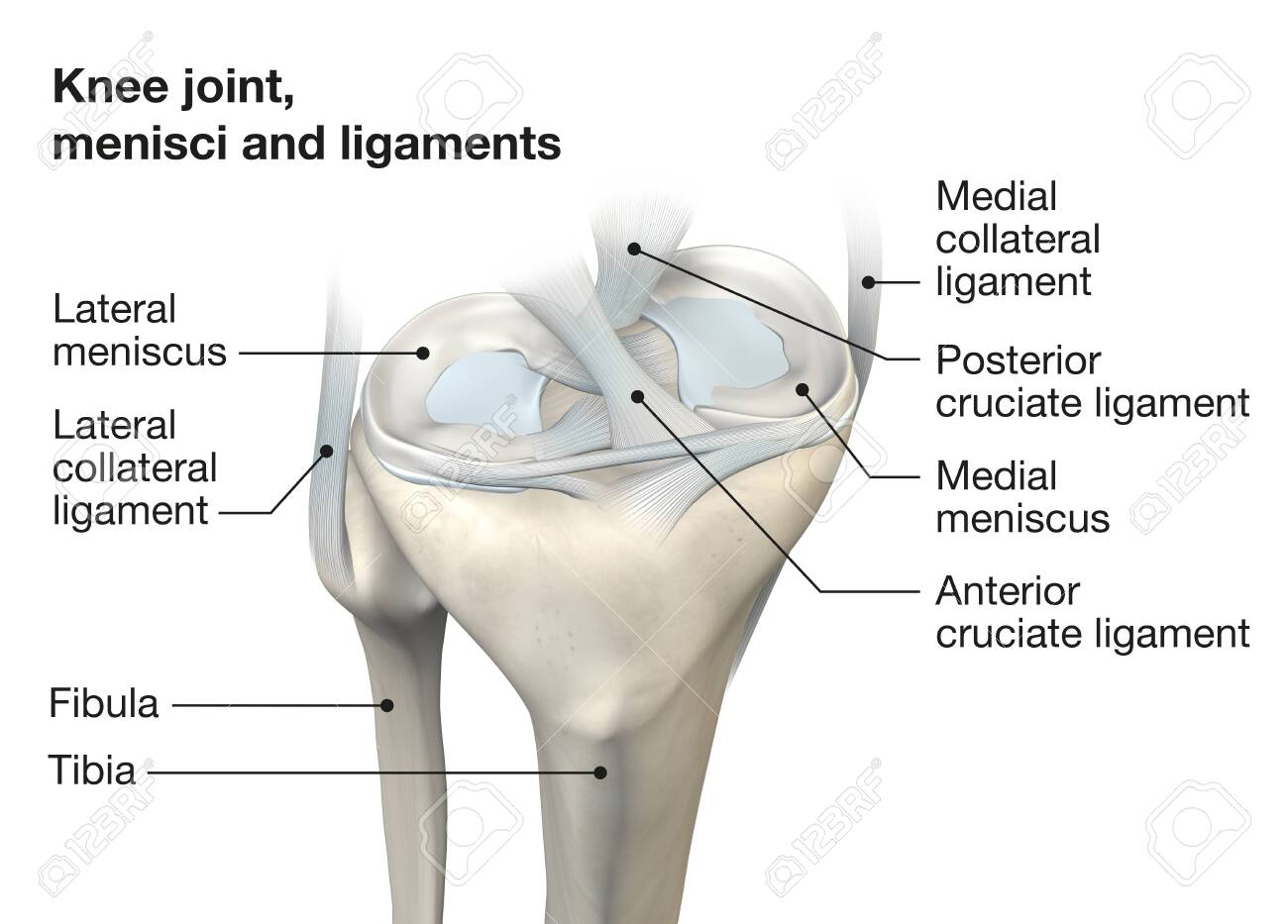
Torn Meniscus Picture Image on MedicineNet.com The medial and lateral meniscus are two thicker wedge-shaped pads of cartilage attached to the leg bone (tibia). Each meniscus is curved in a C-shape, with the front part of the cartilage called the anterior horn and the back part called the posterior horn. If the meniscus is damaged, irritation occurs with each flexion or extension of the knee. Knee Anatomy, Diagram & Pictures | Body Maps Two concave pads of cartilage (strong, flexible tissue) called menisci minimize the friction created at the meeting of the ends of the tibia and femur. There are also several key ligaments, a type... PDF The torn meniscus: Treatment options The meniscus is a small C-shaped piece of tissue in the knee, generally referred to as 'the cartilage,' that lies between your thigh bone (the femur) and your shin bone (the tibia). It acts as a shock absorber within the knee when walking, running and bending. Each knee has an inner (medial) and outer (lateral) meniscus which can tear.
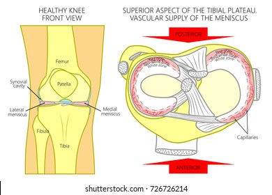
Meniscus diagram of the knee. The Basic Science of Human Knee Menisci Free body diagram of forces acting on the meniscus during loading. As the femur presses down on the meniscus during normal loading, ... changes in kinematics due to medial meniscectomy in the ACL-deficient knee confirm the important role of the medial meniscus in knee stability. Recently, Musahl et al reported that the lateral meniscus plays a role in anterior tibial … Knee Joint Anatomy: Structure, Function & Injuries - Knee ... The knee meniscus is particularly important as it acts as a shock absorber to reduce the forces going through the bones and reduces friction, allowing the bones to move smoothly. The back of the patella is also lined with cartilage, the thickest in the whole body due to the immense forces that go through the kneecap. Bones of the Leg and Foot | Interactive Anatomy Guide 16.07.2019 · The knee is a strong but flexible hinge joint that uses muscles and ligaments to withstand the torques and strains of powerful leg movements. Between the femur and tibia is the meniscus, a layer of tough fibrocartilage that acts as a shock absorber. In the lower leg, the tibia bears most of the body’s weight while the fibula supports the muscles of balance in the lower … Knee Meniscus: Function & Injuries - Knee Pain Explained The knee meniscus is a special layer of cartilage that lines the knee joint. The job of the meniscus is to cushion the knee joint and transfer forces between the tibia and femur, the thigh and shin bones. Most of the joints in our body are lined with a thin layer of articular cartilage, made of collagen and chondroitin. This provides a smooth ...
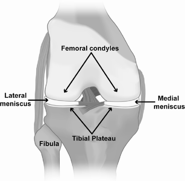
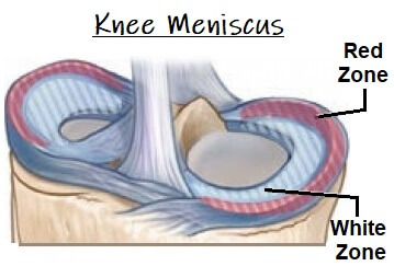
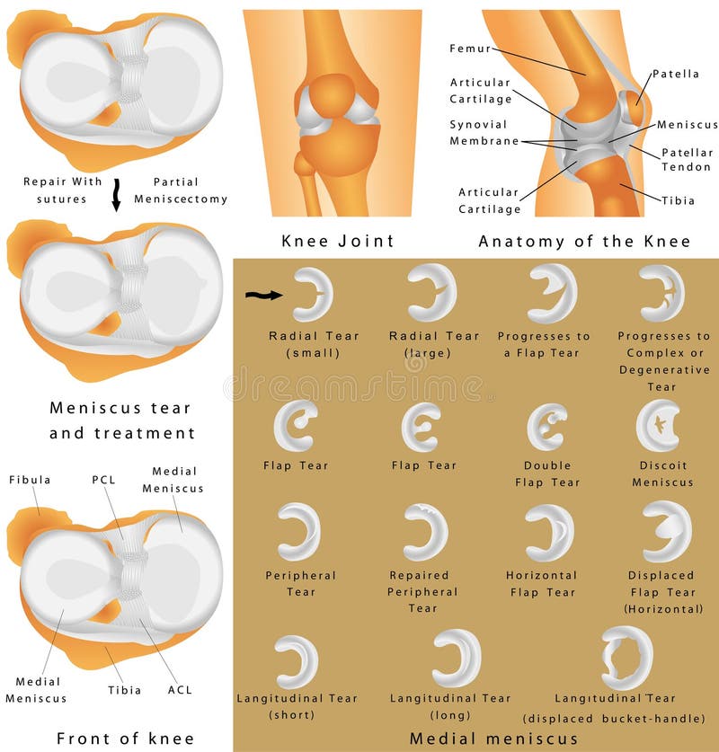
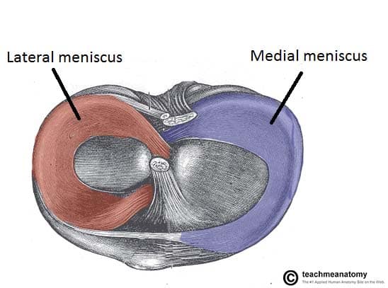
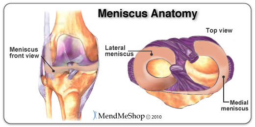
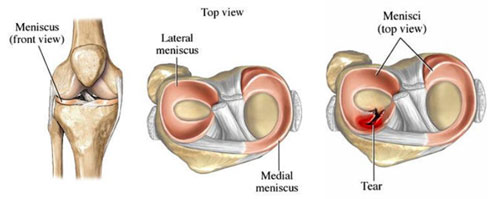
Four-Bar Linkages - University of Illinois Urbana-Champaign The knee connects the femur (the upper leg bone) to the tibia (the larger of the two lower leg bones). These two bones sit next to each other and are free to rotate about a single axis. A mechanism is needed to keep the two legs bones attached to each other, while still allowing rotation. In the case of the human knee this is achieved with a four-bar linkage consisting of … Meniscus anatomy diagram We are pleased to provide you with the picture named Meniscus anatomy diagram.We hope this picture Meniscus anatomy diagram can help you study and research. for more anatomy content please follow us and visit our website: . Anatomynote.com found Meniscus anatomy diagram from plenty of anatomical pictures on the internet.We think this is the most useful anatomy picture that ... What Is a Torn Meniscus in the Knee & How Do I Treat It? The diagram above is a cross section of the knee from the viewpoint looking down at the top of the shin bone (tibia). As you can see, the lateral meniscus on the outside of your knee is C shaped and thicker on the outer rim. The medial meniscus on the inside of your knee is also thicker on the outer rim and has a more oblong shape. 6 Types of Meniscus Tears and Locations - Verywell Health Meniscus tears are injuries that occur in the cartilage of the knee. Sometimes these tears require surgical repair. However, whether they will respond well to surgery depends on the type of tear, the location, and blood flow in the area where the tear occurred.
Schatzker Classification of Tibial Plateau Fractures: Use ... 01.03.2009 · Type IIIA fracture in a 55-year-old woman who fell on ice and injured her knee. (a) Diagram shows a Schatzker type IIIA fracture. (b) Plain radiograph shows depression of the lateral tibial plateau. (c) Coronal CT image shows the lateral tibial plateau depression. The fracture was managed nonoperatively with no weight bearing for 12 weeks. Medial Meniscus Anatomy, Function & Diagram | Body Maps Jan 20, 2018 · Medial meniscus. The medial meniscus is the central band of cartilage attached to the tibia, or shinbone. The band goes around the knee joint in a crescent-shaped path and is located between the ... Anatomy Of The Meniscus - YouTube Dr. Ebraheim's educational animated video describes the Anatomy of the Meniscus.The meniscus is a cushion structure made of cartilage which fits within the k... Osteochondral Lesions of the Knee: Differentiating the ... 17.08.2018 · Several pathologic conditions may manifest as an osteochondral lesion of the knee that consists of a localized abnormality involving subchondral marrow, subchondral bone, and articular cartilage. Although understanding of these conditions has evolved substantially with the use of high-spatial-resolution MRI and histologic correlation, it is impeded by inconsistent …
meniscus - Illustrations et vecteurs libres de droits - Stocklib
Meniscal Tears (Knee Cartilage Injuries) - Patient The areas of articular cartilage can be seen in the side view of the knee joint in the diagram above. How do you tear your meniscal cartilage? The knee is commonly injured in sports, especially rugby, football and skiing. You may tear a meniscus by a forceful knee movement whilst you are weight bearing on the same leg.

The knee – everything about the anatomy of the knee joint | medi
Knee brace calculator - echte-freude-schenken.de 11.03.2022 · Knee brace calculator. email protected] [email protected] [email protected]

Knee anatomy including ligaments, cartilage and meniscus
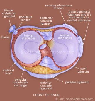
The knee meniscus: structure-function, pathophysiology ... 2.1 Meniscus Anatomy. The knee joint contains the meniscus structure, comprised of both a medial and a lateral component situated between the corresponding femoral condyle and tibial plateau (Figure 1) [].Each is a glossy-white, complex tissue comprised of cells, specialized extracellular matrix (ECM) molecules, and region-specific innervation and vascularization.

Meniscus (anatomy) - Wikipedia
Meniscal tear types diagram | Radiology Case | Radiopaedia.org knee, meniscus, tear, trauma, medial meniscus, lateral meniscus, bucket handle, parrot beak, illustration ... Full screen case with hidden diagnosis + add to new playlist; From the case: Meniscal tear types diagram. Diagram. Loading images... A variety of meniscal tears are illustrated. 1 article features images from this case . Meniscal tear ...

The knee meniscus: Structure–function, pathophysiology ...
Knee (Human Anatomy): Function, Parts, Conditions, Treatments Any form of arthritis or injury may cause a knee effusion. Meniscal tear: Damage to a meniscus, the cartilage that cushions the knee, often occurs with twisting the knee. Large tears may cause the ...
https://cdn-prod.medicalnewstoday.com/content/images/articles/299/299204/human-knee-diagram.jpg
Frozen Shoulder / Adhesive Capsulitis The meniscus is supposed to be smooth to ensure good gliding of the knee when it is bending. With injuries, poor alignment or weak musculature, the meniscus can become bruised and even torn. The outside edges of the meniscus have more blood flow than the inner portions. This means, depending on the area were the damage is located the healing process can be slow.
Common Knee Injuries - OrthoInfo - AAOS
Torn Meniscus | Johns Hopkins Medicine Symptoms of a meniscus tear may be different for each person, but some of the most common symptoms are: Pain in the knee joint: usually on the inside (medial), outside (lateral) or back of the knee. Swelling. Catching or locking of the knee joint. Inability to fully extend or bend the knee joint.

Torn Meniscus – Anatomy and Causes (Video) - Town Center ...
Meniscus (anatomy) - Wikipedia A meniscus is a crescent-shaped fibrocartilaginous anatomical structure that, in contrast to an articular disc, only partly divides a joint cavity. In humans they are present in the knee, wrist, acromioclavicular, sternoclavicular, and temporomandibular joints; in other animals they may be present in other joints.. Generally, the term "meniscus" is used to refer to the cartilage of the knee ...
Meniscus Images, Stock Photos & Vectors | Shutterstock
Knee Pain Location Chart - The Chelsea Knee Clinic Dec 31, 2019 · Knee pain could be the result of a problem with any one of these components, or a combination of several. You may be experiencing knee pain and want to know the possible causes. The diagram, below, is a handy guide to the possible reasons for your pain. Pain at the front above the knee

Knee and meniscus anatomy medical vector illustration ...
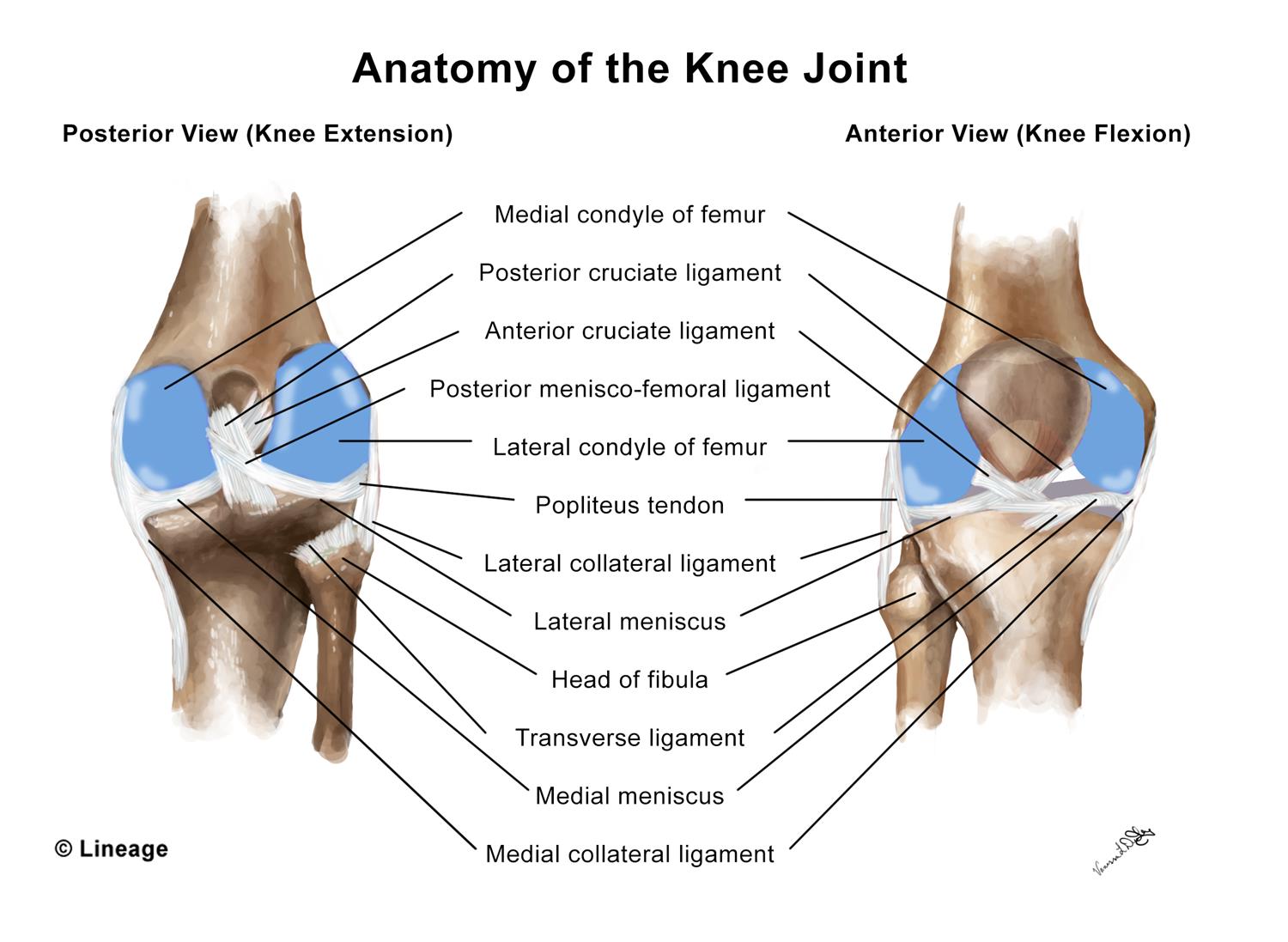
Knee joint: anatomy, ligaments and movements | Kenhub Knee joint (Articulatio genu) The knee joint is a synovial joint that connects three bones; the femur, tibia and patella.It is a complex hinge joint composed of two articulations; the tibiofemoral joint and patellofemoral joint.The tibiofemoral joint is an articulation between the tibia and the femur, while the patellofemoral joint is an articulation between the patella and the femur.

202 Meniscus Illustrations - Getty Images
Knee anatomy including ligaments, cartilage and meniscus There are two meniscal cartilages in the knee that act as shock-absorbers - one on the inner and one on the outer side. They sit between the curved lower part of the thigh bone and the flat upper part of the shin bone. Their job is to evenly distribute the load from the thigh bone to shin bone when walking and to provide knee stability.
The Knee Meniscus: A Complex Tissue of Diverse Cells ...
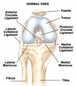
Knee Ligament Injuries - Patient 12.12.2017 · Each knee joint also contains an inner and outer meniscus (a medial and lateral meniscus). The menisci (plural of meniscus) are thick rubbery pads of cartilage tissue. They are C-shaped and become thinner towards the middle of the joint. The meniscal cartilages sit on top of, and are in addition to, the usual thin layer of articular cartilage which covers the top of the …
Knee Meniscus: Function & Injuries - Knee Pain Explained
What Is A Medial Meniscus Tear? — Dr. Bill Sterett And once the medial meniscus is gone, we face a much higher chance of painful arthritis. Lateral Meniscus Tears and Medial Meniscus Tears. A tear to the medial meniscus is the more common injury. The lateral meniscus is on the outside of the knee, while the medial meniscus is on the inside of the knee. Reference the diagram below for a visual ...

Alila Medical Media | Knee meniscus labeled diagram ...
Picture of Medical Anatomy and Illustrations - Torn Meniscus The knee is the joint where the large bones of the upper leg (femur), lower leg (tibia), and kneecap ( patella) meet. Two pieces of "C-shaped" cartilage called the meniscus cushion the ends of the femur and tibia at the knee joint. A tear in this cartilage is called a meniscal tear. Meniscal tears have different names - longitudinal, flap ...

What are the Parts of the Knee Joint? | Systems4Knees™
Organs in the body: Diagram and all you need to know 22.05.2020 · What to know about meniscus injury types Medically reviewed by Angela M. Bell, MD, FACP A meniscus injury is a tear in one of the crescent-shaped pads of cartilage inside each knee joint.
Discoid Meniscus - OrthoInfo - AAOS
What is a Medial Meniscus Tear? (with pictures) A diagram of the knee, showing the medial meniscus. The bones, ligaments, and muscles that meet in the knee joint are protected by a layer of cartilage tissue called the meniscus. The medial meniscus is the section that is deep within the joint, aiding in the flexibility of major ligaments.

Understanding Meniscus Tears
The Knee Joint - Articulations - Movements - Injuries ... 07.02.2022 · The knee joint is a hinge type synovial joint, which mainly allows for flexion and extension (and a small degree of medial and lateral rotation). It is formed by articulations between the patella, femur and tibia. In this article, we shall examine the anatomy of the knee joint – its articulating surfaces, ligaments and neurovascular supply.

Schematic illustrations of the medial meniscus under slight ...
Knee Anatomy Diagram Meniscus - links to knee health sites ... Here are a number of highest rated Knee Anatomy Diagram Meniscus pictures upon internet. We identified it from honorable source. Its submitted by government in the best field. We take on this nice of Knee Anatomy Diagram Meniscus graphic could possibly be the most trending subject when we allocation it in google improvement or facebook.

Knee Anatomy : 2 : Meniscus
Torn meniscus - Symptoms and causes - Mayo Clinic A torn meniscus is one of the most common knee injuries. Any activity that causes you to forcefully twist or rotate your knee, especially when putting your full weight on it, can lead to a torn meniscus. Each of your knees has two C-shaped pieces of cartilage that act like a cushion between your shinbone and your thighbone. A torn meniscus ...

Meniscal Tears, Knee Cartilage Deterioration and Treatment
Meniscus _Tear types stock vector. Illustration of diagram ... Meniscus _Tear types. Royalty-Free Vector. Download preview. Vector illustration. Anatomy of a meniscus in the healthy human knee joint. Types of meniscal tear with cross section of the menisci. For advertising, medical publications. EPS 10. knee replacement,
Anatomy of the Knee stock vector. Illustration of posterior ...
Posterior Horn Medial Meniscus Tear | Knee Specialist ... Posterior Horn of the Medial Meniscus Injury FAQ. The medial meniscus is the cushion that is located on the inside part of the knee. It is generally divided into 3 separate portions, the anterior horn, the mid-body and the posterior horn. The posterior horn is the thickest and most important for overall function of the knee.
The Knee Joint - Articulations - Movements - Injuries ...
PDF The torn meniscus: Treatment options The meniscus is a small C-shaped piece of tissue in the knee, generally referred to as 'the cartilage,' that lies between your thigh bone (the femur) and your shin bone (the tibia). It acts as a shock absorber within the knee when walking, running and bending. Each knee has an inner (medial) and outer (lateral) meniscus which can tear.

NHS Ayrshire & Arran - Knee: Acute Meniscal Tears - Traumatic ...
Knee Anatomy, Diagram & Pictures | Body Maps Two concave pads of cartilage (strong, flexible tissue) called menisci minimize the friction created at the meeting of the ends of the tibia and femur. There are also several key ligaments, a type...

Anatomy Of The Meniscus - Everything You Need To Know - Dr ...
Torn Meniscus Picture Image on MedicineNet.com The medial and lateral meniscus are two thicker wedge-shaped pads of cartilage attached to the leg bone (tibia). Each meniscus is curved in a C-shape, with the front part of the cartilage called the anterior horn and the back part called the posterior horn. If the meniscus is damaged, irritation occurs with each flexion or extension of the knee.
Knee Arthroscopy - Dr Peter Walker

Medial Meniscus Tear Project SPTA
Torn Meniscus | Cedars-Sinai

Menisci of the knee and common tear patterns. | Download ...
Knee Meniscus Anatomy
Lateral meniscus - Physiopedia

Meniscus Anatomy

What Is a Torn Meniscus in the Knee & How Do I Treat It?

Meniscus Repair vs. Removal — The Knee and Shoulder Centers

Meniscus location and anatomy - www.anatomynote.com | Human ...
Knee Joint Anatomy, Side View, Medical 3D Illustration Stock ...
3D Illustration Showing Knee Joint With Transparent Femur And ...
Meniscus Tear - Orthopedics - Medbullets Step 2/3
Meniscal Tears | JointSurgery.in
0 Response to "38 meniscus diagram of the knee"
Post a Comment