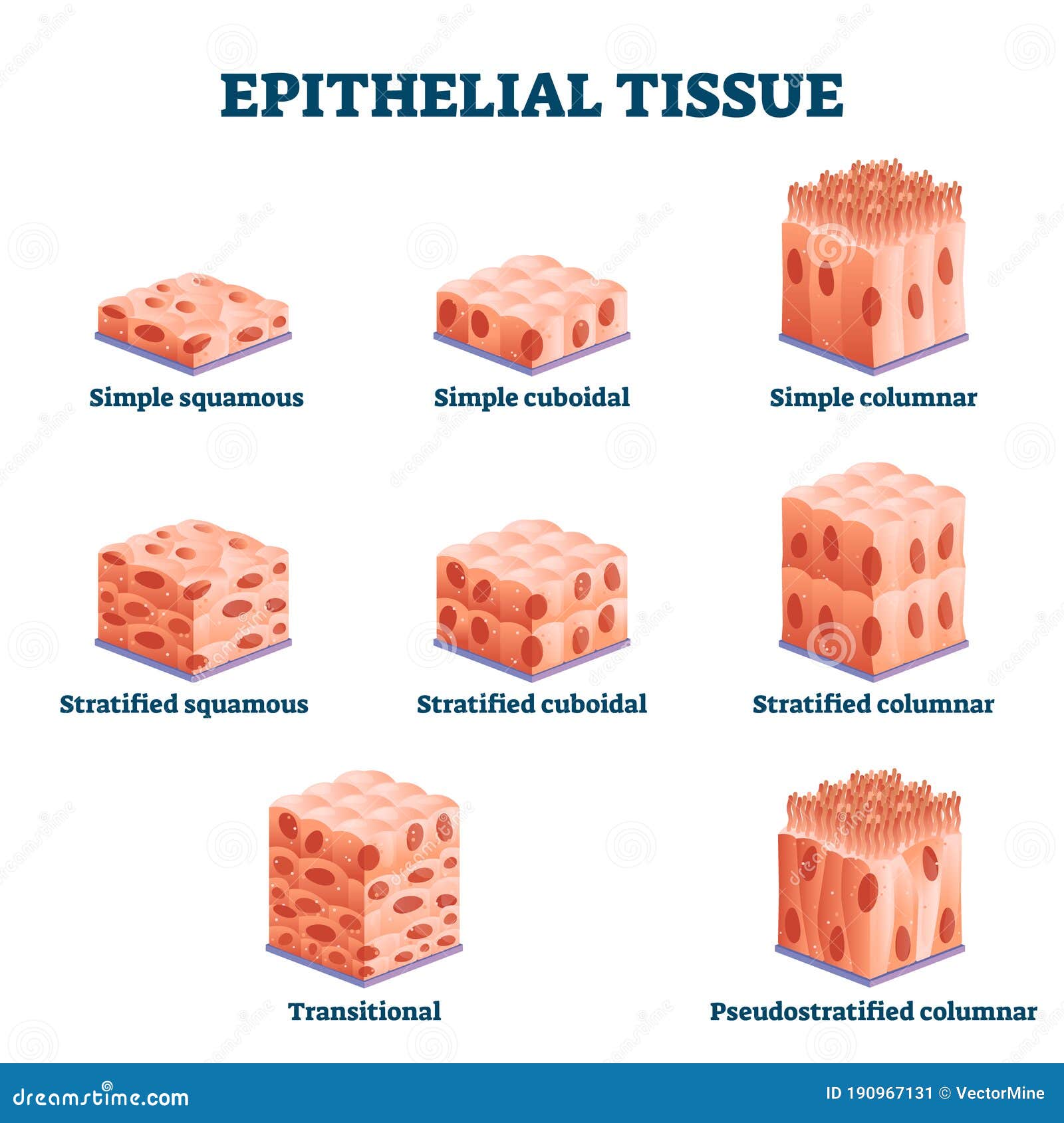
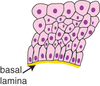
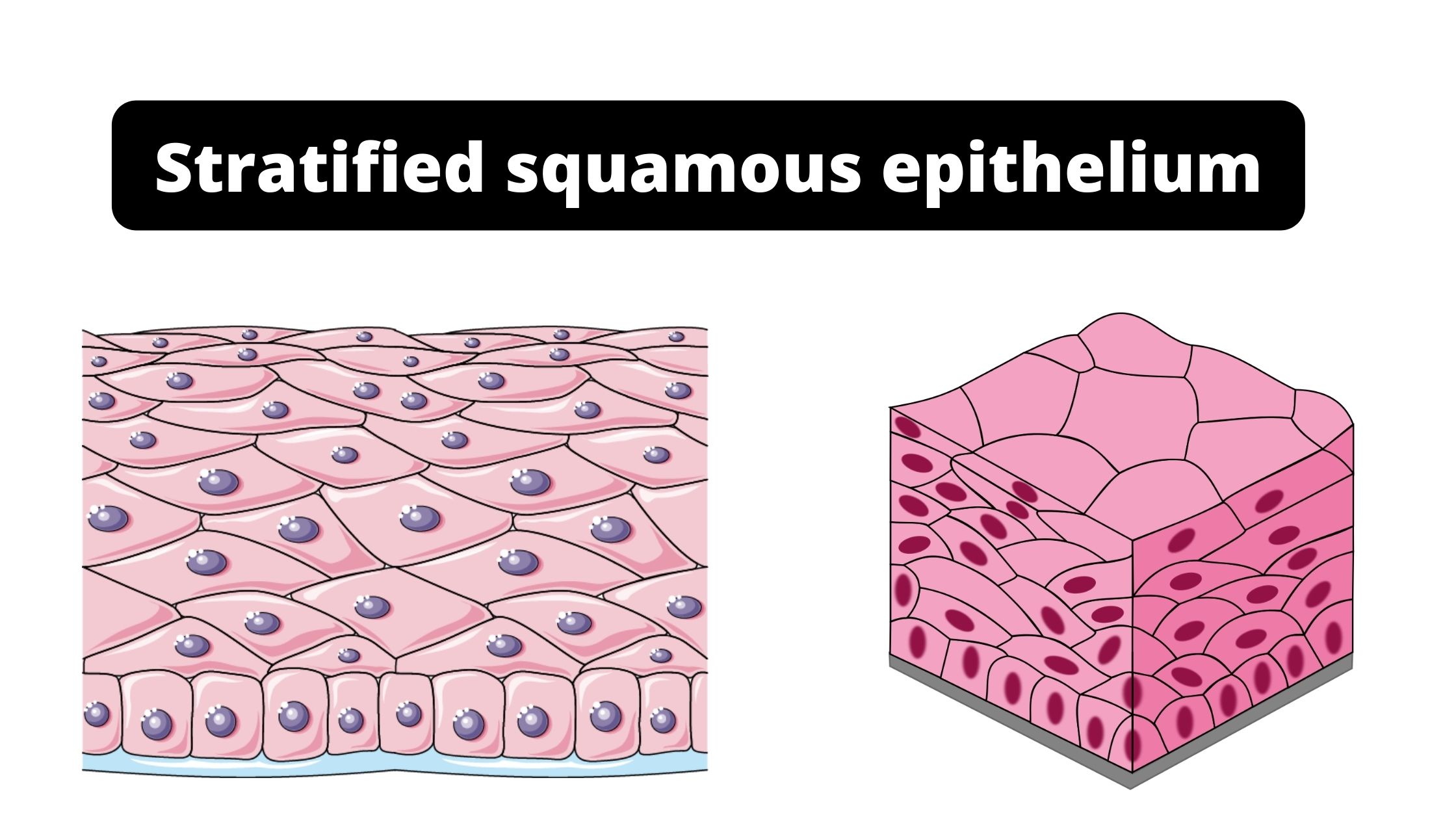
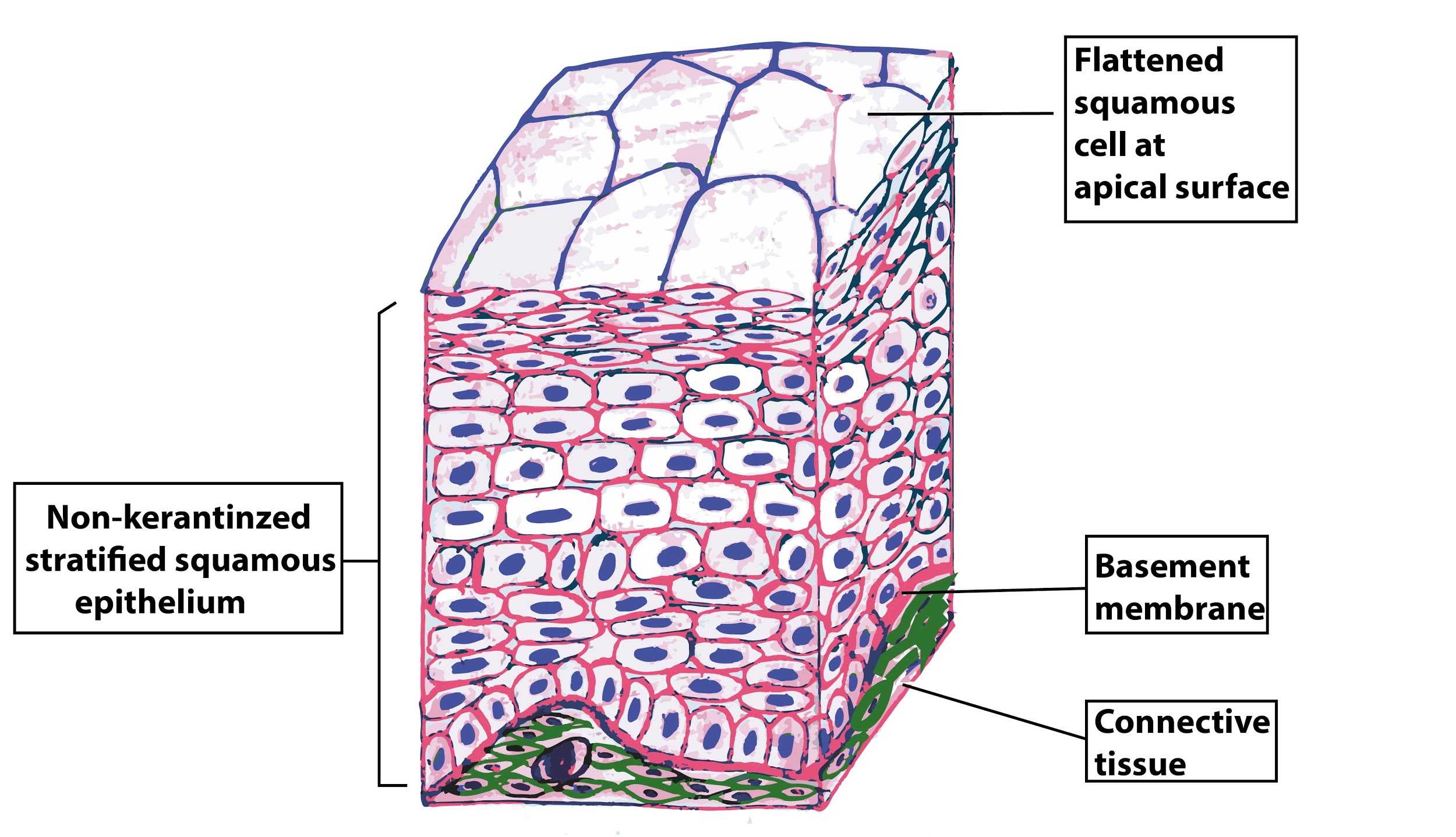
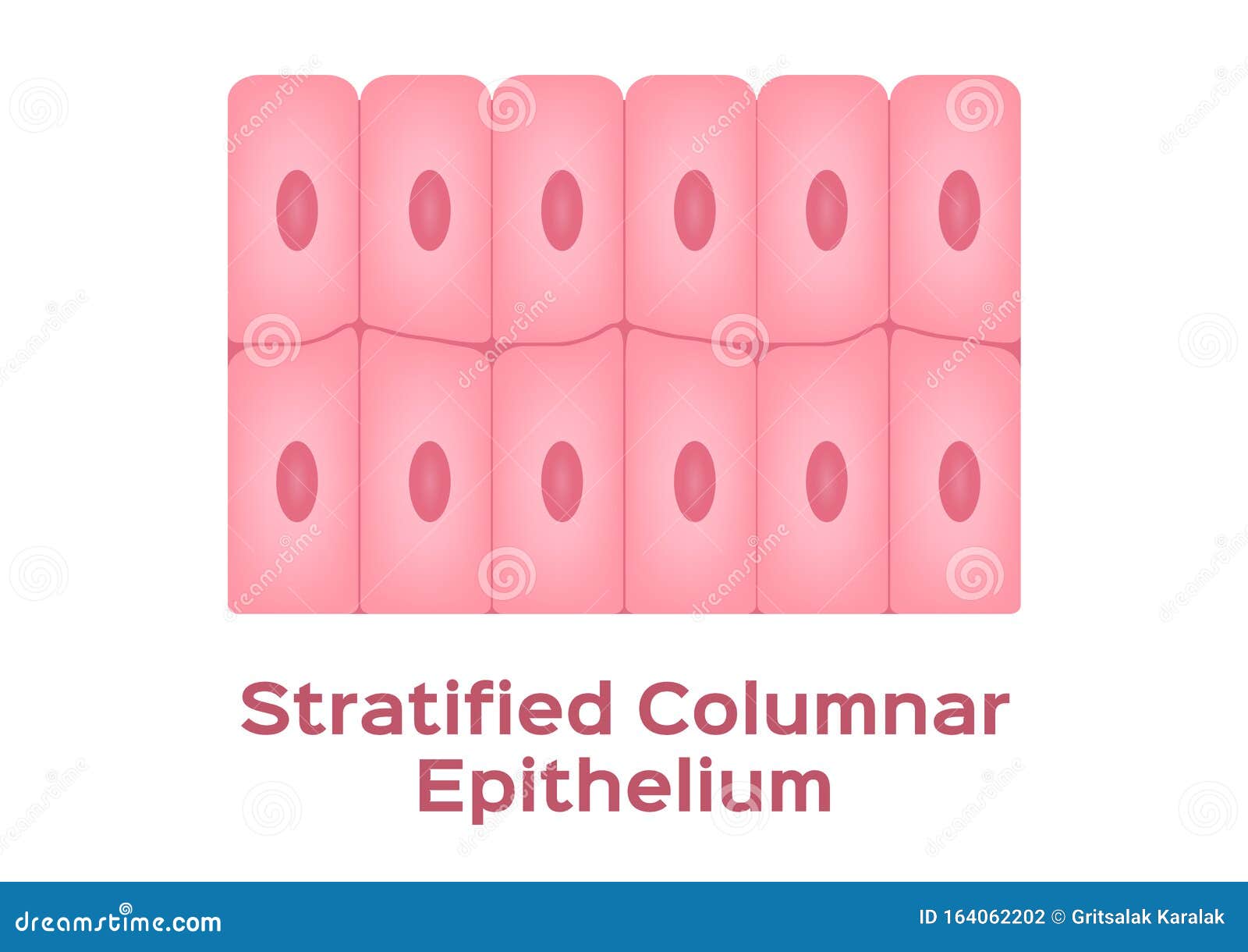
45 stratified columnar epithelium diagram
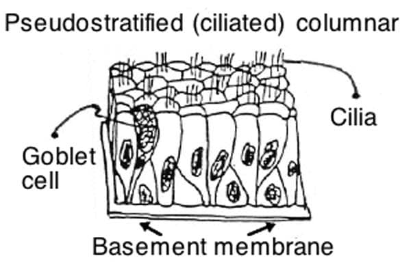
Connective Tissue and Quiz 1 | histology - University of Michigan Slide 40 is also a very good specimen to examine the pseudostratified, ciliated columnar epithelium of the trachea. Note also that thebasement membrane underlying this particular epithelium is especially prominent. Type IV collagen, which does not form fibrils, but rather a fine meshwork, is present in all basement membranes. Epithelial Tissue and Mammary Gland | histology A. Simple columnar epithelium. Slide 29 (small intestine) View Virtual Slide Slide 176 40x (colon, H&E) View Virtual Slide Remember that epithelia line or cover surfaces. In slide 29 and slide 176, this type of epithelium lines the luminal (mucosal) surface of the small and large intestines, respectivel
Simple Squamous Epithelium under a Microscope with a Labeled ... Mar 04, 2022 · Pseudostratified columnar epithelium under a microscope This is not a true stratified epithelium but appears to be stratified. You know the nuclei of the columnar epithelium lie in a row toward the basal part of the cell. But, in the pseudostratified columnar epithelium, the nuclei appear to be arranged into two or more layers.

Stratified columnar epithelium diagram
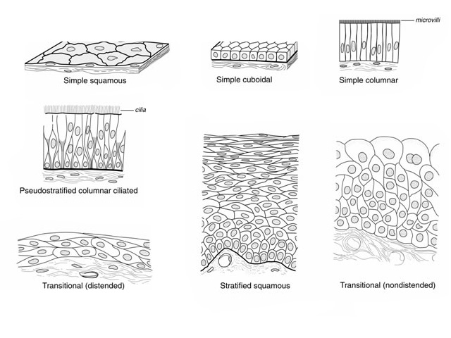
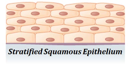
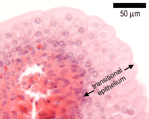
Transitional epithelium - Wikipedia Transitional epithelium also known as urothelium is a type of stratified epithelium. Transitional epithelium is a type of tissue that changes shape in response to stretching (stretchable epithelium). The transitional epithelium usually appears cuboidal when relaxed and squamous when stretched. [1] Epithelial Tissue: Structure with Diagram, Function, Types ... Simple Epithelium- it is composed of one layer of a cell and mostly has a secretory or an absorptive function. Compound (Stratified) Epithelium- it is made up of two or more than two layers of cells and mostly has a protective function. The glandular epithelium is made up of cuboidal or columnar cells. They are specialised for secretion. Respiratory System – Building a Medical Terminology Foundation These bronchi are also lined by pseudostratified ciliated columnar epithelium containing mucus-producing goblet cells (Figure 7.7b). The carina is a raised structure that contains specialized nervous tissue that induces violent coughing if a foreign body, such as food, is present.
Stratified columnar epithelium diagram. Pseudostratified Columnar Epithelium under a Microscope with ... Jun 04, 2022 · The pseudostratified columnar epithelium comprises a single layer of cells but seems to be multilayered. It is because different cellular heights and nuclei are also placed at a different levels. I will show you the pseudostratified columnar epithelium under a light microscope with its identifying points and labeled diagram. Respiratory System – Building a Medical Terminology Foundation These bronchi are also lined by pseudostratified ciliated columnar epithelium containing mucus-producing goblet cells (Figure 7.7b). The carina is a raised structure that contains specialized nervous tissue that induces violent coughing if a foreign body, such as food, is present. Epithelial Tissue: Structure with Diagram, Function, Types ... Simple Epithelium- it is composed of one layer of a cell and mostly has a secretory or an absorptive function. Compound (Stratified) Epithelium- it is made up of two or more than two layers of cells and mostly has a protective function. The glandular epithelium is made up of cuboidal or columnar cells. They are specialised for secretion. Transitional epithelium - Wikipedia Transitional epithelium also known as urothelium is a type of stratified epithelium. Transitional epithelium is a type of tissue that changes shape in response to stretching (stretchable epithelium). The transitional epithelium usually appears cuboidal when relaxed and squamous when stretched. [1]















:watermark(/images/watermark_only_sm.png,0,0,0):watermark(/images/logo_url_sm.png,-10,-10,0):format(jpeg)/images/anatomy_term/simple-cuboidal-epithelium-4/lihfPw1Qnz07Be1pX00Bg_2Simple_cuboidal_epithelium.png)


0 Response to "45 stratified columnar epithelium diagram"
Post a Comment