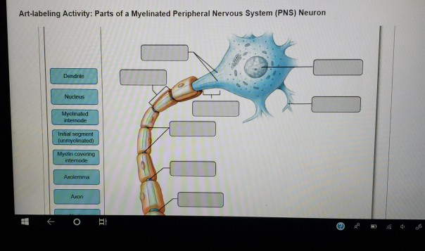
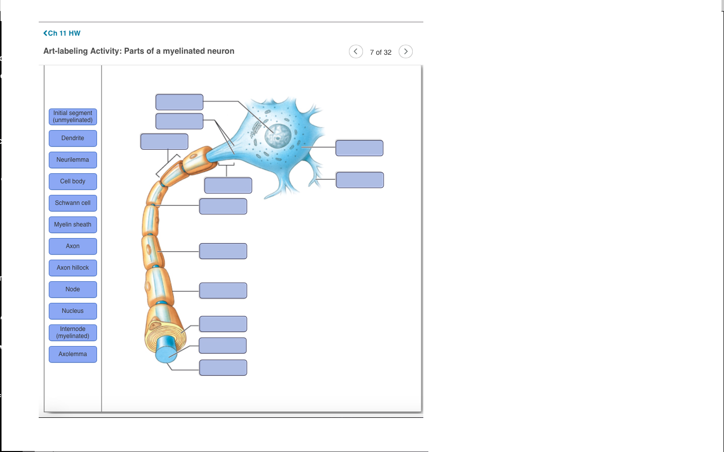
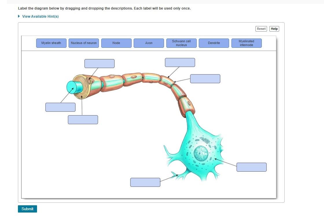
44 drag the labels onto the diagram to identify the parts of a myelinated pns neuron.
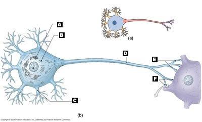
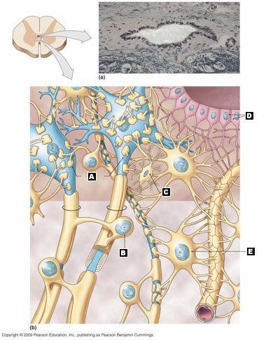
A Labelled Diagram Of Neuron with Detailed Explanations - BYJUS Diagram Of Neuron Diagram Of Neuron A neuron is a specialized cell, primarily involved in transmitting information through electrical and chemical signals. They are found in the brain, spinal cord and the peripheral nerves. A neuron is also known as the nerve cell. HW 7.pdf - HW 7 Due: 11:59pm on Friday, October 27, 2017 To... Part A Drag the labels onto the diagram to identify the parts of a myelinated PNS neuron. ANSWER: Correct Art-labeling Activity: Types of Neuroglia in the Central and Peripheral Nervous Systems Learning Goal: To learn the various types of neuroglia in the central and peripheral nervous systems.
A&P2 Lab and Powerpoints Flashcards | Quizlet Identify the pancreatic structure labeled "I". [Be prepared to identify all labeled pancreatic structures on upcoming exams] islet of Langerhans Name two hormones produced by pancreatic islets that play a critical role in blood glucose homeostasis. insulin and glucagon

Drag the labels onto the diagram to identify the parts of a myelinated pns neuron.
Part a drag the labels onto the diagram to identify - Course Hero See Page 1. Part A Drag the labels onto the diagram to identify the structures of the neuron. ANSWER:cutaneous membrane.serous membrane.mucous membrane.synovial membrane.peritoneal membrane.fibrocartilaginous membranes serous membranes cutaneous membranes mucous membranes synovial membranes. Drag The Labels To Identify Structural Components Of The Heart It Is The Strongest Type Of Cartilage Drag The Labels Onto The Diagram To Identify The Parts Of A Myelinated Pns Neuron Identify 4 The Following Structures… Anatomy, Head And Neck, Skull. Four chambers of the heart and blood circulation. Selecting or hovering over a box will highlight each area in the diagram. A&P2 Lab 13 HW, A&P2 Lab 12 HW, A&P2 Lab 11 HW, A&P2 Lab 10 ... - Quizlet the structure labeled A Identify the structure at the end of the arrow. prostate gland During filtration, anything that is small enough to pass through all three layers of the filtration membrane will become part of the filtrate. Sometimes, the least porous layer of this membrane becomes clogged and then glomerulonephritis may occur.
Drag the labels onto the diagram to identify the parts of a myelinated pns neuron.. Drag The Labels Onto The Diagram To Identify The Structures And ... This diagram here just shows the joint capsule itself. Compact bone drag and drop the labels to the appropriate spots on the knee joint model. Drag the labels onto the diagram to identify the parts of the renal corpuscle. Teres major (movers of the shoulder joint) 3 name the structure and label and describe each number. Levels of organization ... Drag The Labels Onto The Diagram To Identify The Structures And ... / Part A Drag the labels onto the diagram to identify the ... - How would you label the x and y axes?. Drag the labels onto the diagram to the stadium wave climate etc. Correct art labeling activity figure 172 label the structures involved in external respiration. Two pairs of vocal folds are found in the la. Animal Cell Diagram Quizlet : Drag The Labels Onto The Diagram To ... Animal Cell Diagram Quizlet : Drag The Labels Onto The Diagram To Identify The Parts Of ... - Animal cell parts match plant cell parts match..Animal cell anatomy diagram structure with all parts nucleus smooth rough endoplasmic reticulum cytoplasm golgi apparatus. A&P2 Lab 1 HW Flashcards | Quizlet Drag the labels onto the diagram to identify the parts of a myelinated PNS neuron. look at pic Drag the labels to identify the structural components of a typical synapse. look at pic Rabies illustrates a negative consequence to otherwise healthy retrograde flow within axons. Which of the following components will NOT be involved in retrograde flow?
Drag the labels onto the diagram to identify the structures. The gastrocnemius is located on the back of the leg. The rectus femoris is located on the front of the leg. The tensor fascia lata is located on the outer side of the leg. The soleus is located on the back of the leg. The fibularis longus is located on the outer side of the leg. The vastus lateralis is located on the outer side of the thigh. Labeled Neuron Diagram | Science Trends Neurons are a type of cell and are the fundamental constituents of the nervous system and brain. Neurons take in stimuli and convert them to electrical and chemical signals that are sent to our brain. There are 3 major kinds of neurons in the spinal cord: sensory, motor, and interneurons. Neurons communicate vie electrical signals produced by ... Drag The Labels Onto The Diagram To Identify The Structures And ... Drag the labels onto the diagram to the stadium wave climate etc. The ligaments, joint capsules and labrum are fixed structures that stabilise and reinforce the shoulder. Overview of neuron structure and function. Source: Identify, describe and state the functions of the glenoid labrum. NDSU Human Anat I- Exam 2 Flashcards | Quizlet Use the provided ions to correctly complete each sentence about the resting membrane potential. Ions may be used more than once, or not at all. Drag the labels to identify depolarization, repolarization, and hyperpolariztion. Drag the labels onto the diagram to identify the different types of gated ion channels.
Parts of a Myelinated Peripheral Nervous System (PNS) Neuron - Quizlet Parts of a Myelinated Peripheral Nervous System (PNS) Neuron + − Learn Test Match Created by GAChief Terms in this set (9) Axon Hillock ... Nucleus ... Dendrite ... Myelin covering internode ... Axolemma ... Axon ... Nodes ... Initial Segment (unmyelinated) ... Myelinated internode ... The Human Brain (Midsagittal Section) GAChief TEACHER alexak325 Drag the labels onto the diagram to identify the structures. Drag the labels onto the diagram to identify the structures. All tutors are evaluated by Course Hero as an expert in their subject area. This is a diagram showing the posterior muscles of a human. The glutes, hamstrings, calves, erector spinae, lats, and rear shoulder muscles make up the posterior chain muscles, which are located in the back of ... A&P2 Lab 13 HW, A&P2 Lab 12 HW, A&P2 Lab 11 HW, A&P2 Lab 10 ... - Quizlet the structure labeled A Identify the structure at the end of the arrow. prostate gland During filtration, anything that is small enough to pass through all three layers of the filtration membrane will become part of the filtrate. Sometimes, the least porous layer of this membrane becomes clogged and then glomerulonephritis may occur. Drag The Labels To Identify Structural Components Of The Heart It Is The Strongest Type Of Cartilage Drag The Labels Onto The Diagram To Identify The Parts Of A Myelinated Pns Neuron Identify 4 The Following Structures… Anatomy, Head And Neck, Skull. Four chambers of the heart and blood circulation. Selecting or hovering over a box will highlight each area in the diagram.
Part a drag the labels onto the diagram to identify - Course Hero See Page 1. Part A Drag the labels onto the diagram to identify the structures of the neuron. ANSWER:cutaneous membrane.serous membrane.mucous membrane.synovial membrane.peritoneal membrane.fibrocartilaginous membranes serous membranes cutaneous membranes mucous membranes synovial membranes.














0 Response to "44 drag the labels onto the diagram to identify the parts of a myelinated pns neuron."
Post a Comment