45 actin and myosin diagram
Describe the structure of myosin and actin filaments, with ... 6. In sarcomere, myosin tails are arranged to point towards the centre of the sarcomere and the heads point to the sides of the myofilament band. ii. Actin filament: It is a complex type of contractile protein. It is made up of three components: 1. F actin: It forms the backbone of actin filament. F actin is made up of two helical strands. Draw a neat labelled diagram of (a) An actin filament (b ... Draw a neat labelled diagram of (a) An actin filament (b) Myosin. 0. Myofibrils are made up of 1. Myosin and actin 2. Myosin and troponin 3.
Actin vs Myosin- Definition, 14 Major Differences, Examples Myosin is termed a motor protein as it is a type of enzyme that converts chemical energy into mechanical energy. Myosin is an ATPase that moves along actin filaments by connecting the hydrolysis of ATP to conformational changes. All myosins are composed of one or two heavy chains and several light chains.

Actin and myosin diagram
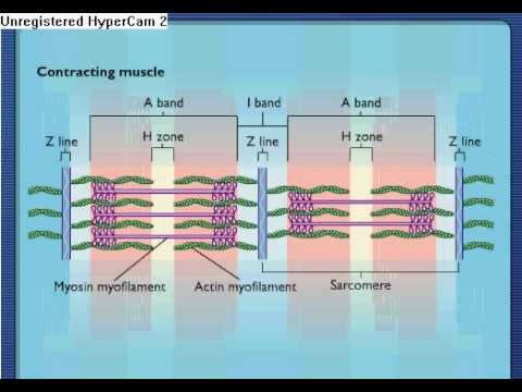
Labeled Sarcomere Diagram The thin filaments Look at the diagram above and realize what happens as a muscle contracts. As will soon be described, the functional unit of a skeletal muscle fiber is the sarcomere, a highly organized arrangement of the contractile myofilaments actin .Play this quiz called Label the Sarcomere and show off your skills. Myosin diagrams. (A) The classic myosin diagram pictures ... Myosin diagrams. (A) The classic myosin diagram pictures two equivalent heads and an a-helical coiled-coil rod domain. Proteolysis at two sites (arrowheads) fragments the molecule into two ... Muscle Contraction & Sliding Filament Theory - TeachPE.com There would be reduced muscle strength because few cross-bridges can form between the actin and myosin. Partially Contracted Muscle The diagram above shows a partially contracted muscle where there is more overlapping of the myosin and actin with lots of potential for cross bridges to form. The I - bands and H - zone is shortened.
Actin and myosin diagram. quizlet.com › 265729985 › mastering-ap-chapter-9Mastering A&P Chapter 9 - Muscle and Muscle Tissue Diagram ... Troponin and tropomyosin are attached to one another, both overlaying actin. When a muscle is relaxed, tropomyosin blocks actin's myosin-binding sites. Calcium binds to troponin, initiating a shape change that removes the blocking action of tropomyosin. This exposes the myosin-binding sites on actin to the myosin heads for cross bridging. Biology Unit 5 Chapter 11 - Muscle Contraction - Quizlet After death, cross bridges between actin and myosin remain firmly bound resulting in rigor mortis. Using information in the diagram, explain what causes the cross bridges to remain firmly bound. (2) no ATP produced; ATP required for separation of actin and myosin PDF The diagram shows part of a muscle myofibril - StudyWise (c) After death, cross bridges between actin and myosin remain firmly bound resulting in rigor mortis. Using information in the diagram, explain what causes the cross bridges to remain firmly bound. Respiration stops so no ATP is produced and ATP is needed for the separation of actin and myosin cross bridges 3. › myosin-atpaseMyosin ATPase - an overview | ScienceDirect Topics Myosin ATPase. The myosin ATPase cycle, partly regulated by intracellular free calcium (Ebashi and Endo, 1968), is therefore the fundamental unit of movement that generates local force (Tyska and Warshaw, 2002) and explains the reiterated interactions with actin during active motion.
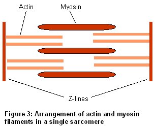
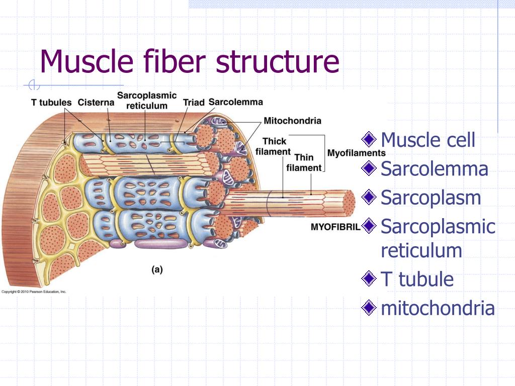
Difference Between Actin and Myosin | Definition ... Actin and myosin proteins form filaments arranged in the myofibrils in a longitudinal manner. The main difference between actin and myosin is that actin forms a thin filament whereas myosin forms a thick filament. The sliding over of the two filaments over one another in a series of repetitive events leads to the contraction of the muscles. Actin and Myosin - Biology Dictionary Myosin forms thick filaments (15 nm in diameter) and actin forms thinner filaments (7nm in diameter). Actin and myosin filaments work together to generate force. This force produces the muscle cell contractions that facilitate the movement of the muscles and, therefore, of body structures. The Structure of Muscles Schematic diagrams of the myosin heavy chains involved in ... Myosin Va (MyoVa) is an actin-based molecular motor that plays key roles in the final stages of secretory pathways, including neurotransmitter release. Several studies have addressed how MyoVa... 3.4: Actin and myosin filaments Diagram | Quizlet What is the structure of myosin and actin filaments? Fibrous molecules with quaternary structures Relaxed state No actin-myosin interaction at binding site Myofilaments overlap a little Contracted state Myosin head pulls actin toward sarcomere center (power stroke) Filaments slide past each other = sliding filament theory
byjus.com › difference-between-actin-and-myosinMajor Differences Between Actin and Myosin - BYJUS Actin and myosin are two protein molecules present in muscles and are mainly involved in the contraction of the muscle in both humans and animals. Both actin and myosin function by controlling the voluntary muscular movements within the body, along with the regulatory proteins known as troponin, tropomyosin and meromyosin. Muscle Contraction & Sliding Filament Theory - TeachPE.com There would be reduced muscle strength because few cross-bridges can form between the actin and myosin. Partially Contracted Muscle The diagram above shows a partially contracted muscle where there is more overlapping of the myosin and actin with lots of potential for cross bridges to form. The I - bands and H - zone is shortened. Myosin diagrams. (A) The classic myosin diagram pictures ... Myosin diagrams. (A) The classic myosin diagram pictures two equivalent heads and an a-helical coiled-coil rod domain. Proteolysis at two sites (arrowheads) fragments the molecule into two ... Labeled Sarcomere Diagram The thin filaments Look at the diagram above and realize what happens as a muscle contracts. As will soon be described, the functional unit of a skeletal muscle fiber is the sarcomere, a highly organized arrangement of the contractile myofilaments actin .Play this quiz called Label the Sarcomere and show off your skills.

Human Physiology Chapter 12 Flashcards | Easy Notecards
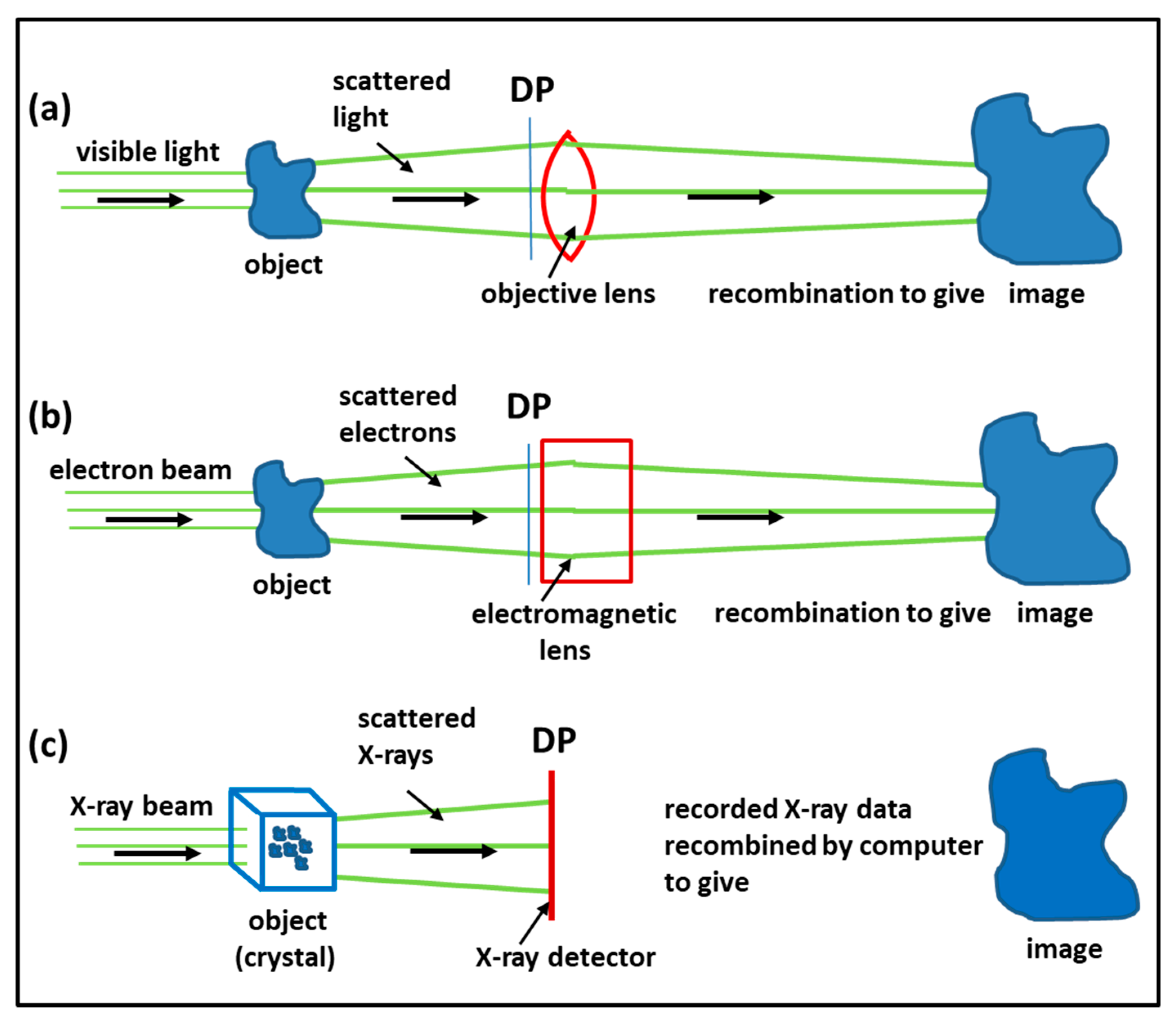
IJMS | Free Full-Text | Special Issue: The Actin-Myosin Interaction in Muscle: Background and ...
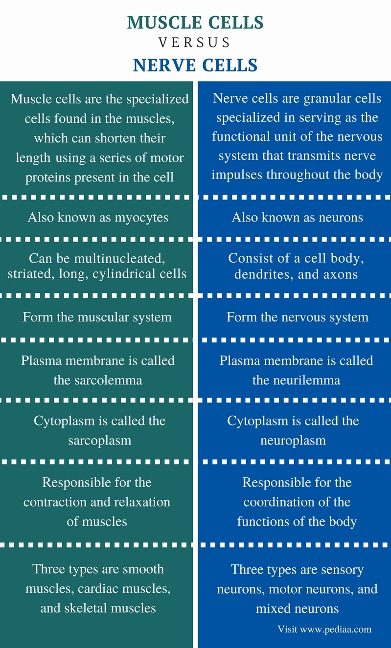
Difference Between Muscle Cells and Nerve Cells | Definition, Structure, Function and Differences

Anatomy/Muscular System - Wiki - Scioly.org
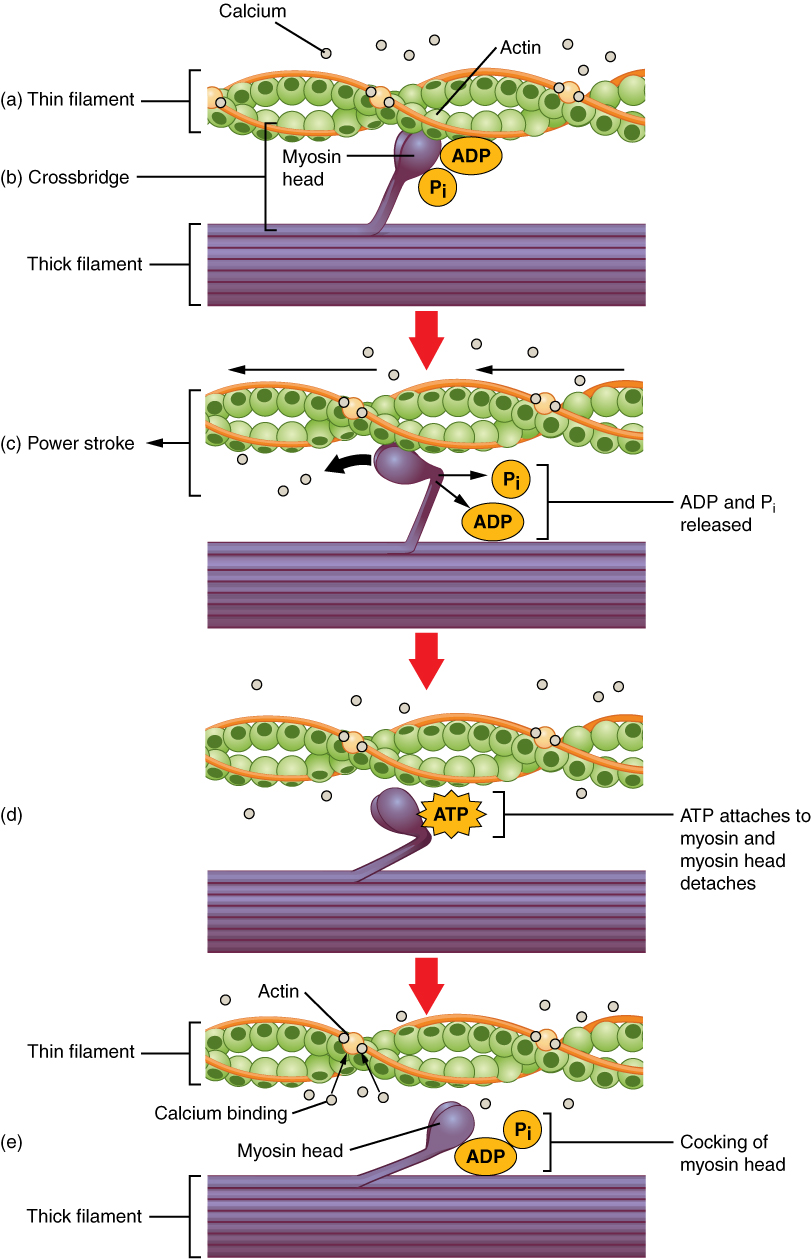
This multipart figure shows the mechanism of skeletal muscle contraction. In the top panel, the ...

Quia - AP Bio - Nervous System and Muscles
.PNG)
Actin and myosin - Presentation Biology

My Side Of The Story

Laura Mandanas | Autostraddle | Page 2
IJMS | Free Full-Text | Special Issue: The Actin-Myosin Interaction in Muscle: Background and ...
How do actin and myosin work during muscle movement? - Quora
Muscle Anatomy & Structure - Sport Fitness Advisor
PPT - Muscle Physiology: The Actions of the Sarcomere. PowerPoint Presentation - ID:2396463
Frontiers | The Actin Cytoskeleton: A Mechanical Intermediate for Signal Integration at the ...

Anatomy & Physiology: Lesson 19 - Myosin and Actin Physiology - WORK THOSE MUSCLES!!!!
Sarcomere Contraction - Process Of Muscle Contraction With Myosin & Actin - YouTube
0 Response to "45 actin and myosin diagram"
Post a Comment