45 diagram of the cell membrane of the axon
en.wikipedia.org › wiki › DendriteDendrite - Wikipedia The cell membrane of neurons covers the axons, cell body, dendrites, etc. The protein channels can differ between chemical species in the amount of required activation voltage and the activation duration. Action potentials in animal cells are generated by either sodium-gated or calcium-gated ion channels in the plasma membrane. These channels ... Labeled Neuron Diagram| EdrawMax Template The following labeled diagram shows the parts of a neuron. In order to make it more understandable to the students, we have added all the functions of the Neuron in the labeled diagram. The major parts of the Neuron are Dendrites, Cell Body, Cell Membrane, Axon Hillock, Node of Ranvier, Schwann Cell, Axon Terminal, Myelin Sheath, Axon, and Nucleus.
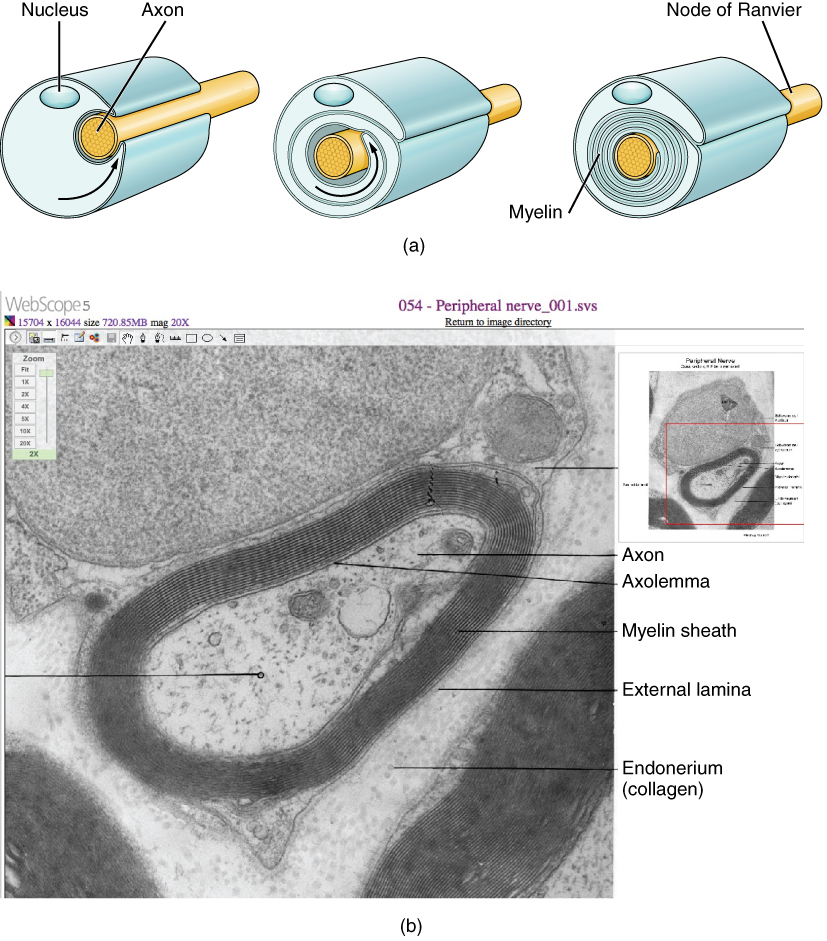
Histology, Axon - PubMed Myelin forms by the concentric wraps of the plasma membrane of neuroglia cells around the axon. These cells are the Schwann cells (or neurolemmocytes) in the PNS and oligodendrocytes in the CNS. As a general rule, oligodendrocytes myelinate multiple adjacent axons, while Schwann cells myelinate only one axon.
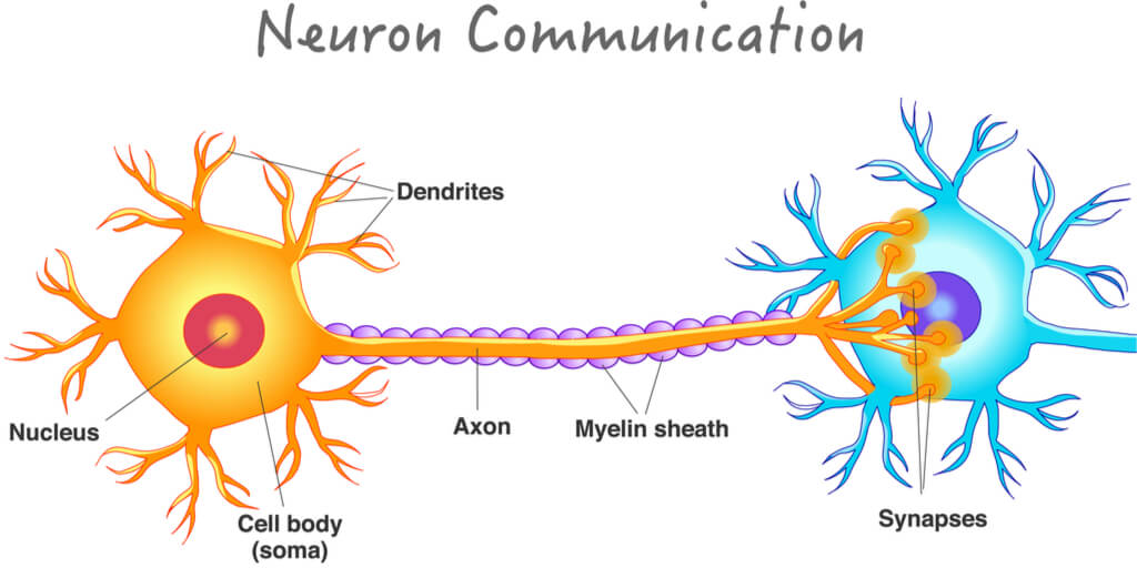
Diagram of the cell membrane of the axon
Synapse | Its Structure, Types, Function and Transmission Steps 29/03/2021 · The cell body (soma) contains the nucleus and cytoplasm. It has organelles like Nissl granules, Golgi apparatus, lysosomes, etc. Dendrites are the numerous short extensions from the cell body. They receive the incoming signals and transmit them to the cell body. Axon is the long tubular process that transmits output signals. The axon ends at ... Schwann Cells and Unmyelinated Axons | GetBodySmart A series of Schwann cells covers the length of each axon. Abutting Schwann cells are tightly joined and nodes of Ranvier do not form. Each Schwann cell typically contains several axons (up to 20), which are often brought into the cell by invaginations of the plasma membrane. The internalized axons are not myelinated by the Schwann cell. 1. 2. Axon: Structure and function | Kenhub Axon. Axons are processes from the cell body (soma) or from the axon hillock (a specialized part of the cell body) of a neuron that conduct impulses away from cell body. Unlike dendrites that are a series of processes in the vicinity of the cell body which receive information, axons are typically a single, longer, cylinderical process that ...
Diagram of the cell membrane of the axon. Module 1.1 Cells of the Nervous System Flashcards | Quizlet Axon Thing fiber of constant diameter; the neuron's information sender Blood-brain barrier Mechanism that excludes most chemicals from the brain Cell body (soma) Structure containing the nucleus, ribosomes, and mitochondria Dendrites Branching fibers from a neuron that receive information from other neurons Dendritic spines A Labelled Diagram Of Neuron with Detailed Explanations - BYJUS Axon–Axon is a tube-like structure that functions by carrying an electrical impulse from the cell body to the axon terminals for passing the impulse to another neuron. Synapse– This structure functions by permitting the entry of a neuron to move an electrical or chemical signal from one neuron to another neuron. cell membrane | Definition, Function, & Structure | Britannica The chemical structure of the cell membrane makes it remarkably flexible, the ideal boundary for rapidly growing and dividing cells. Yet the membrane is also a formidable barrier, allowing some dissolved substances, or solutes, to pass while blocking others. Lipid-soluble molecules and some small molecules can permeate the membrane, but the lipid bilayer effectively repels the many large ... Neurons Flashcards | Quizlet Describe the two channels which affect permeability 1. Non-gated channels ie leak channels which are open at rest 2. Gated channels ie they're are either voltage, ligand or mechanically gated Explain the ratio of K+ leak channels to Na+ leak channels in neurons There are many more K+ leak channels than Na+ leak channels it's a 40:1 ratio
Nodes of Ranvier: Function and Definition - Study.com The axon is coated in a fatty substance called myelin, which is secreted by cells called oligodendricytes or Schwann cells. Below is a diagram of the parts of a neuron. Below is a diagram of the ... byjus.com › biology › diagram-of-neuronA Labelled Diagram Of Neuron with Detailed Explanations - BYJUS Axon–Axon is a tube-like structure that functions by carrying an electrical impulse from the cell body to the axon terminals for passing the impulse to another neuron. Synapse– This structure functions by permitting the entry of a neuron to move an electrical or chemical signal from one neuron to another neuron. Multipolar Nerve Cell | Function, Structure, Features & Summary Benefits. When considering the structural features of a nerve cell, it is common to speak of a multipolar type of neuron. It possesses a nerve cell body, the perikaryon, from which, as mentioned above, a number of dendritic extensions and one axonal extension originate. As a result, this is the key feature of these nerve cells - a single axon ... Histology, Schwann Cells - StatPearls - NCBI Bookshelf Structure. Each Schwann cell makes up a single myelin sheath on a peripheral axon, with each ensuing myelin sheath made by a different Schwann cell, such that numerous Schwann cells are needed to myelinate the length of an axon. ... While the nucleus remains fixed, the inner turn of the glial cell membrane spirals around the axon to add ...
What Is a Neuron? Diagrams, Types, Function, and More - Healthline It's enclosed by a membrane that both protects it and allows it to interact with its immediate surroundings. Axon. An axon is a long, tail-like structure. It joins the cell body at a specialized ... › books › NBK554382Histology, Cell - StatPearls - NCBI Bookshelf May 08, 2022 · The cell is the fundamental organizational unit of life. All living things are composed of cells, which then further subdivide based on the presence or absence of the nucleus, into two types: eukaryotic cells (Greek, Eu=true, karyo=nut, nucleus) - these cells are present in all the human, animal and plants with a clear, distinct nucleus. Prokaryotic cells are some bacteria and blue-green algae ... Animal Cell Anatomy & Diagram - Enchanted Learning Cells are covered by a cell membrane and come in many different shapes. The contents of a cell are called the protoplasm. Glossary of Animal Cell Terms: Cell Membrane The thin layer of protein and fat that surrounds the cell. The cell membrane is semipermeable, allowing some substances to pass into the cell and blocking others. Neuromuscular Junction | Structure, Function, Summary & Clinical Neuromuscular junction is a microstructure present at the junction of motor neurons and the skeletal muscle fibers. It acts as a bridge connecting the skeletal system and the nervous system. The neuromuscular junction is a chemical synapse. The presynaptic terminal is the axonal terminal of. motor neuron containing synaptic vesicles.
Axon Terminal - The Definitive Guide | Biology Dictionary
Neuron Diagram, Structure & Function | What Is a Neuron? - Video ... A detailed diagram of a neuron, showing the synapse, cell body, axon, and dendrites. Dendrites One unique feature of neurons is that they have extensions known as dendrites that extend outward from...
12.5 The Action Potential – Anatomy & Physiology
Axon - Structure and Functions | GetBodySmart The initial segment of an axon has the highest concentration of these reactive proteins. this region is an axon's trigger zone, where action potentials are first produced if electrochemical events in the dendrites and/or cell body reach a threshold level. Axons vary in length from a few millimeters to just over a meter.

Modeling of inhomogeneous electromagnetic fields in the ...
biologydictionary.net › axon-terminalAxon Terminal - The Definitive Guide | Biology Dictionary May 22, 2020 · The axon terminal, also known as the synaptic bouton and terminal bouton, is the most distal portion of a neuron’s axon and is critical for neural communication. When action potentials reach the axon terminal, calcium floods the neuron, allowing synaptic vesicles to fuse with the membrane and release stored neurotransmitters to target cells.

16.2 How Neurons Communicate – Concepts of Biology – 1st ...
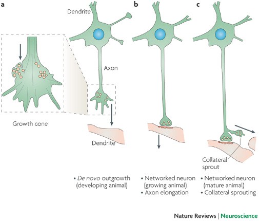
May the force be with you: Mechanosensitivity shapes dendrite ... Navigational instruction is partly encoded in the properties and distribution of guidance cues, which are diffusible or membrane-bound proteins that steer axon and dendrite growth by signaling through their cognate receptors at the neuronal membrane. This conceptual framework has underpinned axon guidance research over the past three decades (

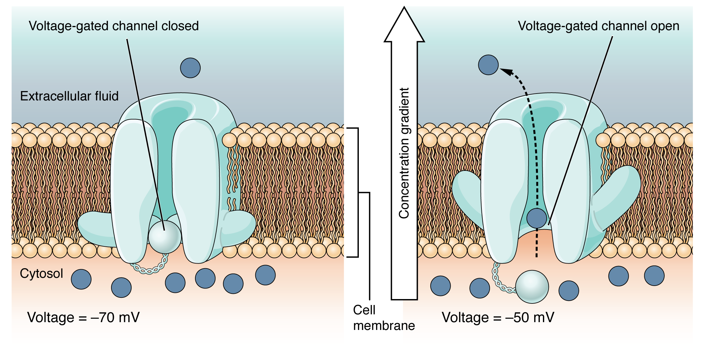
The voltage-gated channel of the axon cell membrane ...
Unmyelinated and Myelinated Axons | GetBodySmart 1 2 3 The sheaths consist of a series of myelinated segments, each extending about 1 millimeter along the outer surface of the axon. The segments are separated by small gaps (about 1 micrometer) called nodes of Ranvier (enlarged for this illustration). 1 2
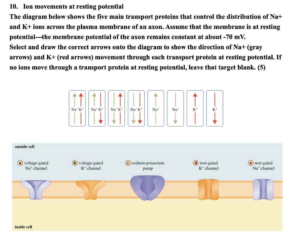
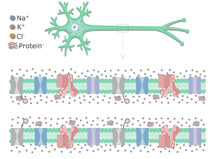
Solved 10. Ion movements at resting potential The diagram ...
Histology, Cell - StatPearls - NCBI Bookshelf 08/05/2022 · The cell is the fundamental organizational unit of life. All living things are composed of cells, which then further subdivide based on the presence or absence of the nucleus, into two types: eukaryotic cells (Greek, Eu=true, karyo=nut, nucleus) - these cells are present in all the human, animal and plants with a clear, distinct nucleus. Prokaryotic cells are some bacteria …

The Action Potential
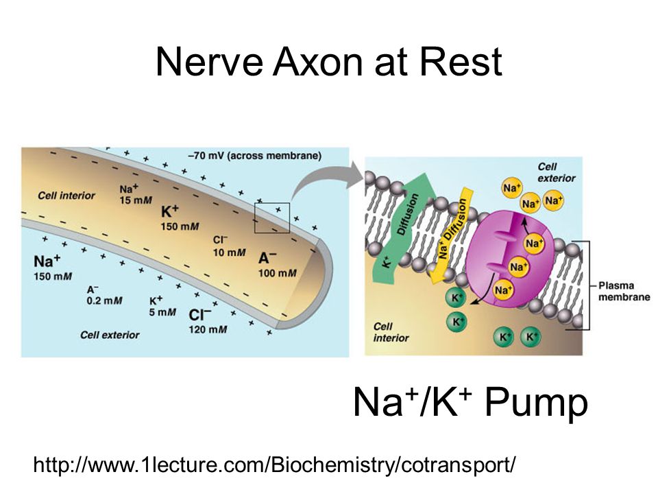
Distribution of Ions In Resting Axons | GetBodySmart The ion concentrations in the extracellular fluid (ECF) and intracellular fluid (ICF) of a resting axon can be measured. 1 2 These concentrations are often expressed in millimolar (mM) units. Molarity: Molarity (M) is the number of moles of a solute in one liter of solution. One Mole of a solute contains 6.023 x 10²³ particles.

Histology of the Peripheral Nerves and Light Microscopy ...
Why Do Action Potentials Travel In One Direction? - The Classic Wanderer The axon is surrounded by a membrane that is selectively permeable, meaning that it allows some substances to cross it while keeping others out. This membrane is also electrically charged.When a neuron is at rest, the charge on the outside of the membrane is more positive than the charge on the inside.
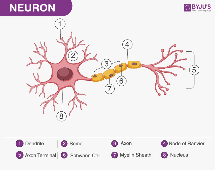
A Labelled Diagram Of Neuron with Detailed Explanations
Action potential - Definition, Steps, Phases | Kenhub Learn the structure and the types of the neurons with the following study unit. ... An action potential propagates along the cell membrane of an axon until it reaches the terminal button. Once the terminal button is depolarized, it releases a neurotransmitter into the synaptic cleft. The neurotransmitter binds to its receptors on the ...

Schwann Cells Function | Simply Psychology
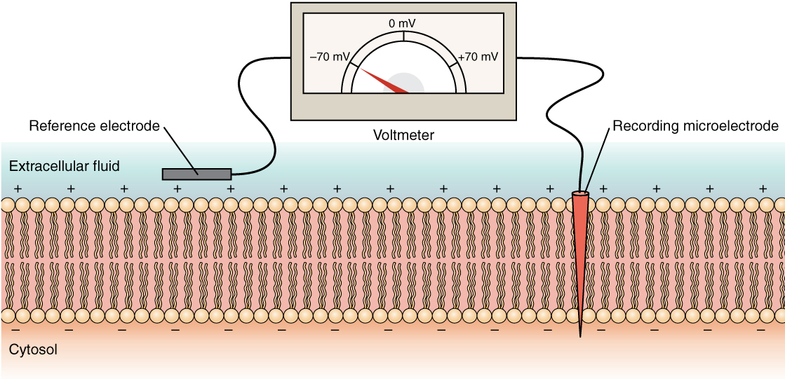
› en › libraryMembrane potential: Definition, equilibrium, ions | Kenhub Jul 06, 2022 · A resting membrane potential is the difference between the electric potential in the intracellular and extracellular matrices of the cell when it isn’t excited. Every cell of the body has its own membrane potential, but only excitable cells - nerves and muscles - are capable to change it and generate an action potential .
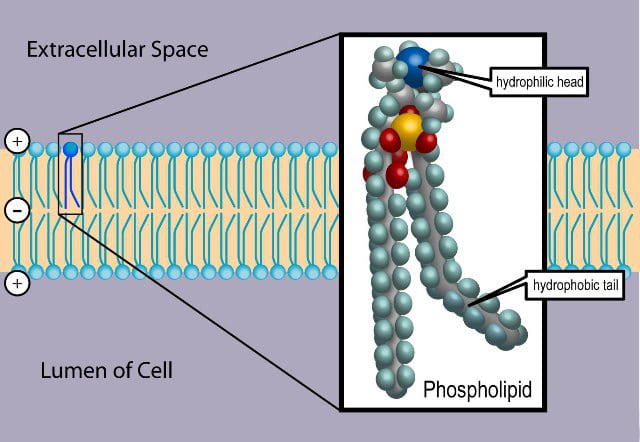
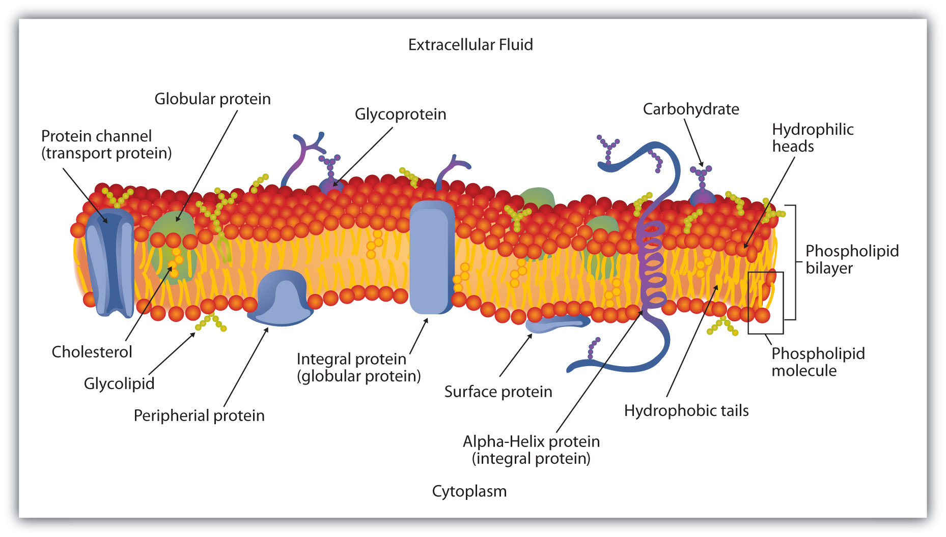
The Cell Membrane - Structure - Phospholipids - TeachMePhysiology
Ionic Currents Across an Axon Membrane Under Voltage Clamp Figure 7-5 A diagram of the current injected by a voltage-clamp amplifier into an axon in response to a voltage step from the normal resting membrane potential to the sodium equilibrium potential. The initial sodium current is absent because there is no driving force for sodium current when Em equals ENa. by a maintained outward current.
12.5 The Action Potential – Anatomy & Physiology
Measuring the Resting Membrane Potential | GetBodySmart Several factors play a role in creating the resting membrane potential. Na+/K+ pumps (or Na+/K+-ATPases) move Na+ and K+ ions to opposite sides of the membrane. 1. 2. Each pump protein uses one molecule of ATP to transfer 3 Na+ ions out of the cell and 2 K+ ions in. As a result, Na+ ions are concentrated outside the axon membrane and K+ ions ...

Axon - Structure and Functions | GetBodySmart
Axon Terminal (Location + Function of the Brain) The process behind the influx of calcium ions is the electrical depolarization of the membrane (at the synapse). These calcium ions trigger the vesicles to fuse against the axon terminal's membrane. These calcium ions also trigger the various proteins within the (synaptic) vesicle to form fusion pores along the axon terminal/vesicle membrane.

Animal Cell- Definition, Structure, Parts, Functions, Labeled ...
Membrane potential: Definition, equilibrium, ions | Kenhub 06/07/2022 · Definition. Resting membrane potential (EM) originates from the different concentrations of ions (expressed in mmol/l) at the inner and outer surface of the cell membrane. There are four excitable tissues in our body, and all of them have different EM values: Skeletal muscle cell = -90 millivolts (mV) ; Smooth muscle cell = -55mV; Cardiac muscle cell = …
Nervous Tissue – Anatomy & Physiology
Bioflix Activity How Neurons Work Action Potential Events An action potential arrives at the synaptic terminal. . This process which occurs during the firing of the neurons allows a nerve cell to transmit an electrical signal down the axon a portion of the neuron that carries nerve impulses away from the cell body. To review the events that occur during an action potential watch this BioFlix animation.
The cell is the smallest unit of life. All organisms are ...
Neuron under Microscope with Labeled Diagram - AnatomyLearner This initial segment is the narrow part of the other part of the plasma membrane of the cell body of a neuron. From this initial segment of a cell body, the myelin sheath begins. You will also find the plasma membrane around the axon, known as the axolemma. The axon branches at the right angle in the axon course and give the collateral branches.

hillis2e_ch34
Function and Purpose of the Axon Hillock - Study.com The axon is a singular, long projection of plasma membrane on the opposite side of the cell body. The axon is permeable to ions that allow for electrical impulses to be transmitted. The axon...

A Gate Keeper for Axonal Transport: Cell
Axon Terminal - The Definitive Guide | Biology Dictionary 22/05/2020 · The more Na⁺ ions that enter the cell, the more depolarized the cell becomes, and therefore the more Na⁺ channels that open sequentially down the axon. Because Na⁺ channels go through a refractory stage after they close where they are unlikely to open again for a short time period, the action potential can only continue in the direction of the axon terminal and synapse.

Structure of the Cell Membrane | Biology for Majors I
Myelin sheath: Myelination, function, clinical relations | Kenhub 27/09/2022 · Each Schwann cell myelinates only one axon, where one peripheral axon will have multiple Schwann cells myelinating its length as one Schwann cell wraps a lipid-rich membrane layer around approximately 1 mm of an axon’s length. However, in a different arrangement, a Schwann cell can enclose many (up to 20) unmyelinated axons. In this way, the unmyelinated …
Activity 2.2.2: Student Response Sheet
Myelin sheath: Myelination, function, clinical relations | Kenhub Each Schwann cell myelinates only one axon, where one peripheral axon will have multiple Schwann cells myelinating its length as one Schwann cell wraps a lipid-rich membrane layer around approximately 1 mm of an axon's length. However, in a different arrangement, a Schwann cell can enclose many (up to 20) unmyelinated axons.

Cell types. Neuron. Atlas of Plant and Animal Histology
Dendrite - Wikipedia Dendrites (from Greek δένδρον déndron, "tree"), also dendrons, are branched protoplasmic extensions of a nerve cell that propagate the electrochemical stimulation received from other neural cells to the cell body, or soma, of the neuron from which the dendrites project. Electrical stimulation is transmitted onto dendrites by upstream neurons (usually via their axons) via …
The Cell Membrane - Structure - Phospholipids - TeachMePhysiology
› Subjects › animalsAnimal Cell Anatomy & Diagram - Enchanted Learning Cells are covered by a cell membrane and come in many different shapes. The contents of a cell are called the protoplasm. Glossary of Animal Cell Terms: Cell Membrane The thin layer of protein and fat that surrounds the cell. The cell membrane is semipermeable, allowing some substances to pass into the cell and blocking others.
Activity 2.2.2: Student Response Sheet
Myelin structure - Schwann Cells - GUWS Medical Like other cell membranes, myelin has a high electrical resistance and low capacitance, which is essential for rapid saltatory conduction from node to node. Figure 8.4 Myelinating Schwann cells. There is a 1:1 relationship between a myelinating Schwann cell and the axon it myelinates. Consecutive Schwann cells are separated by nodes of Ranvier ...

The cell - Knowledge @ AMBOSS
PDF The Cell: Cell Membrane | Concise Medical Knowledge A cell membrane (also known as the plasma membrane or plasmalemma) is a biological membrane that separates the cell contents from the outside environment. A cell membrane is composed of a phospholipid bilayer and proteins that function to protect cellular DNA and mediate the exchange of ions and molecules.

Action potential velocity (article) | Khan Academy
› synapseSynapse | Its Structure, Types, Function and Transmission Steps Mar 29, 2021 · The cell body (soma) contains the nucleus and cytoplasm. It has organelles like Nissl granules, Golgi apparatus, lysosomes, etc. Dendrites are the numerous short extensions from the cell body. They receive the incoming signals and transmit them to the cell body. Axon is the long tubular process that transmits output signals.

Neurons | Organismal Biology
Axon: Structure and function | Kenhub Axon. Axons are processes from the cell body (soma) or from the axon hillock (a specialized part of the cell body) of a neuron that conduct impulses away from cell body. Unlike dendrites that are a series of processes in the vicinity of the cell body which receive information, axons are typically a single, longer, cylinderical process that ...

3,015 Cell Membrane Stock Illustrations, Cliparts and Royalty ...
Schwann Cells and Unmyelinated Axons | GetBodySmart A series of Schwann cells covers the length of each axon. Abutting Schwann cells are tightly joined and nodes of Ranvier do not form. Each Schwann cell typically contains several axons (up to 20), which are often brought into the cell by invaginations of the plasma membrane. The internalized axons are not myelinated by the Schwann cell. 1. 2.

Cell Membranes - ScienceDirect
Synapse | Its Structure, Types, Function and Transmission Steps 29/03/2021 · The cell body (soma) contains the nucleus and cytoplasm. It has organelles like Nissl granules, Golgi apparatus, lysosomes, etc. Dendrites are the numerous short extensions from the cell body. They receive the incoming signals and transmit them to the cell body. Axon is the long tubular process that transmits output signals. The axon ends at ...
Plasma membrane expansion: a neuron's Herculean task | Nature ...

Dendrite - Wikipedia
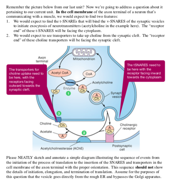
Solved Remember the picture below from our last unit? Now ...
Cells | Free Full-Text | The Role of Lipids, Lipid Metabolism ...

Axon - Wikipedia

Entropy | Free Full-Text | Spiking Neural P Systems with ...

Measuring the Resting Membrane Potential | GetBodySmart
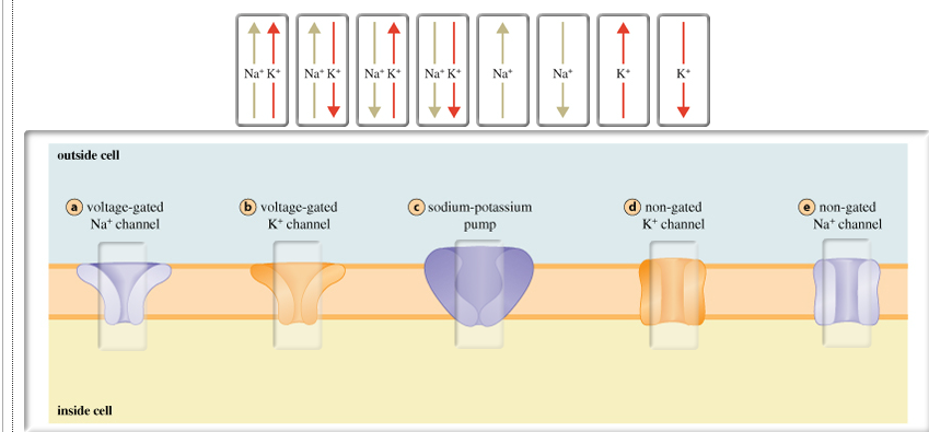
Solved The diagram below shows the five main transport ...
Membranes and Membrane Lipids

Organization of Cell Types (Section 1, Chapter 8 ...
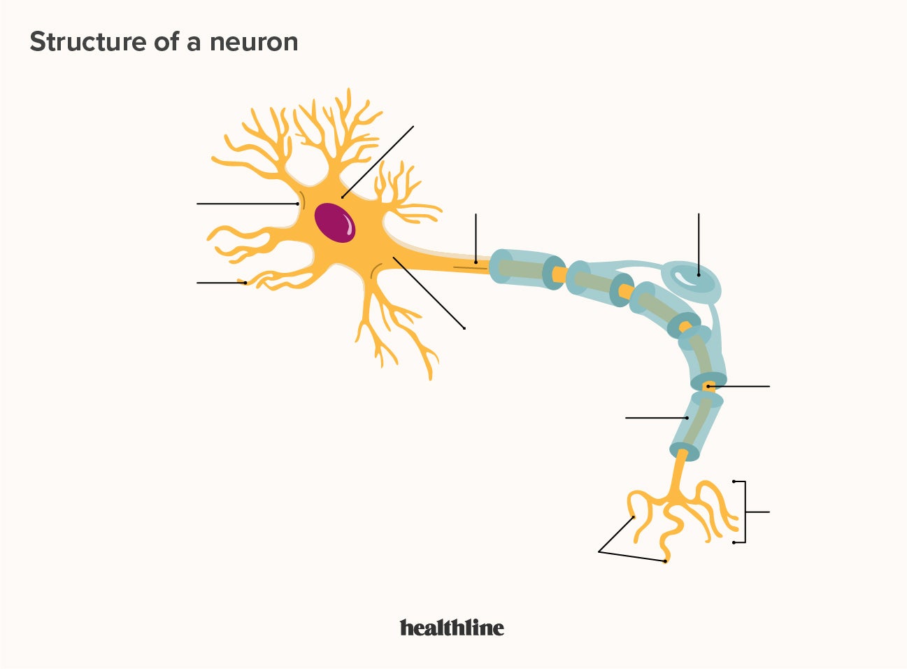
What Is a Neuron? Diagrams, Types, Function, and More
Introduction to Axons at Rest | GetBodySmart

Histology of the Nervous System (The Neuron) Part 1 | Neurons ...
ACTION POTENTIALS

a) Sketch of nerve cell with a myelinated axon, b) signal ...
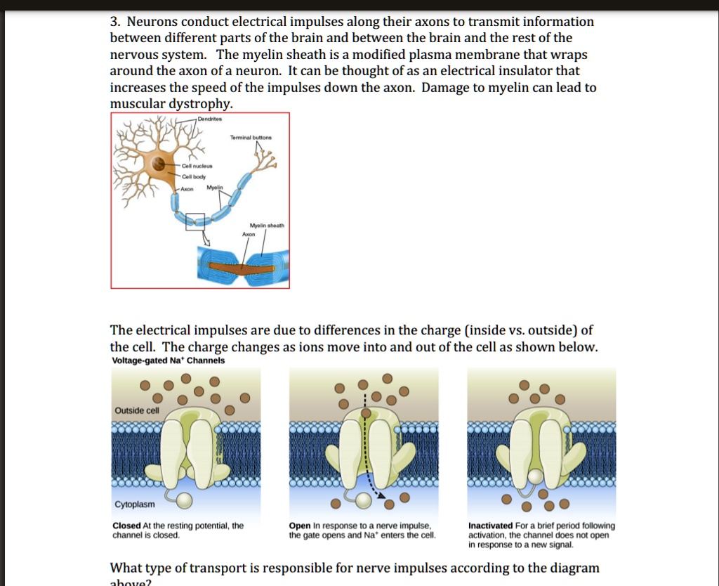
SOLVED: Neurons conduct electrical impulses along their axons ...
Axon - Wikipedia
0 Response to "45 diagram of the cell membrane of the axon"
Post a Comment