39 eye to brain connection diagram
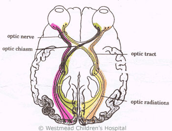
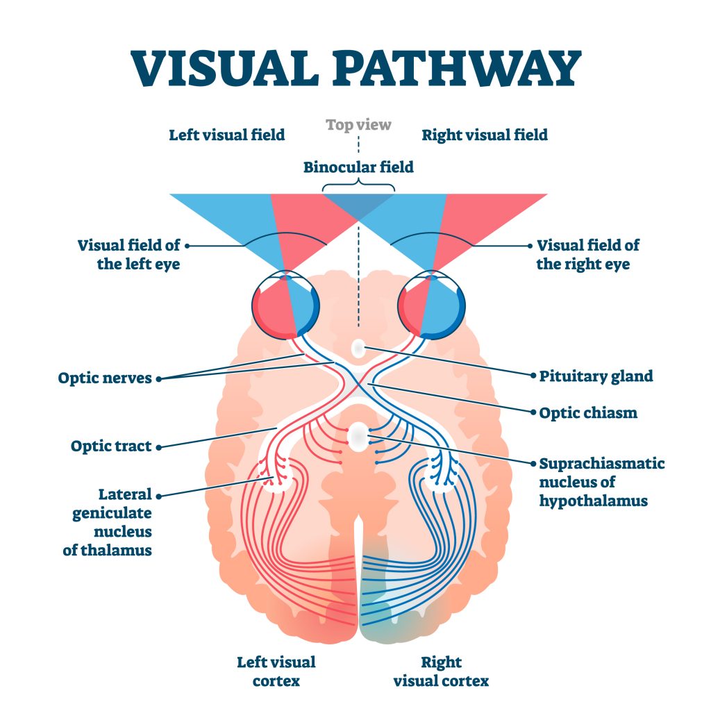
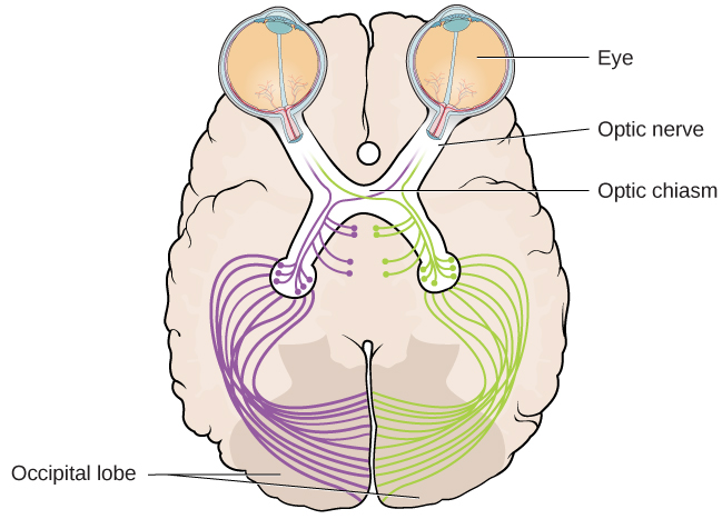
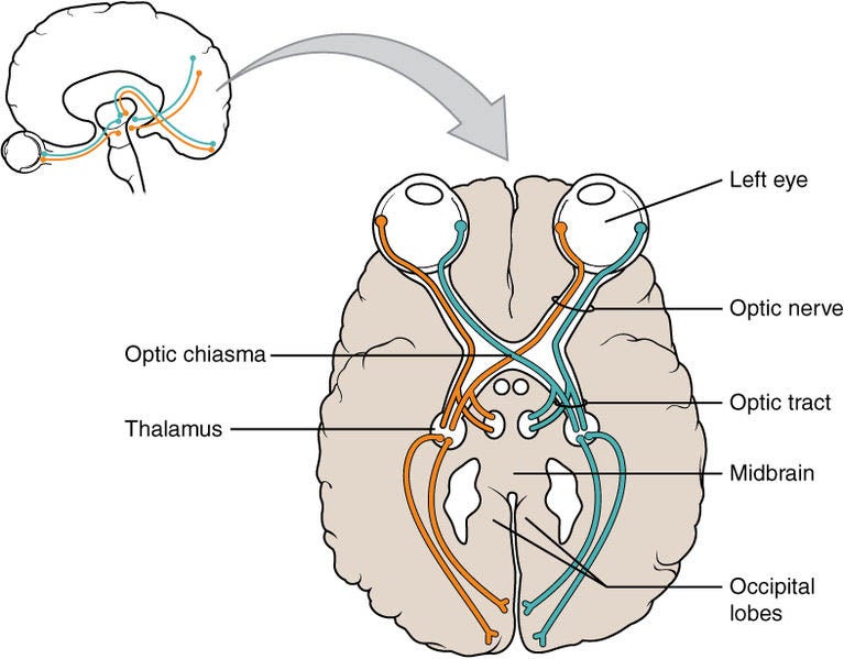
Eye Diagram Stock Photos, Pictures & Royalty-Free Images - iStock How eye work medical illustration, eye - brain diagram, eye structure and connection with brains. Vector EPS10 HUman Eye The human eye is an organ that reacts to light and has several purposes. As a conscious sense organ, the mammalian eye allows vision. Parts of the eye, labeled vector illustration diagram Our eyes and brain. Relationship status: It's complicated. These nerves serve as the main "connectors" between the retina and the brain. Each optic nerve starts at its corresponding eye. However, midway through their journey to the brain, some of the axons in the optic nerves cross over at a specific location, called the optic chiasm (cue my sister's confusion).
Circuit Diagrams | Brain Initiative BRAIN Initiative: Engineering and optimization of molecular technologies for functional dissection of neural circuits (UM1 Clinical Trial Not Allowed) This BRAIN Initiative FOA is to further develop molecular tools of high impact that are targetable to brain cell types for the monitoring and manipulation of neural circuits in experimental animals.

Eye to brain connection diagram
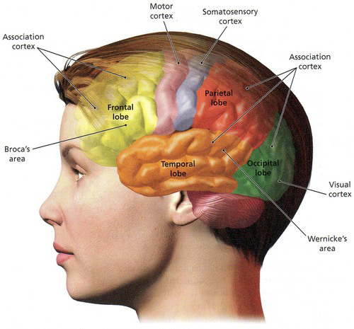
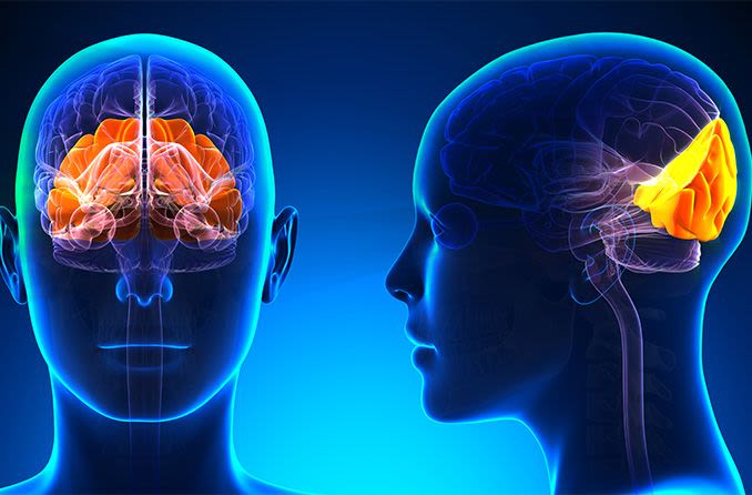
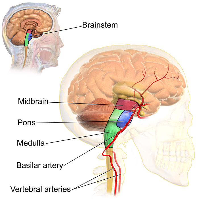
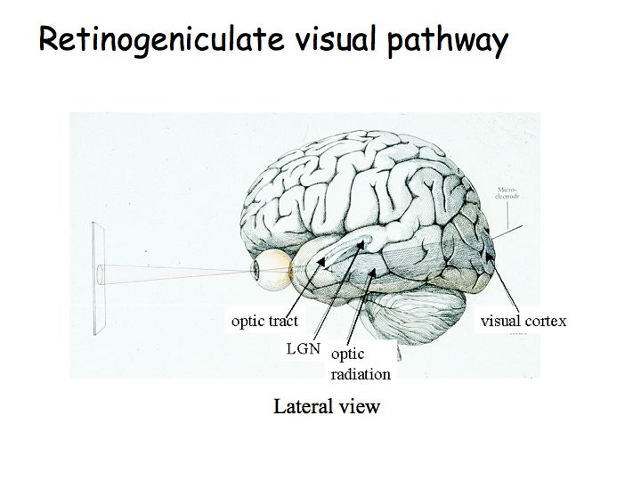
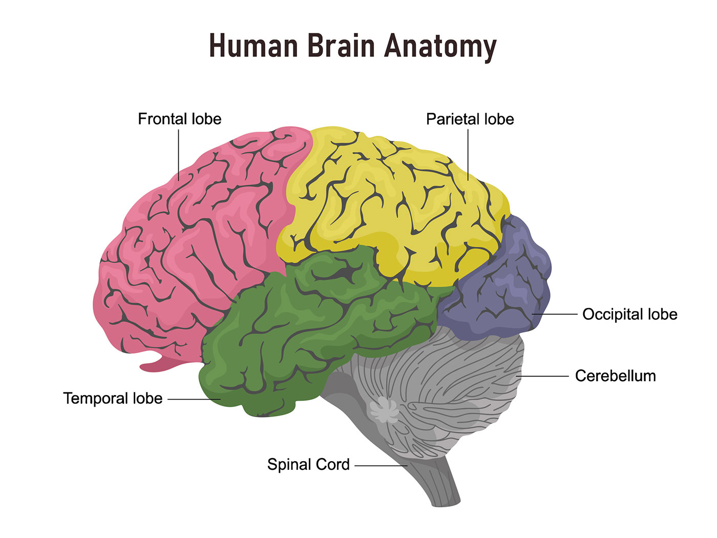
What Part of the Brain Controls Vision? - All About Vision The brain consists of four main segments called lobes. The frontal lobe up front, the parietal lobe on top, the temporal lobe on bottom and the occipital lobe pulling up the rear. All of our senses, thoughts and actions start in one of these lobes. Most visual functions are controlled in the occipital lobe, a small section of the brain near the ... Pathways: From the eye to the brain | Welcome to Bio-X Courtesy of Shatz laboratory: A diagram of electrical activity measured by an electrode array attached to retinal neurons. Each blue dot represents a neuron's approximate location. The size of each red dot represents the amount of electrical activity recorded over one-half second at the corres- ponding neuron. Diagram Of Brain with their Labelings and Detailed Explanation - BYJUS Midbrain. The midbrain is also called as Mesencephalon. The midbrain is the smallest region of the brain, found at the centre of the brain, between cerebral cortex and hindbrain. It comprises tectum, cerebral peduncle, tegmentum, cerebral aqueduct, substantia nigra, several nuclei and fasciculi. The midbrain is responsible for hearing, vision ...
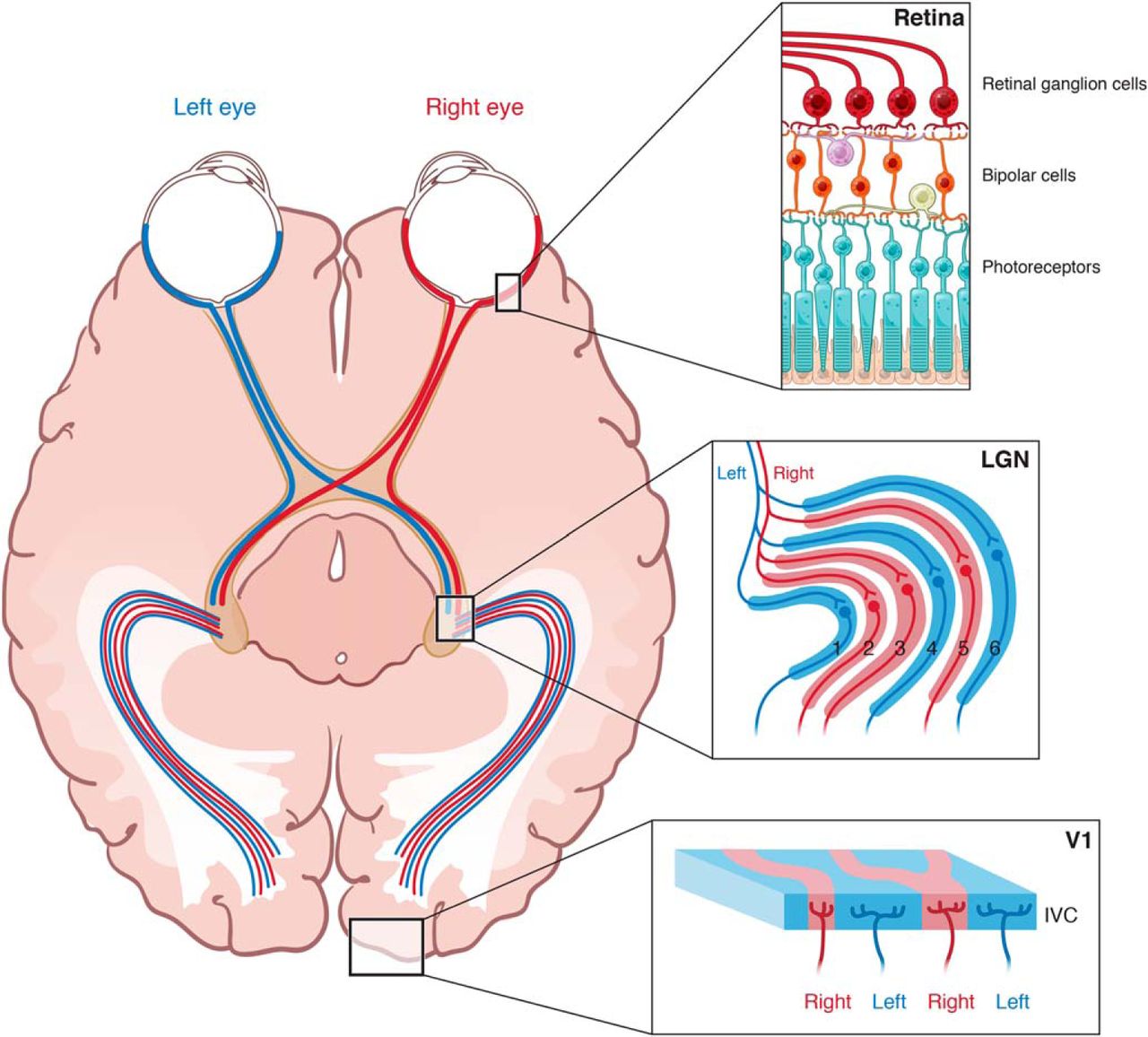
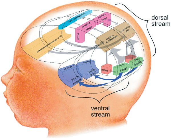
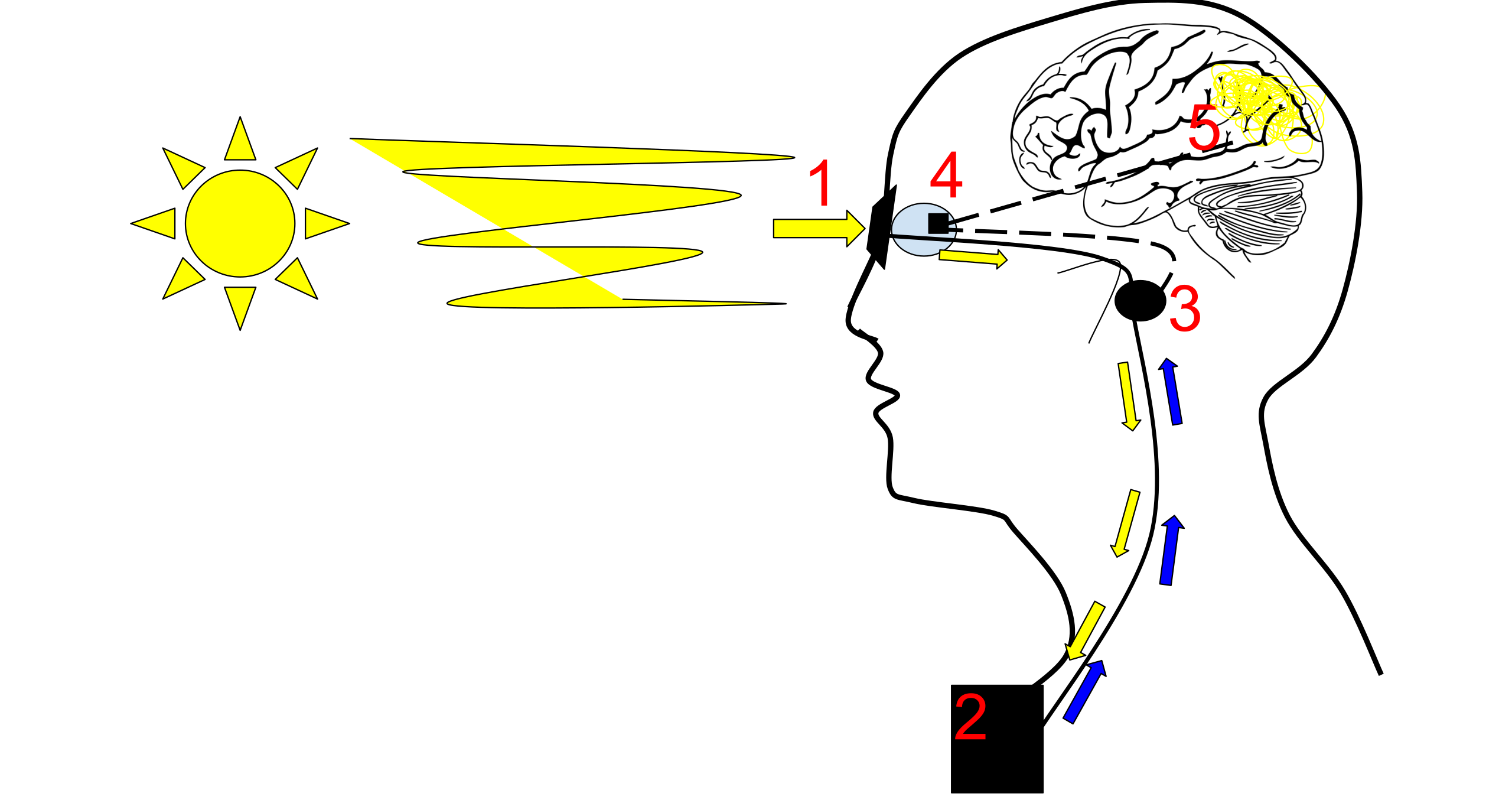
Eye to brain connection diagram. Making connections in the eye: Wiring diagram of retinal neurons is ... Citation: Making connections in the eye: Wiring diagram of retinal neurons is first step toward mapping the human brain (2013, August 7) retrieved 19 September 2022 from ... The Brain and the Eye - How They Work Together - Discovery Eye Foundation A bundle of more than a million nerve fibers carrying visual messages from the retina to the brain. Your brain actually controls what you see, since it combines images. Also the images focused on the retina are upside down, so the brain turns images right side up. This reversal of the images Is a lot like what a mirror does in a camera. Making connections in the eye | MIT News - Massachusetts Institute of ... Tracing connections Neurons in the retina are classified into five classes: photoreceptors, horizontal cells, bipolar cells, amacrine cells and ganglion cells. Within each class are many types, classified by shape and by the connections they make with other neurons. The visual pathway from the eye to the brain Visual cortex: This is where images received from your retina begin to get processed. The visual cortex has six layers and is the very beginning of your brain's process of interpreting and recognizing what you see. Within these layers, depth perception is processed, and form, color, and motion are perceived.
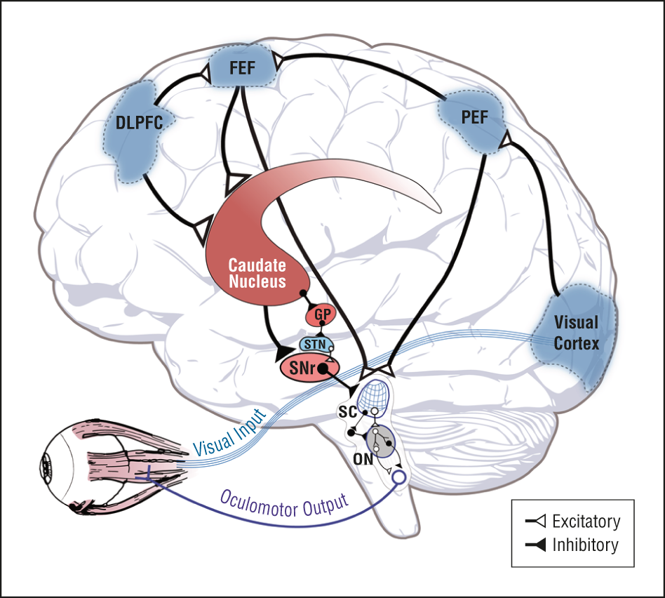
3: Diagram of the human brain. Arrows indicate the connection between ... Arrows indicate the connection between the eye and the principal areas in the brain involved in the visual attention process: frontal eye fields and posterior parietal cortex, which guide spatial... How Eye Work Medical Illustration, Eye - Brain Diagram, Eye Structure ... Picture of How eye work medical illustration, eye - brain diagram, eye structure and connection with brains. stock photo, images and stock photography. Image 94286109. Discover millions of stock images, photos, video and audio. Anatomy, Head and Neck, Eye Nerves - StatPearls - NCBI Bookshelf The eyes are a set of sensory organs that play a crucial role in the visual system. The eyes are responsible for detecting light that enters the eyes. Then, the light gets converted into an image in the brain. The sensory and motor innervation of the eyes originate from six paired cranial nerves. These nerves work in sync to manifest movements, reflexes, and vision. A Neurologist Weighs in on the Eye-Brain Connection - Well+Good "Much of our brain is devoted to social processing: interpreting the behavior of others (aka mentalization), conversing, and emotional attunement," says Dr. Choi. "Staying social keeps our brains...
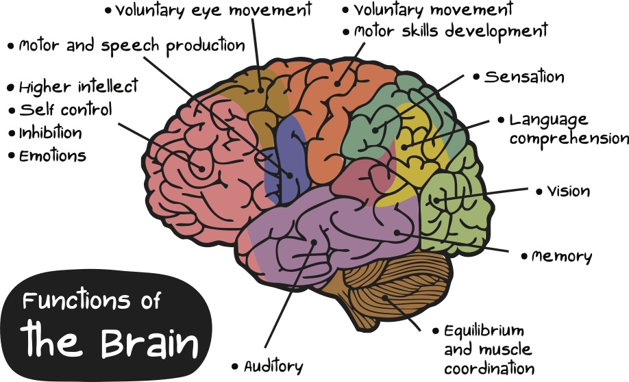
Diagram of the Brain and its Functions - Bodytomy Functions. The frontal lobe is involved with the main executive functions of the brain, which include: Judgment, that is, the ability to recognize future consequences resulting from ongoing actions. This activity mostly occurs in the pre-frontal area. Analytical and critical reasoning. How Eye Work Medical Illustration Eye Brain Diagram Eye Structure And ... Description How eye work medical illustration, eye - brain diagram, eye structure and connection with brains. Vector EPS10 1 credit Essentials collection for this image $4 with a 1-month subscription (10 Essentials images for $40) Continue with purchase View plans and pricing Includes our standard license. Add an extended license. Diagram of the Eye - Lions Eye Institute Instructions Click the parts of the eye to see a description for each. Hover the diagram to zoom. Need any help? If you would like to know more about us, or want to make an appointment, please don't hesitate to get in touch. (08) 9381 0777 carecentre@lei.org.au Request an appointment Customer Care Centre (08) 9381 0777 The Eye-Brain Connection - BrainFacts Light-sensing cells in your retinas transform light and color into electrical signals. But, to turn that information into a complete picture of the world around you, those signals need to be relayed to multiple areas of the brain quickly and accurately. That's the job of cells like these — retinal ganglion cells.
Right Or Left - Does One Side Of Your Brain Control Your Vision?
How eye work medical illustration, eye - brain diagram - Dreamstime Illustration about How eye work medical illustration, eye - brain diagram, eye structure and connection with brains. Vector EPS10. Illustration of disc, fovea, info - 108512838
Kids Health Information : Brain injury - Eyes and vision
Visualizing the Connections Between Eye and Brain In this study published May 31 in the journal Cell, Mark Andermann, PhD, Chinfei Chen, MD, PhD, and colleagues developed a means of tracking the activity of the far-reaching ends of retinal neurons (called boutons) as they deliver visual information to the thalamus, a brain region involved in image processing.
Activity Shapes Neural Circuit Form and Function: A ...
The Eye & Brain Connection - Chadwick Optical, Inc. The signals may START OUT on the straight and narrow, with signals from the right eye going to the right side of the brain and signals from the left side going to the left side of the brain. But about midway through their journey, some of the axons change course at a point called the optic chiasm. Big word, we know.
Eye connected to brain hi-res stock photography and images ...
How Does the Eye Work? - Optometrists.org Step 1: Light enters the eye through the cornea When we look at an object, the light that is reflected off of the object enters the eye through the clear front layer of the eye, called the cornea. The cornea bends the light before it passes through a watery substance that fills the area behind the cornea, called the aqueous humor.

366 Eye Brain Diagram Stock Photos, Pictures & Royalty-Free ...
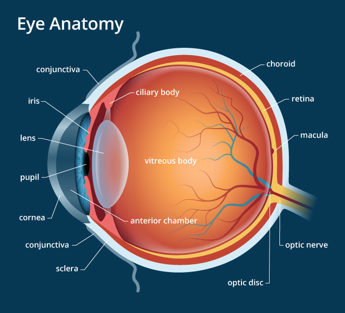
Eye Anatomy: 16 Parts of the Eye & Their Functions - Vision Center The following are parts of the human eyes and their functions: 1. Conjunctiva. The conjunctiva is the membrane covering the sclera (white portion of your eye). The conjunctiva also covers the interior of your eyelids. Conjunctivitis, often known as pink eye, occurs when this thin membrane becomes inflamed or swollen.

The brain and spinal cord | Canadian Cancer Society
The Optic Nerve And Its Visual Link To The Brain - Discovery Eye Foundation The Optic Nerve And Its Visual Link To The Brain Discovery Eye Foundation March 12, 2015 Anatomy of the Eye, Eye Health, Glaucoma The optic nerve, a cable-like grouping of nerve fibers, connects and transmits visual information from the eye to the brain. The optic nerve is mainly composed of retinal ganglion cell (RGC) axons.

A child lost a sixth of his brain, then made an amazing ...
Eye wiring - creation.com Normally, half the nerves of each eye go to each side of the brain, so that each eye is mapped to both the left hemisphere and the right hemisphere (see wiring diagram), but scans on the German girl showed that retinal nerve fibres that should go to the right hemisphere of the brain diverted to the left.
The Visual Experience: Reading 2014
Structure And Function Of The Eye - Vision - MCAT Content - Jack Westin The human eye is an organ that reacts with light and allows light perception, color vision, and depth perception. The photoreceptive cells of the eye, where transduction of light to nervous impulses occurs, are located in the retina (shown in Figure 1) on the inner surface of the back of the eye. But light does not impinge on the retina unaltered.

human eye | Definition, Anatomy, Diagram, Function, & Facts ...
The Brain From Top to Bottom The optic nerve is the pathway that carries the nerve impulses from each eye to the various structures in the brain that analyze these visual signals. The optic nerves of the two eyes emerge from their optics discs and intersect at the optic chiasm just in front of the pituitary gland.
What Part of the Brain Controls Vision?
Making connections in the eye: Wiring diagram of retinal neurons is ... Making connections in the eye: Wiring diagram of retinal neurons is first step toward mapping the human brain Date: August 7, 2013 Source: Massachusetts Institute of Technology
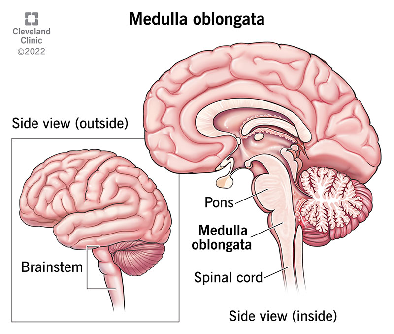
Medulla Oblongata: What It Is, Function & Anatomy
Simple Anatomy of the Retina - Webvision - NCBI Bookshelf The retina is approximately 0.5 mm thick and lines the back of the eye. The optic nerve contains the ganglion cell axons running to the brain and, additionally, incoming blood vessels that open into the retina to vascularize the retinal layers and neurons (Fig. 2). A radial section of a portion of the retina reveals that the ganglion cells (the output neurons of the retina) lie innermost in ...
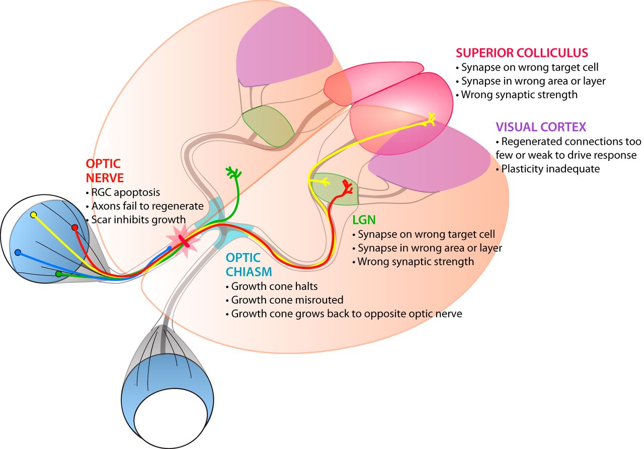
Reconnecting Eye to Brain | Journal of Neuroscience
Diagram Of Brain with their Labelings and Detailed Explanation - BYJUS Midbrain. The midbrain is also called as Mesencephalon. The midbrain is the smallest region of the brain, found at the centre of the brain, between cerebral cortex and hindbrain. It comprises tectum, cerebral peduncle, tegmentum, cerebral aqueduct, substantia nigra, several nuclei and fasciculi. The midbrain is responsible for hearing, vision ...
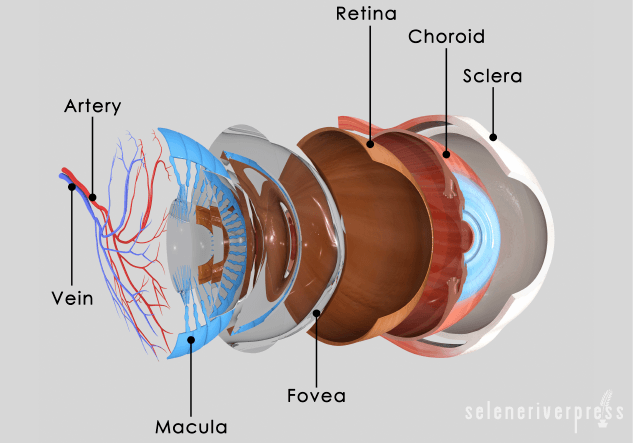
The Eye-Brain Connection - Selene River Press
Pathways: From the eye to the brain | Welcome to Bio-X Courtesy of Shatz laboratory: A diagram of electrical activity measured by an electrode array attached to retinal neurons. Each blue dot represents a neuron's approximate location. The size of each red dot represents the amount of electrical activity recorded over one-half second at the corres- ponding neuron.
Vision – Introduction to Psychology & Neuroscience
What Part of the Brain Controls Vision? - All About Vision The brain consists of four main segments called lobes. The frontal lobe up front, the parietal lobe on top, the temporal lobe on bottom and the occipital lobe pulling up the rear. All of our senses, thoughts and actions start in one of these lobes. Most visual functions are controlled in the occipital lobe, a small section of the brain near the ...

366 Eye Brain Diagram Stock Photos, Pictures & Royalty-Free ...
The Eye

366 Eye Brain Diagram Stock Photos, Pictures & Royalty-Free ...
Cognitive vision, its disorders and differential diagnosis in ...
Blood vessels and nerves of the eye: Anatomy | Kenhub

How eye work medical illustration, eye - brain diagram canvas print

Neural pathway - Wikipedia
The midbrain - Queensland Brain Institute - University of ...
File:The Process of Bionic Eyes for the Visually Impaired.svg ...
Perception Lecture Notes: LGN and V1
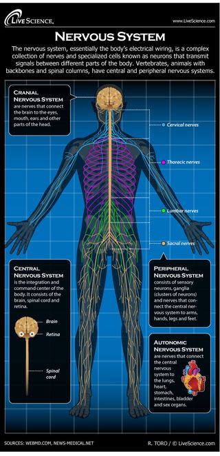
Human Nervous System - Diagram - How It Works | Live Science

How the Eye and the Brain Work Together - AbilityPath

Brain: Function and Anatomy, Conditions, and Health Tips
How We See | Introduction to Psychology

Neuroscience for Kids - Motion, form and depth
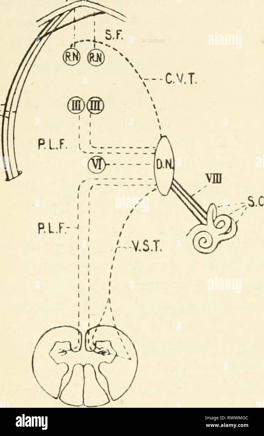
The Human Balance System Part 1 - Arizona Chiropractic Neurology
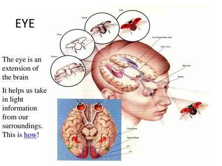
EYE The eye is an extension of the brain. Eye brain proxomity ...

optic nerve | anatomy | Britannica
Vision | Cochlea
Free Brain Diagram, Download Free Brain Diagram png images ...
Brain Anatomy and How the Brain Works | Johns Hopkins Medicine
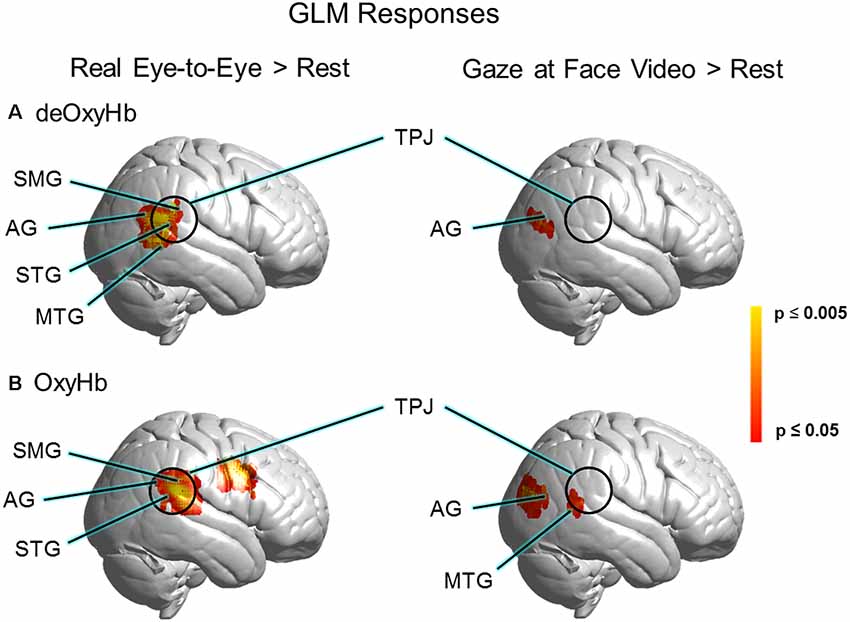
Frontiers | Real-Time Eye-to-Eye Contact Is Associated With ...
14.3 The Brain and Spinal Cord – Anatomy & Physiology
How Vision Works: Our Sense of Sight | Ask A Biologist
A standardized protocol for quantification of saccadic eye ...
0 Response to "39 eye to brain connection diagram"
Post a Comment