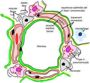
45 on the diagram below identify alveolar epithelium
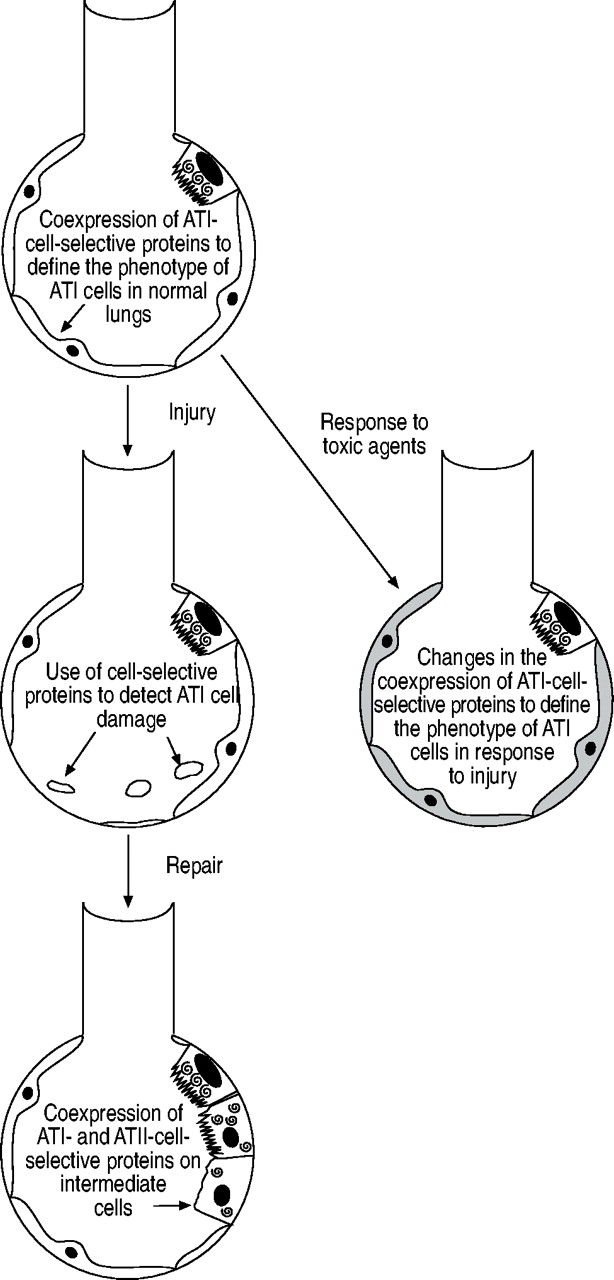
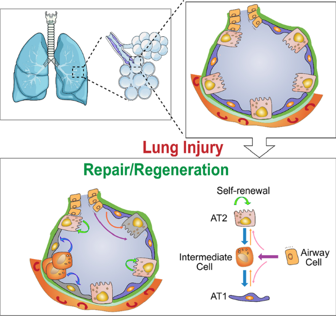
-Alveolar epithelial repair. Following injury, the alveolar epithelium ... Alveolar Type 1 epithelial cells cover 95-98% of the internal lung surface area and are vulnerable to oxygen toxicity even at low levels of inhaled oxygen (Harris et al., 1991; McElroy and Kasper ... 20200413_085950.jpg - 302 Review Sheet 23 14 On the diagram below ... View 20200413_085950.jpg from UNKNOWN 8061771C2 at Blackhawk Technical College. 302 Review Sheet 23 14 On the diagram below, identify alveolar epithelium, capillary endothelium, alveoli, and red. Study Resources. Main Menu; by School; by Literature Title ... epithelial cells of the alveolus; University of Regina • KIN 268. Kin 68 10.docx. 1 ...
Respiratoryanatomy - description - ####### NAME ... On the diagram below identify alveolar epithelium, capillary endothelium, alveoli, and red blood cells. Bracket the ##### respiratory membrane. Demonstrating Lung Inflation in a Sheep Pluck ##### 14. Does the lung inflate part by part or as a whole, like a balloon? ##### 15. What happened when the pressure was released?
On the diagram below identify alveolar epithelium
Respiratory: The Histology Guide - University of Leeds This diagram shows a diagram of an alveolar sac, showing how the organisation of the alveoli, and the network of blood capillaries that surround the alveoli (in red). The epithelium of the alveoli, contains two main types of cells: Alveoli: Anatomy, function and clinical points | Kenhub Histological slides illustrating type I pneumocytes (left) and type II pnemocytes (right) Type II pneumocytes The type II alveolar cells (also known as type II pneumocytes) have two functions: (1) to repair the alveolar epithelium when squamous cells are damaged, and (2) to secrete pulmonary surfactant. Surfactant is composed of phospholipids and protein, and coats the alveoli and smallest ... Histology, Alveolar Cells - StatPearls - NCBI Bookshelf Regeneration of alveolar epithelium after injury [3] [4] Cytoplasm stains pink Nuclei stains blue Collagen appears blue Cytoplasm appears pink Nuclei are dark brown to black. Incubation with diluted primary antibodies for 60 minutes at room temperature Fluorochrome-conjugated secondary antibodies are added for 60 minutes at room temperature.
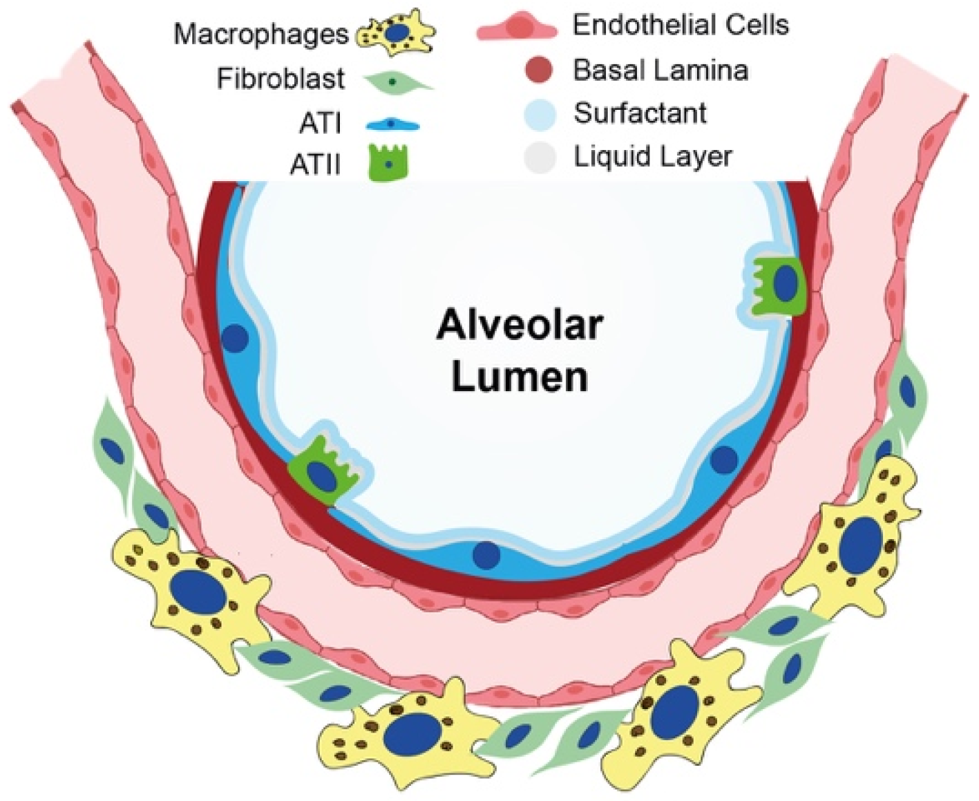
On the diagram below identify alveolar epithelium. Schematic diagram of the alveolar epithelium. Tight (in orange) and ... Download scientific diagram | Schematic diagram of the alveolar epithelium. Tight (in orange) and adherens junctions (in red) on adjacent epithelial cells provide a restrictive barrier in order to ... Alveolar Epithelium - an overview | ScienceDirect Topics The alveolar epithelium is composed of two types of epithelial cells, named alveolar type I and type II cells. Type I cells are large flat cells that comprise about 95% of the alveolar surface. Type II cells are small cuboidal cells with characteristic lamellar inclusions and apical microvilli and cover about 5% of the alveolar surface. Epithelium Diagram | Quizlet Start studying Epithelium. Learn vocabulary, terms, and more with flashcards, games, and other study tools. ... Can be acinar (alveolar), tubular or tubuloacinar. A gland is compound if the duct divides one or more times on its way to the gland cells. Sets with similar terms. Epithelium & Glands. 82 terms. kaitlyn_palmer6. Human anatomy ... Alveoli Flashcards | Quizlet - The epithelial lining of alveoli consists mainly of type I alveolar cells (also known as type I pneumocytes). - These are large, flat, squamous cells with few organelles and thin cytoplasm. - They cover about 93% of alveolar surface area. - Their primary purpose is air-blood gas exchange. - The junctions between these cells are narrow (1nm).
Answered: 14. On the diagram below, identify… | bartleby On the diagram below, identify alveolar epithelium, capillary endothelium, alveoli, and red blood cells. Bracket the respiratory membrane. Elastic - fiber Connective-tissue fibers Monocyte Connective-tissue cell Question Transcribed Image Text: 14. 23-4.jpg - 300 Review She 14. On the diagram below, identify aveolar ... View 23-4.jpg from A&P 201A at National University. 300 Review She 14. On the diagram below, identify aveolar epithelium, capillary endothelium, alveoli, and red blood cells. Bracket the Solved iagram below, identify alveolar epithelium, capillary - Chegg Question: iagram below, identify alveolar epithelium, capillary endothelium, alveoli, and red blood cells. Bracket the brane respira Elastic fiber Connective tissue fibers Monocyte This problem has been solved! See the answer Show transcribed image text Expert Answer 100% (3 ratings) Ans. 1- Alveolar epithelium 2- Capil … View the full answer Solved 13. On the diagram below, identify the alveolar duct, - Chegg On the diagram below, identify the alveolar duct, respiratory bronchioles, terminal bronchiole, alveoli, and alveolar saci Examining Prepared Slides of Tracheal and Lung Tissue 14. The tracheal epithelium is ciliated and has goblet cells. What is the function of each of these modifications? cilia: goblet cells: 15.
Solved > 14.On the diagram below identify aveolar epithelium, capillary ... 14.On the diagram below identify aveolar epithelium, capillary endothelium, alveoli, and red blood cells. Bracket the respiratory membrane. Step-By-Step Solution Chapter 23, Problem 14 View Solution View Sample Solution View All Complete Guide Complete Guide Complete Guide Alveoli Definition, Location, Anatomy, Function, Diagrams Structure and Anatomy of the Alveoli The balloon-shaped alveoli cover approximately 70 square meter area within the lungs, with this wide surface area contributing towards a more efficient gas exchange [6]. Alveoli Alveolar Epithelium The one-cell thick walls of the alveoli are composed of two distal airway epithelium cell types (pneumocytes) [7]. Epithelial Tissue: Structure with Diagram, Function, Types and ... - BYJUS Epithelial cells form membranes. The epithelial membrane consists of a layer of epithelial tissue and has underlying connective tissue. There are two types of epithelial membranes, mucous membrane and serous membrane. Mucous membrane: It is also known as mucosa. There are goblet cells present, which secrete mucus. Histology, Alveolar Cells - StatPearls - NCBI Bookshelf Regeneration of alveolar epithelium after injury [3] [4] Cytoplasm stains pink Nuclei stains blue Collagen appears blue Cytoplasm appears pink Nuclei are dark brown to black. Incubation with diluted primary antibodies for 60 minutes at room temperature Fluorochrome-conjugated secondary antibodies are added for 60 minutes at room temperature.
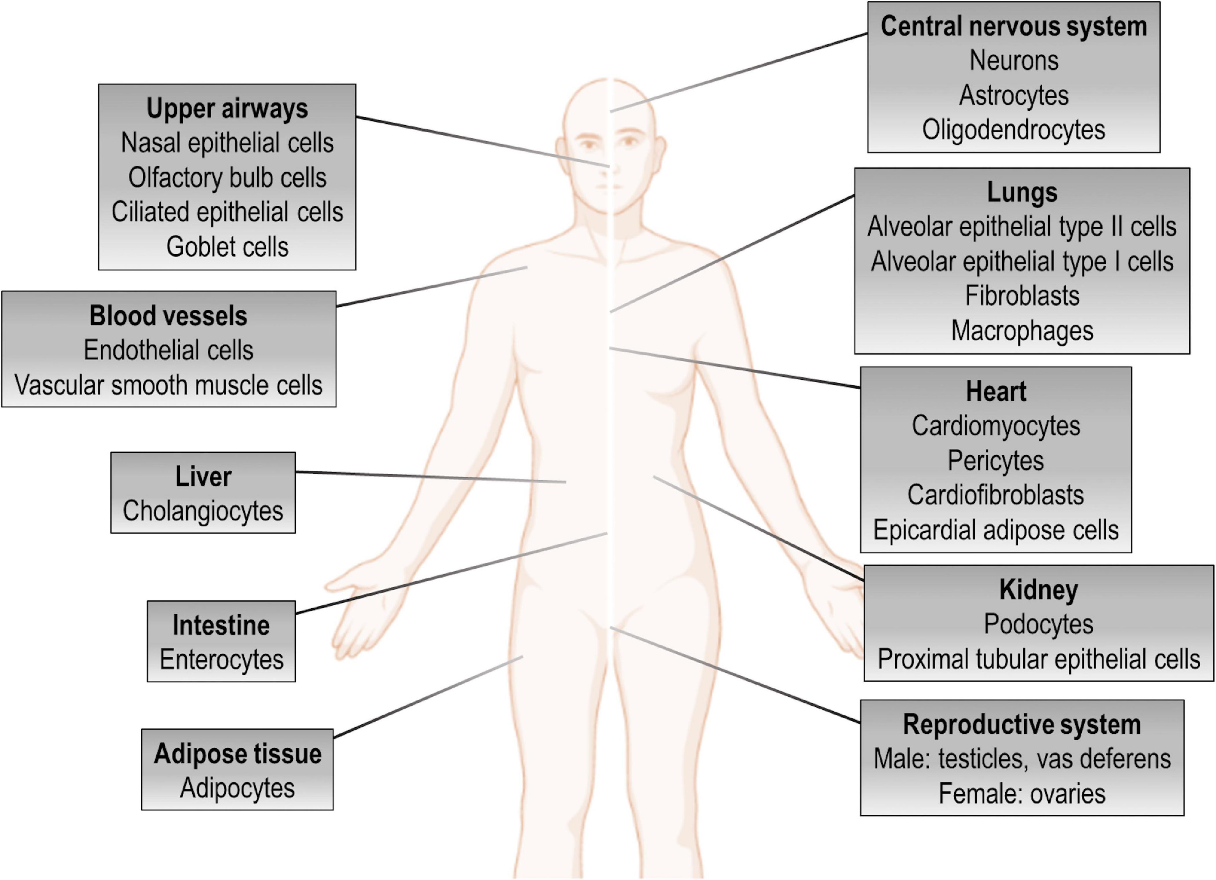
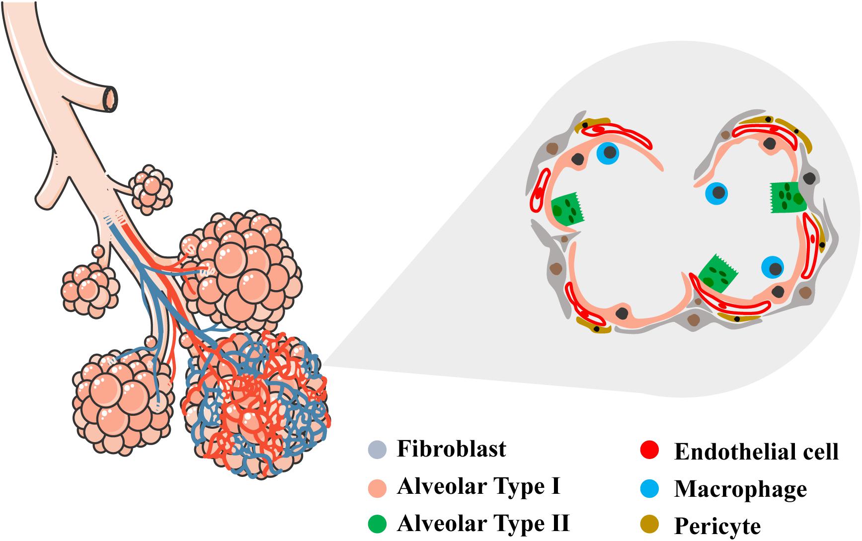
Human microphysiological models of airway and alveolar epithelia
Alveoli: Anatomy, function and clinical points | Kenhub Histological slides illustrating type I pneumocytes (left) and type II pnemocytes (right) Type II pneumocytes The type II alveolar cells (also known as type II pneumocytes) have two functions: (1) to repair the alveolar epithelium when squamous cells are damaged, and (2) to secrete pulmonary surfactant. Surfactant is composed of phospholipids and protein, and coats the alveoli and smallest ...

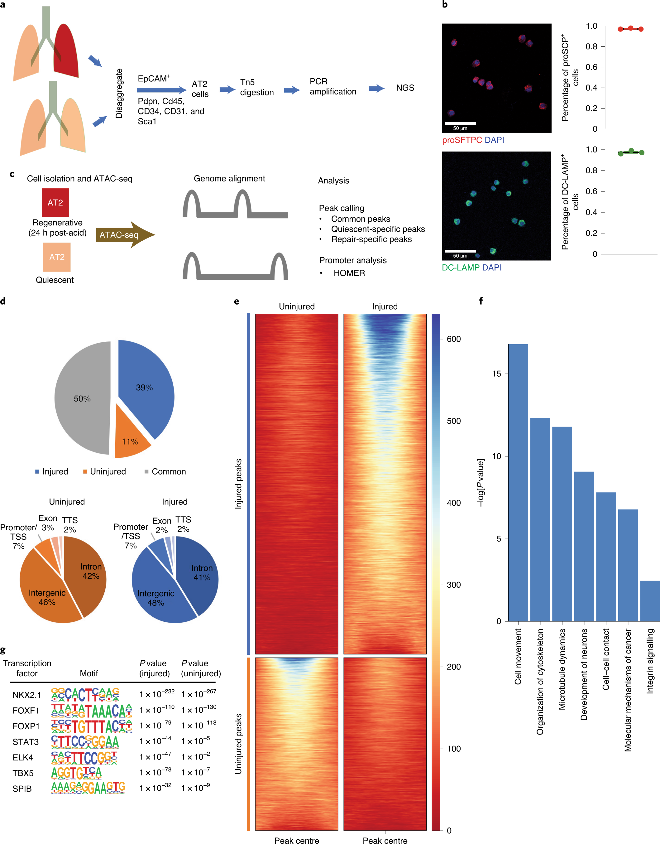
Comprehensive epigenomic profiling of human alveolar ...
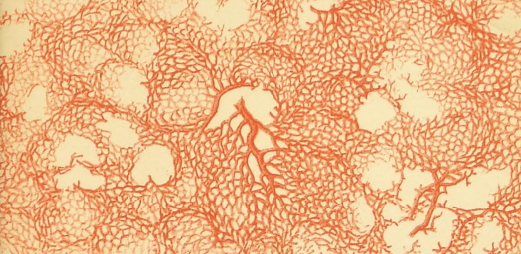
Respiratory: The Histology Guide - University of Leeds This diagram shows a diagram of an alveolar sac, showing how the organisation of the alveoli, and the network of blood capillaries that surround the alveoli (in red). The epithelium of the alveoli, contains two main types of cells:
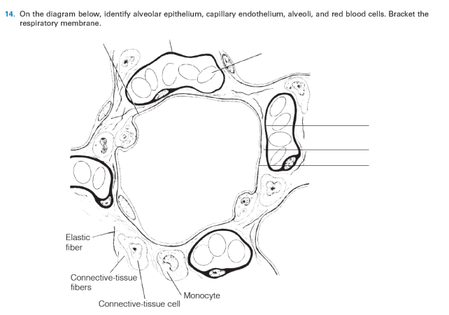
Answered: 14. On the diagram below, identify… | bartleby

3D Lung-on-Chip Model Based on Biomimetically Microcurved ...

Allogeneic Human Mesenchymal Stem Cells Restore Epithelial ...
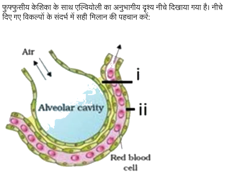
i. Basement membrane , ii: Endothelium
Alveoli - Physiopedia
The use of alveolar epithelial type I cell-selective markers ...
Frontiers | Pathogenesis of Multiple Organ Injury in COVID-19 ...

The impact of the lung environment on macrophage development ...
STAT3–BDNF–TrkB signalling promotes alveolar epithelial ...

Alveolar wars: The rise of in vitro models to understand ...

Organs and Structures of the Respiratory System | Anatomy and ...

20200413_085950.jpg - 302 Review Sheet 23 14 On the diagram ...

20200413_085950.jpg - 302 Review Sheet 23 14 On the diagram ...

SARS-CoV-2 Infection of Pluripotent Stem Cell-Derived Human ...
Respiratory: The Histology Guide

Age-dependent alveolar epithelial plasticity orchestrates ...

Uncategorized Archives - Page 2 of 8 - Respiratory Learning

Tumor-polarized GPX3+ AT2 lung epithelial cells promote ...

20200413_085950.jpg - 302 Review Sheet 23 14 On the diagram ...

IJMS | Free Full-Text | Alveologenesis: What Governs ...

Alveoli form directly by budding led by a single epithelial ...

20200413_085950.jpg - 302 Review Sheet 23 14 On the diagram ...

Respiratory system - Wikipedia
Histology at SIU
Frontiers | Bioengineering of Pulmonary Epithelium With ...

Innate immune responses in lung infections. Coronavirus ...

Patient-specific iPSCs carrying an SFTPC mutation reveal the ...

Organs and Structures of the Respiratory System | Anatomy and ...
Cells | Free Full-Text | Airway-On-A-Chip: Designs and ...

Hedgehog-responsive PDGFRa(+) fibroblasts maintain a unique ...
Microorganisms | Free Full-Text | Immunomodulatory Effects of ...

Mechanisms of airway and alveolar epithelial repair ...

Acquisition of cellular properties during alveolar formation ...
Lab 9 Quiz Study Flashcards - Cram.com

Real-time imaging of asthmatic epithelial cells identifies ...
Function of epithelial stem cell in the repair of alveolar injury
Cells | Free Full-Text | Regeneration or Repair? The Role of ...

Journey to a Receptor for Advanced Glycation End Products ...

Biology Notes for A level: #82 Question 5
Function of epithelial stem cell in the repair of alveolar ...

Organs and Structures of the Respiratory System | Anatomy and ...
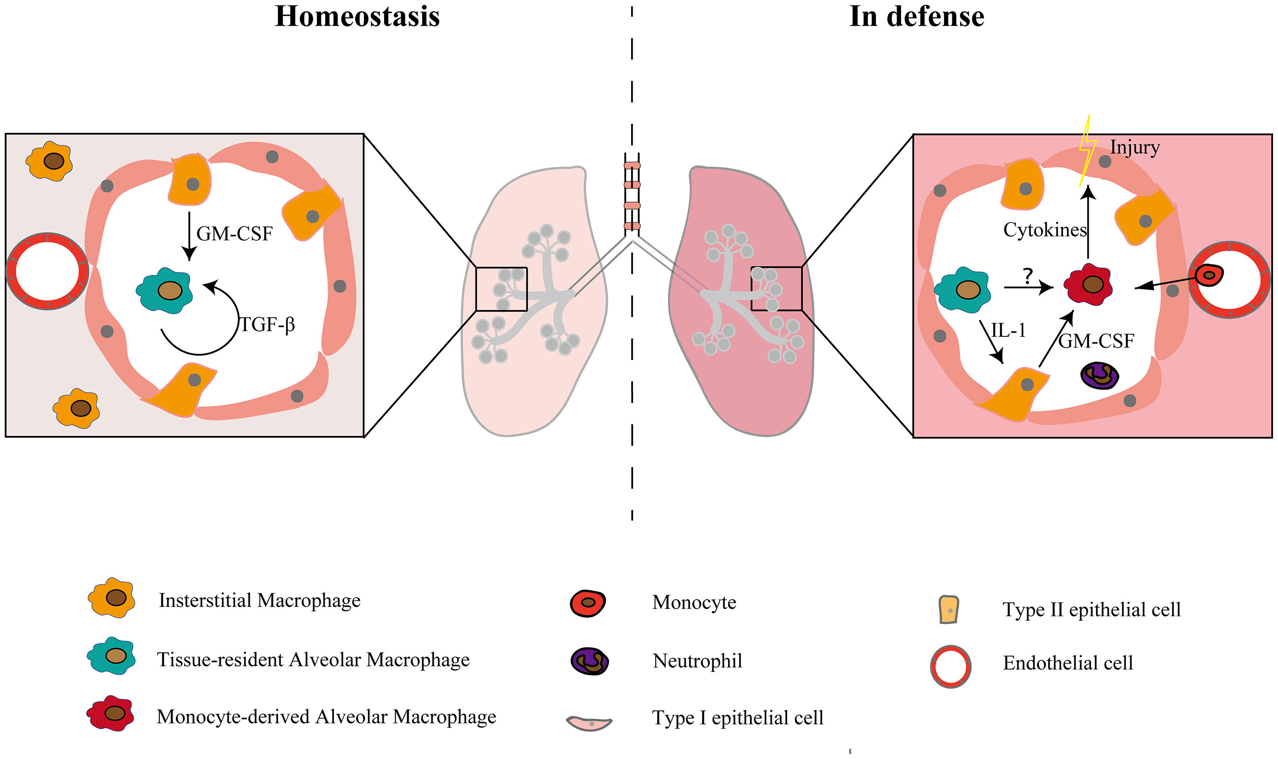
Frontiers | Diversity of Macrophages in Lung Homeostasis and ...
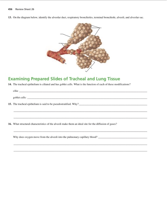
Solved 456 Review Sheet 26 13. On the diagram below, | Chegg.com
0 Response to "45 on the diagram below identify alveolar epithelium"
Post a Comment