41 cardiac muscle tissue diagram
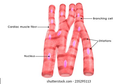
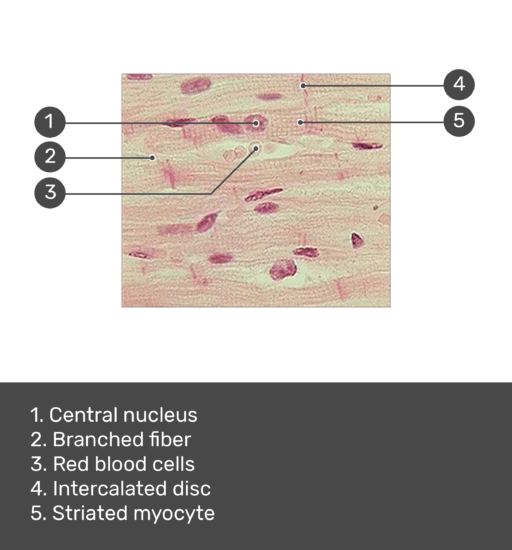
How To Draw Skeletal, Smooth and Cardiac Muscle Diagram - YouTube In this video I have shown the simplest way of drawing Muscle drawing. It is the pen diagram of Skeletal, Smooth and Cardiac Muscle for class 10, 11 and 12. ... Cardiac muscle tissue: function and labeled diagram | GetBodySmart The cardiac muscle cell or fiber. 1 2 They are relatively short, branched fibers that measure approximately 10 to 10 micrometers in diameter and 50 to 100 micrometers in length. The cardiac muscle tissue consists of short branched fibers. 1 2 Typically each cardiac myocyte contains a single nucleus, which is centrally positioned.
cardiac muscle tissue diagram muscle electron cardiac micrograph muscular transmission heart sarcomere intercalated disc structure cells longitudinal showing section which wellcome cell span throughout The Heart Diagrams Labeled And Unlabeled unlabeled labeled diagrams circulatory unlabelled JO IB SEHS YR I: 1B Muscular System mysciencesquad.weebly.com

Cardiac muscle tissue diagram
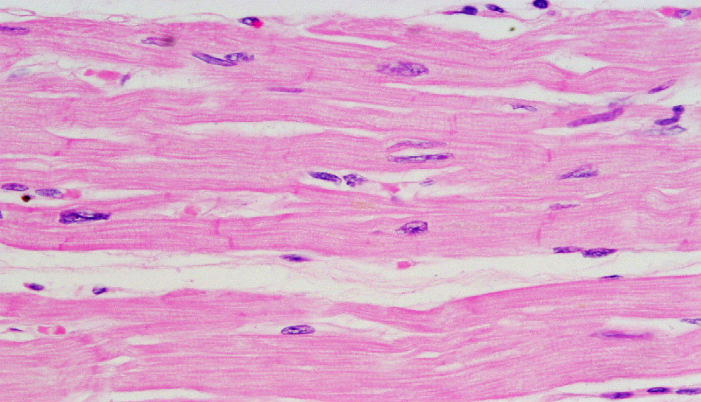
Cardiac Muscle Histology and Slide Identification Points Cardiac muscle histology I will try to show you these histological features from the real slide images and the diagram. #1. Longitudinal section of cardiac muscle #2. Cross section of cardiac muscle #3. Cardiac muscle fibers from both longitudinal and cross section #4. The nucleus of cardiac muscle fiber (single or binucleated) #5. Cardiac muscle tissue: Definition, function, and structure Cardiac muscle tissue, or myocardium, is a specialized type of muscle tissue that forms the heart. This muscle tissue, which contracts and releases involuntarily, is responsible for... Muscle Tissue of Animals: Origin and Functions (With Diagram) The cardiac muscles are found in the wall of the heart and in the wall of large veins (e.g., pulmonary veins and superior vena cava) where these veins enter the heart. Structure: These fibres show the characters of both un-striped and striped muscle fibres. Each fibre is a long and cylindrical structure which has a definite sarcolemma.
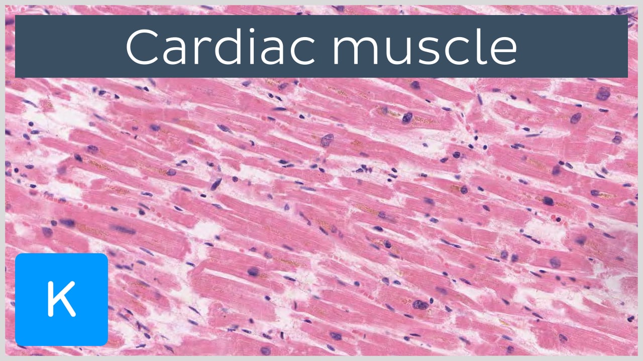
Cardiac muscle tissue diagram. Physiology, Cardiac Muscle - StatPearls - NCBI Bookshelf Cardiac muscle, also called the myocardium, is one of three major categories of muscles found within the human body, along with smooth muscle and skeletal muscle. Cardiac muscle, like skeletal muscle, is made up of sarcomeres that allow for contractility. However, unlike skeletal muscle, cardiac muscle is under involuntary control. cardiac muscle tissue Diagram | Quizlet the muscular tissue of the heart endocardium the thin, smooth membrane that lines the inside of the chambers of the heart and forms the surface of the valves. Composed of simple squamous and connective tissue. heart wall epicardium, myocardium, endocardium Recommended textbook solutions Fundamentals of Biochemistry: Life at the Molecular Level Cardiac Muscle - Definition, Function and Structure - Biology Dictionary Cardiac muscle, also known as heart muscle, is the layer of muscle tissue which lies between the endocardium and epicardium. These inner and outer layers of the heart, respectively, surround the cardiac muscle tissue and separate it from the blood and other organs. Cardiac muscle is made from sheets of cardiac muscle cells. Cardiac muscle tissue histology | Kenhub Cardiac muscle tissue, also known as myocardium, is a structurally and functionally unique subtype of muscle tissue located in the heart, that actually has characteristics from both skeletal and muscle tissues. It is capable of strong, continuous, and rhythmic contractions that are automatically generated.
Cardiac Muscle Tissue | Anatomy and Physiology I | | Course Hero Figure 1. Cardiac Muscle Tissue. Cardiac muscle tissue is only found in the heart. LM × 1600. (Micrograph provided by the Regents of University of Michigan Medical School © 2012) Cardiac muscle tissue is only found in the heart. Highly coordinated contractions of cardiac muscle pump blood into the vessels of the circulatory system. cardiac tissue diagram Muscle Tissue. 9 Images about Muscle Tissue : Physiology Glossary: Cardiac Muscle Cell | Draw It to Know It, Tissues Class 9 ppt and also Myology - Introduction (Skeletal, Cardiac, Smooth Muscles) - YouTube. Muscle Tissue histologydrawings.blogspot.com. cardiac histology skeletal hematoxylin eosin. Tissues Class 9 Ppt . tissues Cardiac Muscle Tissue Diagram | Quizlet Start studying Cardiac Muscle Tissue. Learn vocabulary, terms, and more with flashcards, games, and other study tools. Cardiac Muscle Tissue Diagram | Quizlet Start studying Cardiac Muscle Tissue. Learn vocabulary, terms, and more with flashcards, games, and other study tools. ... Among the differences with the other striated muscle, skeletal muscle, cardiac myocytes have nuclei wihich are _____ placed compared to skeletal muscle myocytes which have _____ nuclei placed on the _____ of the cell ...
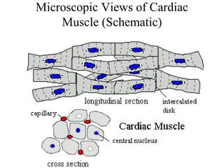
Muscle: The Histology Guide - University of Leeds Muscle. : Cardiac Muscle. Cardiac muscle is striated, like skeletal muscle, as the actin and myosin are arranged in sarcomeres, just as in skeletal muscle. However, cardiac muscle is involuntary. Cardiac muscle cells usually have a single (central) nucleus. The cells are often branched, and are tightly connected by specialised junctions. cardiac muscle tissue diagram muscle cardiac section tissue paradigm longitudinal. 3. Cardiac Muscle Tissue . sectional labeled flowchart. Jackie's 2015 A&P: Cardiac Muscle Cells jhan496.blogspot.com. cardiac muscle cells heart tissue jackie types. Muscle Tissue Chart | Biocam WP-13 prolabscientific.com. tissue biocam. Human Cardiac Muscle Cells Stock ... Cardiac Muscle Tissue | Interactive Anatomy Guide - Innerbody Cardiac muscle tissue is made up of many interlocking cardiac muscle cells, or fibers, that give the tissue its properties. Each cardiac muscle fiber contains a single nucleus and is striated, or striped, because it appears to have light and dark bands when seen through a microscope. Basic Properties of Cardiac Muscle (With Diagram) - Biology Hence, cardiac muscle can be divided into four groups: i. The smallest fibres with least glycogen at the nodes. ii. The broader fibres with more glycogen in the ventricles. iii. The still broader fibres with more glycogen in the atria. iv. The broadest fibres with abundant glycogen in the Purkinje fibres, bundle of His and its branches.
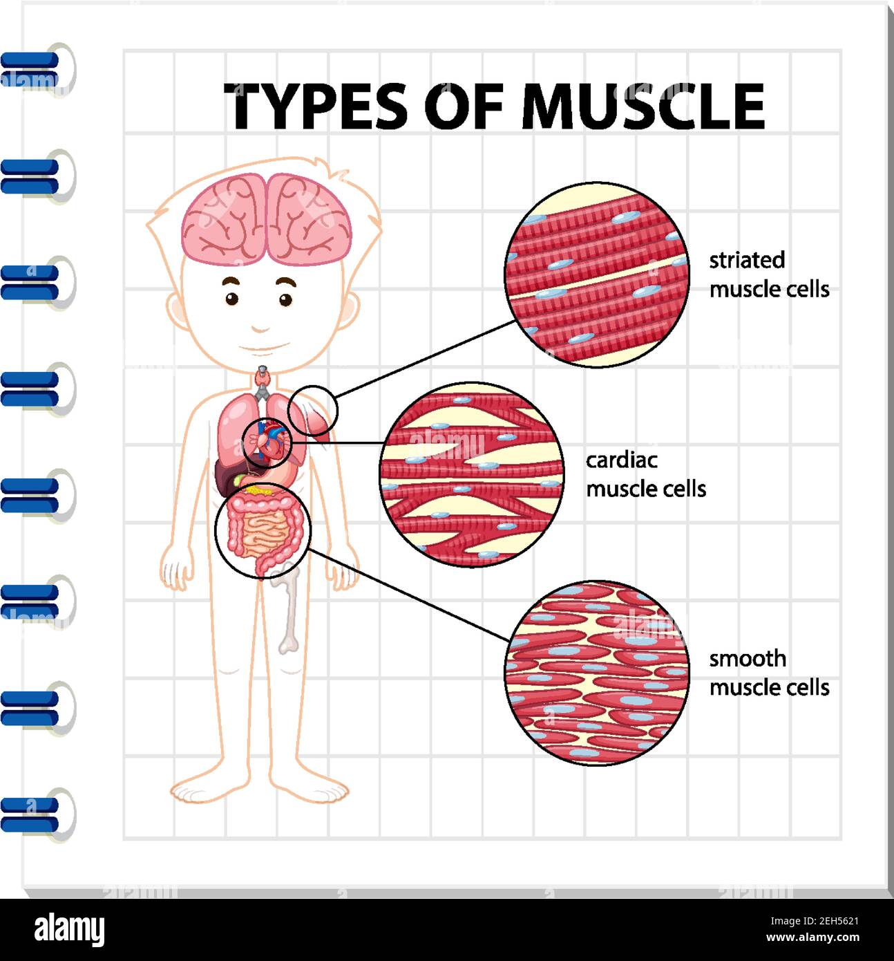
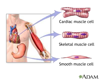
Premium Vector | Diagram showing types of muscle cells
Heart Muscle Diagram The diagram below represents the different phases of the cardiac cycle. Source: courses.lumenlearning.com. A coating of fluid separates the two layers of membrane letting the heart move as it beats. Source: . Cardiac muscle also known as heart muscle is the layer of muscle tissue which lies between the endocardium and epicardium.

Muscle Tissue Diagram | Quizlet
Cardiac Muscle Tissue Diagram | Quizlet Terms in this set (11) striations, intercalated disc, branched fibers and nucleus. Describe the features of cardiac muscle tissue. cardiac muscle tissue. Identify the tissue. cardiac muscle tissue. Identify the tissue. cardiac muscle tissue. Identify the tissue.

cardiac muscle | Definition, Function, & Structure | Britannica
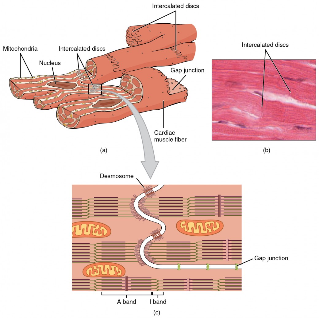
Cardiac Muscle Tissue - Anatomy & Physiology - University of Hawaiʻi Cardiac muscle fibers cells also are extensively branched and are connected to one another at their ends by intercalated discs. An intercalated disc allows the cardiac muscle cells to contract in a wave-like pattern so that the heart can work as a pump. Cardiac Muscle Tissue Cardiac muscle tissue is only found in the heart. LM × 1600.

619 Cardiac Muscle Tissue Photos and Premium High Res ...
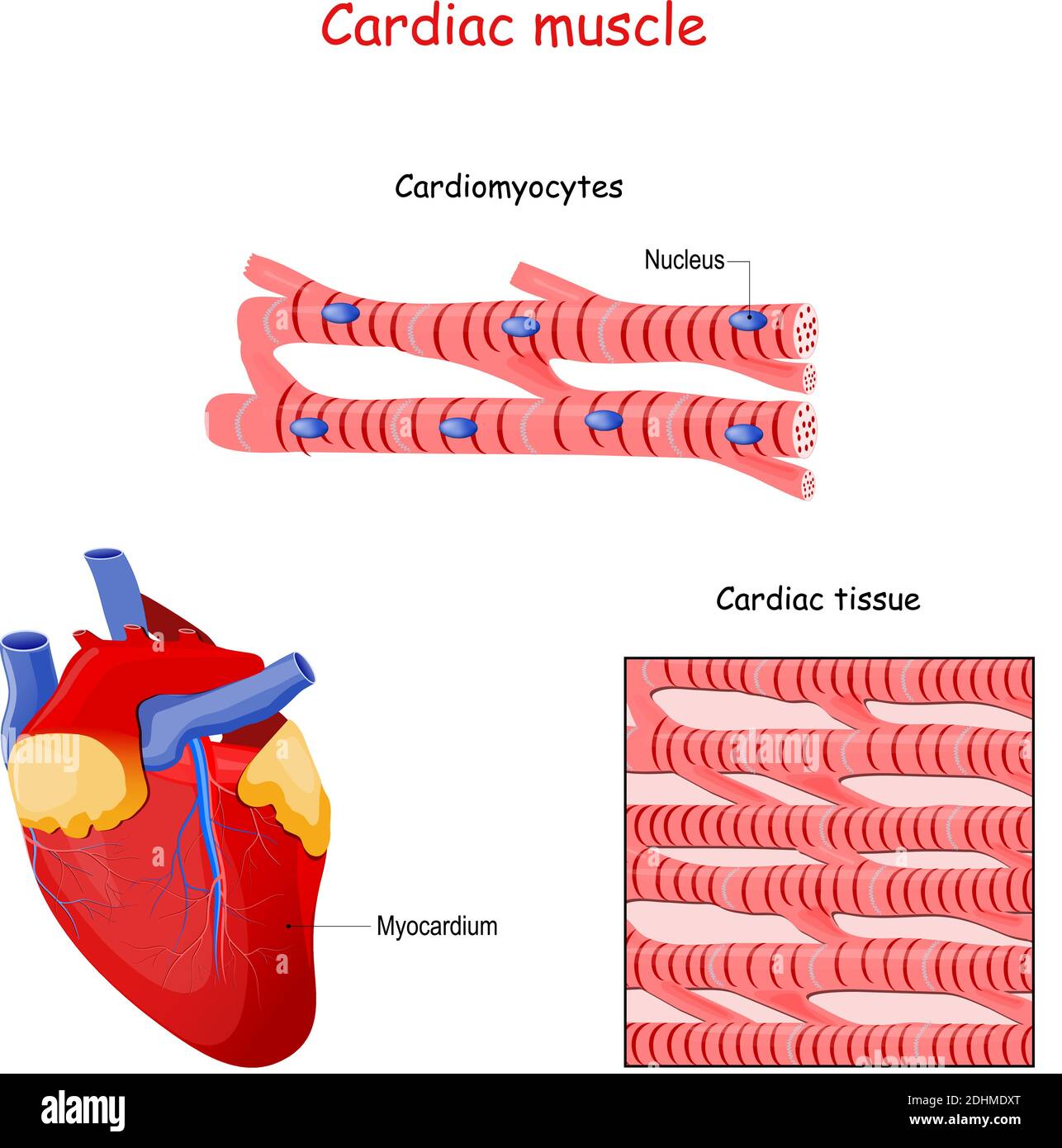
Cardiac Muscle Diagram - byjus.com The cardiac muscle or the myocardium forms the musculature of the heart. These are striated and involuntary muscles that are supplied by autonomic nerve fibres. They form the middle layer of the heart wall and are composed of cardiac muscle fibres. The other two layers are the pericardium (outer layer) and the endocardium (inner layer).

Cardiac Muscle Tissue Diagram | Quizlet
Human Heart under the Microscope Human Cardiac Muscle under Microscope. The human cardiac muscle (or heart muscle) is an involuntary, striated muscle found in the walls of the heart. There are 3 major types of muscle: Skeletal muscle (covering the skeleton and giving the body shape - these are the type of muscles most often thought of when the term "muscle" is used).

Cardiac Muscle Tissue Diagram | Quizlet
cardiac muscle tissue Diagram | Quizlet Start studying cardiac muscle tissue. Learn vocabulary, terms, and more with flashcards, games, and other study tools.
Muscle tissue with smooth, striated and cardiac examples outline diagram
cardiac muscle tissue diagram - Microsoft cardiac muscle tissue diagram Muscle tissue skeletal cell structure single connective which muscles corresponds chapter following striated unit smallest structural figure questions above characteristic. Microscopic muscle anatomy and muscle physiology week #10 flashcards.

loadBinary_012.gif
Structure of Cardiac Muscle - Myopathy - TeachMePhysiology Fig 2 - Diagram showing the overall structure of cardiac muscle and highlighting the position of gap junctions. Clinical Relevance - Cardiac Hypertrophy Hypertrophy refers to the thickening of muscle, in this case cardiac muscle, through an increase in muscle cell size. This occurs in response to pressure overload.

Muscular Tissue Quiz - Human Physiology Quiz | Muscle and ...
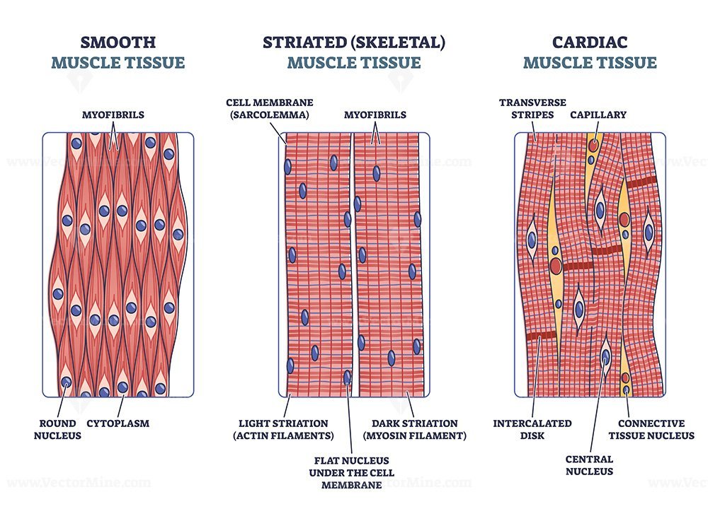
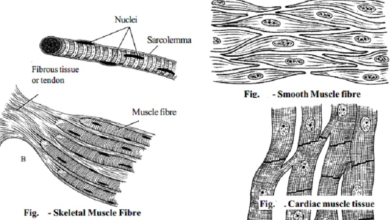
Muscular tissue: skeletal, smooth and cardiac muscle Types of muscle. skeletal muscle. smooth muscle. cardiac muscle. 1. Skeletal muscle: It acquires its name because most of the muscles involved are attached to skeleton, and make it move. Also known as Striated muscle -because it cell (fibre) are composed of alternating light and dark band (stripe). Also known as voluntary muscle.

Cardiac muscle under microscope | Image + structure
Cardiac Muscle Tissue: Function, Structure, Conditions, and Pictures Use this interactive 3-D diagram to explore the movement of cardiac muscle tissue. What are heart muscles made of? Intercalated discs Intercalated discs are small connections that join...
Muscle: The Histology Guide
Human Heart - Diagram and Anatomy of the Heart - Innerbody The myocardium is the muscular middle layer of the heart wall that contains the cardiac muscle tissue. Myocardium makes up the majority of the thickness and mass of the heart wall and is the part of the heart responsible for pumping blood. Below the myocardium is the thin endocardium layer. Endocardium. Endocardium is the simple squamous ...

Draw a labelled diagram of the cardiac muscles.
Muscle Tissue of Animals: Origin and Functions (With Diagram) The cardiac muscles are found in the wall of the heart and in the wall of large veins (e.g., pulmonary veins and superior vena cava) where these veins enter the heart. Structure: These fibres show the characters of both un-striped and striped muscle fibres. Each fibre is a long and cylindrical structure which has a definite sarcolemma.
15,086 Cardiac Muscle Images, Stock Photos & Vectors ...
Cardiac muscle tissue: Definition, function, and structure Cardiac muscle tissue, or myocardium, is a specialized type of muscle tissue that forms the heart. This muscle tissue, which contracts and releases involuntarily, is responsible for...

Cardiac Muscle Tissue Diagram | Quizlet
Cardiac Muscle Histology and Slide Identification Points Cardiac muscle histology I will try to show you these histological features from the real slide images and the diagram. #1. Longitudinal section of cardiac muscle #2. Cross section of cardiac muscle #3. Cardiac muscle fibers from both longitudinal and cross section #4. The nucleus of cardiac muscle fiber (single or binucleated) #5.

Cardiac Muscle Stock Illustrations – 4,047 Cardiac Muscle ...
Types of muscle cell diagram illustration Stock Vector Image ...

Cardiac Muscle Cell - Physiology Flashcards | Draw it to Know it
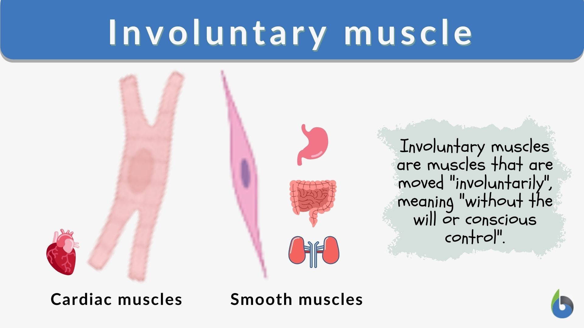
Involuntary muscle - Definition and Examples - Biology Online ...
Cardiac muscle tissue: function and labeled diagram ...

Cardiomyocytes (Cardiac Muscle Cells) - Structure, Function ...

muscle tissue. Skeletal muscle, smooth (in a gastrointestinal ...
Cardiac Muscle and Electrical Activity | Anatomy and ...
Cardiac muscle hi-res stock photography and images - Alamy

Write important functional differences between striated and ...

Cardiac muscle tissue: Definition, function, and structure

10): Cardiac muscle cells. (Reece J.B. ,Urry L.A., Cain M.L. ...
Advancements in Cardiac Tissue Engineering - Aurora Scientific

Muscular Tissue Diagram | Quizlet

Muscle Cell - Comparison in 2022 | Types of muscles, Muscle ...
Cardiac muscle

2.2.6 Muscles - Siyavula: Life Sciences Grade 10 - OpenStax CNX
Types of muscle tissue: MedlinePlus Medical Encyclopedia Image
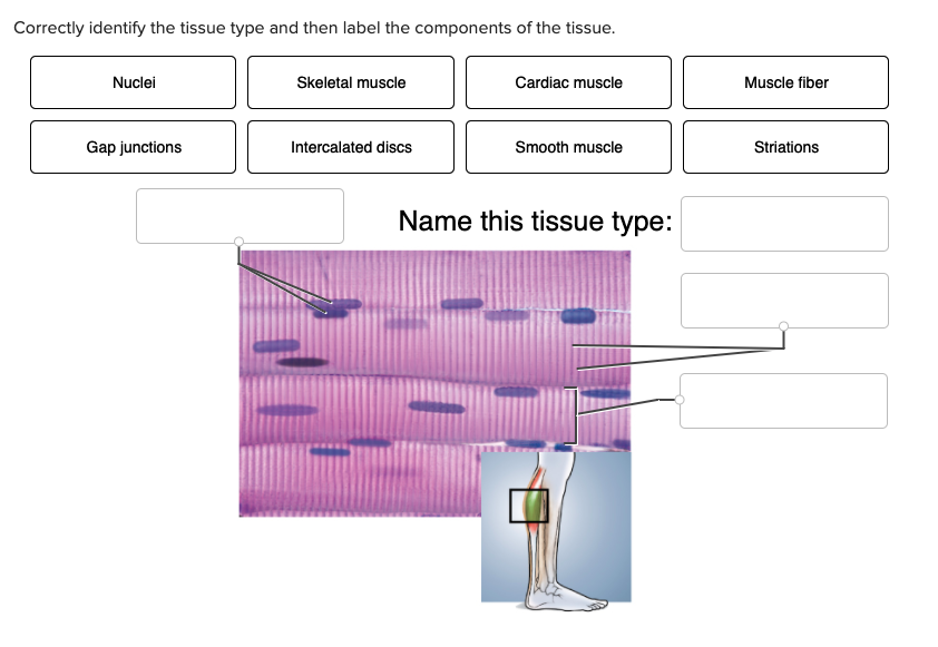
Solved Correctly identify the tissue type and then label the ...
Types of muscle tissues

3d Image Render Of Anatomy Of Cardiac Muscle Of Human For ...

Cardiac Muscle Diagram | Quizlet

Cardic Muscle - Anatomy and Physiology

Cardiac Muscle Tissue labeled Diagram | Quizlet

Premium Vector | Types of muscle cell diagram

Cardiac Muscle Tissue Diagram Diagram | Quizlet
3. cardiac muscle tissue
0 Response to "41 cardiac muscle tissue diagram"
Post a Comment